. 2011 Sep 18;478(7368):260-3.
doi: 10.1038/nature10430.

Mechanical strain in actin networks regulates FilGAP and integrin binding to filamin A

Affiliations

Mechanical strain in actin networks regulates FilGAP and integrin binding to filamin A

A J Ehrlicher et al. Nature. .

Abstract

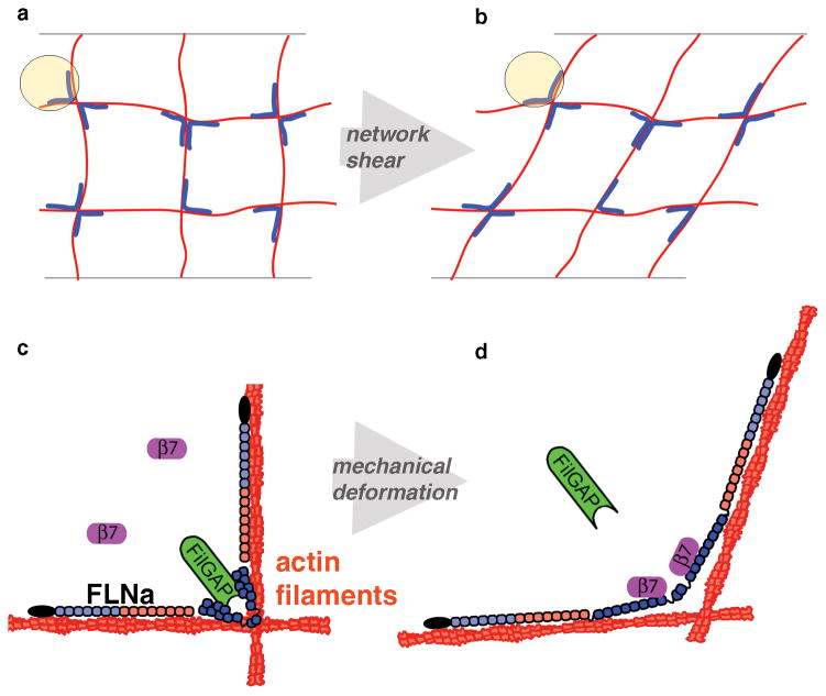
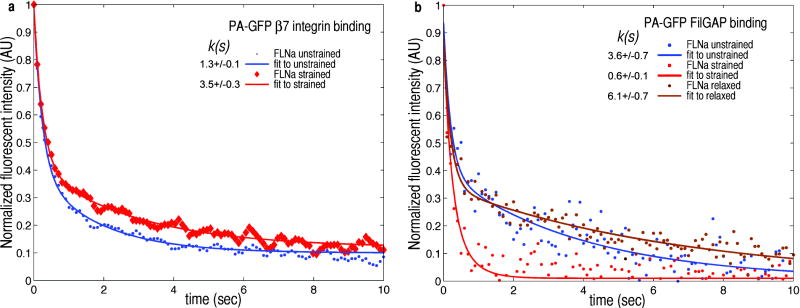
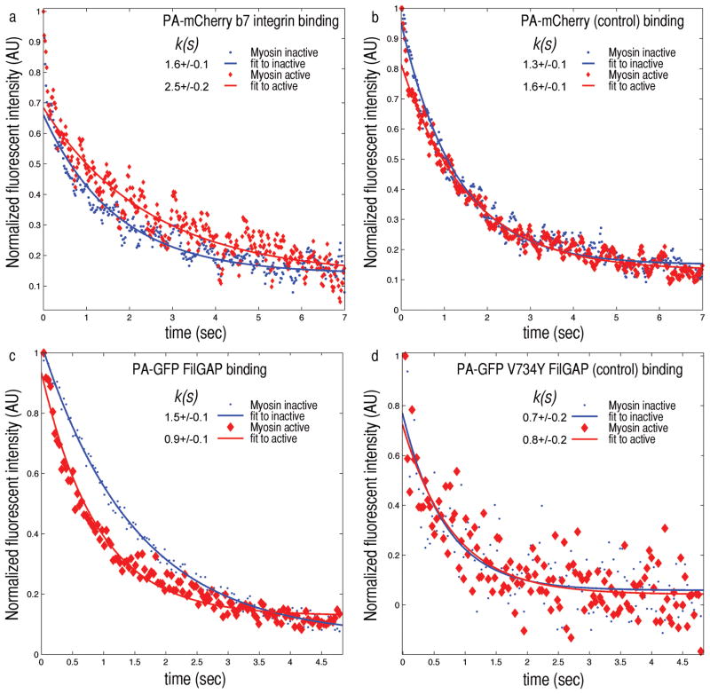
Mechanical stresses elicit cellular reactions mediated by chemical signals. Defective responses to forces underlie human medical disorders such as cardiac failure and pulmonary injury. The actin cytoskeleton's connectivity enables it to transmit forces rapidly over large distances, implicating it in these physiological and pathological responses. Despite detailed knowledge of the cytoskeletal structure, the specific molecular switches that convert mechanical stimuli into chemical signals have remained elusive. Here we identify the actin-binding protein filamin A (FLNA) as a central mechanotransduction element of the cytoskeleton. We reconstituted a minimal system consisting of actin filaments, FLNA and two FLNA-binding partners: the cytoplasmic tail of β-integrin, and FilGAP. Integrins form an essential mechanical linkage between extracellular and intracellular environments, with β-integrin tails connecting to the actin cytoskeleton by binding directly to filamin. FilGAP is an FLNA-binding GTPase-activating protein specific for RAC, which in vivo regulates cell spreading and bleb formation. Using fluorescence loss after photoconversion, a novel, high-speed alternative to fluorescence recovery after photobleaching, we demonstrate that both externally imposed bulk shear and myosin-II-driven forces differentially regulate the binding of these partners to FLNA. Consistent with structural predictions, strain increases β-integrin binding to FLNA, whereas it causes FilGAP to dissociate from FLNA, providing a direct and specific molecular basis for cellular mechanotransduction. These results identify a molecular mechanotransduction element within the actin cytoskeleton, revealing that mechanical strain of key proteins regulates the binding of signalling molecules.


Conflict of interest statement

The authors declare no competing financial interests.

Figures

Figure 1
Figure 1. Differential mechanotransduction in FLNa occurs through spatial separation of binding sites and opening cryptic sites
a) A Filamin (blue) crosslinked actin (red) gel forms an orthogonal network. b) When this network is strained, crosslinks are deformed. c) The actin-binding domain of FLNa is shown in black, followed by repeats 1–7 (light blue) and 8–15 (red), which form the linear rod 1 region. Repeats 16–23 (dark blue) form the compact rod 2 region. FilGAP (green) binds repeats 23 and the cytoplasmic domain of β7 integrin (purple) is unbound. d) When FLNa is mechanically deformed, the cryptic integrin site on repeat 21 is exposed allowing β7 integrin to bind, while repeats 23 are spatially separated, preventing FilGAP from binding both.
Figure 2
Figure 2. External bulk shear on F-actin-FLNa networks alters FLNa’s binding affinity for β7 integrin and FilGAP
a) Fluorescence intensity in time of PA-GFP β7 integrin after photoactivation. When unstrained (blue) fluorescence of β7 integrin decays with a characteristic time constant k(s) of 1.3 seconds. Following the application of γ=0.28 shear strain, the time constant increases to 3.5 seconds, as the integrin dissociates more slowly from FLNa (n=18). b), Fluorescence intensity in time of PA-GFP FilGAP after photoactivation. Unstrained (blue) FilGAP’s fluorescence decay time k is 3.6 s. A 4% strain (red) decreased k to 0.6 s from its unstrained decay of 3.6 s. This behavior is reversible, and after allowing the network to relax strain for 10 minutes, k increases to 6.1 s (brown) (n=10).
Figure 3
Figure 3. Myosin II forces applied to F-actin-FLNa networks changes FLNa’s binding affinity to β7 integrin and FilGAP
a) When depleted of ATP, myosin is in a rigor state. The FLNa within the network is not stressed and PA-GFP β7 integrin fluorescence decays with a characteristic time constant k(s) of 1.6 seconds (blue). After caged ATP is released myosin reactivates, straining FLNa crosslinks. The decay time constant increases to 2.5 seconds as the integrin dissociates more slowly from FLNa under stress. b) PA-mCherry alone as a control shows no significant difference in the unstrained or strained state. c) Fluorescence intensity in time of PA-GFP FilGAP after photoactivation. In the ATP depleted state FilGAP’s fluorescence decay time k is 1.5 s, and after releasing the caged ATP (red) k decreases to 0.9 s. PA-GFP V734Y FilGAP, a non-FLNa binding mutant as a control, shows no significant difference in the decay times of unstrained (0.7 s) or strained state (0.8 s) (n=20).

References

    1. Ingber DE. Mechanobiology and diseases of mechanotransduction. Ann Med. 2003;35:564–577. - PubMed
    1. Discher DE, Janmey P, Wang YL. Tissue cells feel and respond to the stiffness of their substrate. Science (New York, N Y. 2005;310:1139–1143. - PubMed
    1. Moore SW, Roca-Cusachs P, Sheetz MP. Stretchy proteins on stretchy substrates: the important elements of integrin-mediated rigidity sensing. Developmental cell. 2010;19:194–206. doi: 10.1016/j.devcel.2010.07.018. S1534-5807(10)00346-1 [pii] - DOI - PMC - PubMed
    1. Schwartz MA. Integrins and extracellular matrix in mechanotransduction. Cold Spring Harb Perspect Biol. 2010;2:a005066. doi: 10.1101/cshperspect.a005066. cshperspect.a005066 [pii] - DOI - PMC - PubMed
    1. Kruger M, Linke WA. Titin-based mechanical signalling in normal and failing myocardium. J Mol Cell Cardiol. 2009;46:490–498. S0022-2828(09)00021-2 [pii] - PubMed

Publication types