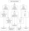
Differentiation between benign and malignant solid thyroid nodules using an US classification system
- PMID: 21927557
- PMCID: PMC3168797
- DOI: 10.3348/kjr.2011.12.5.559
Differentiation between benign and malignant solid thyroid nodules using an US classification system
Abstract
Objective: To evaluate the diagnostic accuracy of a new ultrasound (US) classification system for differentiating between benign and malignant solid thyroid nodules.
Materials and methods: In this study, we enrolled 191 consecutive patients who received real-time US and subsequent US diagnoses for solid thyroid nodules, and underwent US-guided fine-needle aspiration. Each thyroid nodule was prospectively classified into 1 of 5 diagnostic categories by real-time US: "malignant," "suspicious for malignancy," "borderline," "probably benign," and "benign". We evaluated the diagnostic accuracy of thyroid US and the cut-off US criteria by comparing the US diagnoses of thyroid nodules with cytopathologic results.
Results: Of the 191 solid nodules, 103 were subjected to thyroid surgery. US categories for these 191 nodules were malignant (n = 52), suspicious for malignancy (n = 16), borderline (n = 23), probably benign (n = 18), and benign (n = 82). A receiver-operating characteristic curve analysis revealed that the US diagnosis for solid thyroid nodules using the 5-category US classification system was very good. The sensitivity, specificity, positive and negative predictive values, and accuracy of US diagnosis were 86%, 95%, 91%, 92%, and 92%, respectively, when benign, probably benign, and borderline categories were collectively classified as benign (negative).
Conclusion: The diagnostic accuracy of thyroid US for solid thyroid nodules is high when the above-mentioned US classification system is applied.
Keywords: Classification; Fine-needle aspiration; Malignancy; Solid, Ultrasound; Thyroid nodule.
Figures


References
-
- Kim EK, Park CS, Chung WY, Oh KK, Kim DI, Lee JT, et al. New sonographic criteria for recommending fine-needle aspiration biopsy of nonpalpable solid nodules of the thyroid. AJR Am J Roentgenol. 2002;178:687–691. - PubMed
-
- Cappelli C, Castellano M, Pirola I, Cumetti D, Agosti B, Gandossi E, et al. The predictive value of ultrasound findings in the management of thyroid nodules. QJM. 2007;100:29–35. - PubMed
-
- Salmaslioglu A, Erbil Y, Dural C, Issever H, Kapran Y, Ozarmagan S, et al. Predictive value of sonographic features in preoperative evaluation of malignant thyroid nodules in a multinodular goiter. World J Surg. 2008;32:1948–1954. - PubMed
-
- Horvath E, Majlis S, Rossi R, Franco C, Niedmann JP, Castro A, et al. An ultrasonogram reporting system for thyroid nodules stratifying cancer risk for clinical management. J Clin Endocrinol Metab. 2009;94:1748–1751. - PubMed
-
- Papini E, Guglielmi R, Bianchini A, Crescenzi A, Taccogna S, Nardi F, et al. Risk of malignancy in nonpalpable thyroid nodules: predictive value of ultrasound and color-Doppler features. J Clin Endocrinol Metab. 2002;87:1941–1946. - PubMed
Publication types
MeSH terms
LinkOut - more resources
Full Text Sources
Medical

