Review
. 2011 Aug;3(8):1501-31.
doi: 10.3390/v3081501.

Intracellular events and cell fate in filovirus infection

Affiliations
Review

Intracellular events and cell fate in filovirus infection

Judith Olejnik et al. Viruses. 2011 Aug.

Abstract

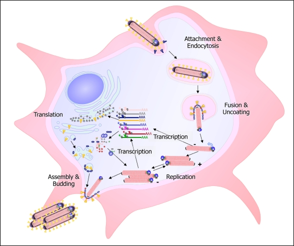
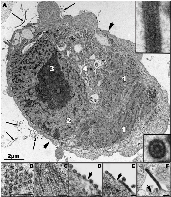
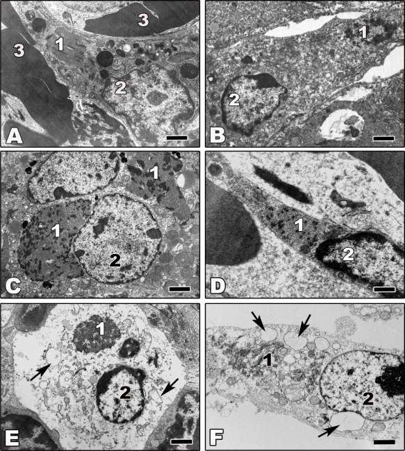
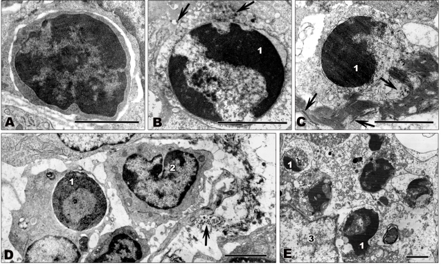
Marburg and Ebola viruses cause a severe hemorrhagic disease in humans with high fatality rates. Early target cells of filoviruses are monocytes, macrophages, and dendritic cells. The infection spreads to the liver, spleen and later other organs by blood and lymph flow. A hallmark of filovirus infection is the depletion of non-infected lymphocytes; however, the molecular mechanisms leading to the observed bystander lymphocyte apoptosis are poorly understood. Also, there is limited knowledge about the fate of infected cells in filovirus disease. In this review we will explore what is known about the intracellular events leading to virus amplification and cell damage in filovirus infection. Furthermore, we will discuss how cellular dysfunction and cell death may correlate with disease pathogenesis.

Keywords: Ebola Virus; Marburg Virus; animal models; bystander apoptosis; cell death; filoviruses; target cells; ultrastructural analysis; viral replication cycle; virus-cell interaction.


Figures

Figure 1.
Figure 1.
Scheme of the filovirus infection cycle. Filoviruses enter the cell by receptor-mediated endocytosis or macropinocytosis. After fusion of the viral and cellular membrane, the nucleocapsid is released into the cytoplasm and serves as a template for transcription and replication. The replicated RNA is encapsidated by the nucleocapsid proteins. The newly synthesized nucleocapsids are transported to the sites of viral release, where budding takes place.
Figure 2.
Figure 2.
Morphological characteristics of Zaire ebolavirus (ZEBOV) replication. (A) Ultrathin section of a ZEBOV-infected Vero cell containing large viral inclusions. The inclusions are composed of granular material (1; also shown in (C)) and rod-like nucleocapsids. Released virions are indicated by long arrows and are shown in the inserts. (B) Cross section of viral inclusion containing nucleocapsids. (C) Longitudinal section of viral inclusion containing nucleocapsids. (D–F) Budding of viral particles. In the initial step of budding a particle can be positioned parallel (D and E, cross and longitudinal sections), or perpendicular (F) to the membrane and then subsequently is enveloped. Short arrows indicate the cellular plasma membrane. 2—nucleus, 3—nucleolus, 4—Golgi zone. Bars in Figures 2B–F correspond to 250 nm. Thick part of frame around cross-sectioned virion corresponds to 120 nm, and thick part of frame around longitudinal section corresponds to 160 nm. Transmission electron microscopy. Cells were fixed at 16 h p.i.
Figure 3.
Figure 3.
Transmission electron microscopy of ultrathin sections of tissues from animals experimentally infected with filoviruses. Tissues were fixed at day 5 or 6 p.i. (A) Ultrathin section of liver tissue from ZEBOV-infected rhesus monkey showing an infected macrophage. (B,C) Ultrathin sections of liver tissue from Marburg virus (MARV)-infected guinea pig. Shown are an infected macrophage (B) and an infected hepatocyte (C). (D) Ultrathin section of spleen tissue from ZEBOV-infected African green monkey showing an infected endothelial cell. (E) Ultrathin section of lymphatic node tissue from ZEBOV-infected African green monkey showing a necrotic infected macrophage. (F) Ultrathin section of a necrotic ZEBOV-infected Vero cell showing an infected macrophage. Arrows show vacuolization of endoplasmic reticulum cisterns in cells undergoing non-apoptotic cell death. 1—filovirus inclusions; 2—nucleus; 3—erythrocyte. Bars correspond to 2 μm.
Figure 4
Figure 4
Transmission electron microscopy of ultrathin sections of lymph node tissue from filovirus-infected African green monkeys infected with ZEBOV (A,C–E) or MARV (B). Tissues were fixed at 4 days p.i. (A) Small lymphocyte showing normal nuclear morphology with large areas of heterochromatin. (B,C) Apoptotic lymphocytes. Typical signs of apoptosis such as chromatin condensation and marginal location of chromatin are visible. (D,E) Apoptotic lymphocytes being engulfed by macrophages. (D) shows initial stages of phagocytosis. The lymphocyte is engulfed by a macrophage. The macrophage shown in (E) contains several destroyed apoptotic lymphocytes. Arrows show filoviral particles. 1—highly condensed heterochromatin; 2—monocyte showing normal nuclear morphology; 3—nucleus of macrophage. Bars correspond to 2 μm.

References

    1. Kuhn JH. Filoviruses. A compendium of 40 years of epidemiological, clinical, and laboratory studies. Arch Virol Suppl. 2008;20:13–360. - PubMed
    1. Mohamadzadeh M, Chen L, Schmaljohn AL. How Ebola and Marburg viruses battle the immune system. Nat Rev Immunol. 2007;7:556–567. - PubMed
    1. Bradfute SB, Swanson PE, Smith MA, Watanabe E, McDunn JE, Hotchkiss RS, Bavari S. Mechanisms and consequences of ebolavirus-induced lymphocyte apoptosis. J Immunol. 2010;184:327–335. - PubMed
    1. Ksiazek TG, West CP, Rollin PE, Jahrling PB, Peters CJ. ELISA for the detection of antibodies to Ebola viruses. J Infect Dis. 1999;179:S192–S198. - PubMed
    1. Onyango CO, Opoka ML, Ksiazek TG, Formenty P, Ahmed A, Tukei PM, Sang RC, Ofula VO, Konongoi SL, Coldren RL, et al. Laboratory diagnosis of Ebola hemorrhagic fever during an outbreak in Yambio, Sudan, 2004. J Infect Dis. 2007;196:S193–S198. - PubMed

Publication types

MeSH terms

LinkOut - more resources