Review
. 2012 Mar 15;16(6):496-505.
doi: 10.1089/ars.2011.4281. Epub 2011 Dec 22.

Redox-based regulation of apoptosis: S-glutathionylation as a regulatory mechanism to control cell death

Affiliations
Review

Redox-based regulation of apoptosis: S-glutathionylation as a regulatory mechanism to control cell death

Vikas Anathy et al. Antioxid Redox Signal. .

Abstract

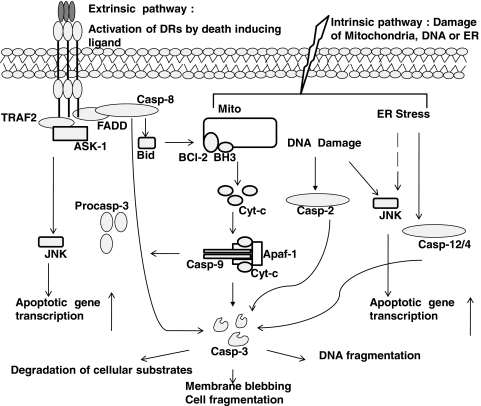
Significance: Redox-based signaling governs a number of important pathways in tissue homeostasis. Consequently, deregulation of redox-controlled processes has been linked to a number of human diseases. Among the biological processes regulated by redox signaling, apoptosis or programmed cell death is a highly conserved process important for tissue homeostasis. Apoptosis can be triggered by a wide variety of stimuli, including death receptor ligands, environmental agents, and cytotoxic drugs. Apoptosis has also been implicated in the etiology of many human diseases.

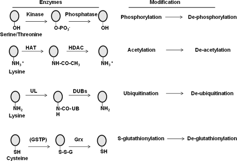
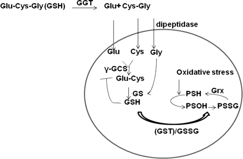
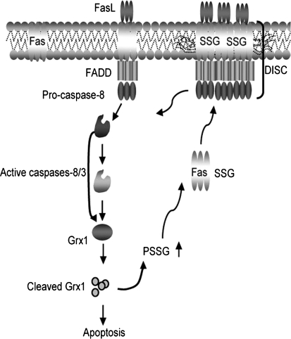
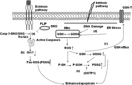
Recent advances: Recent discoveries demonstrate that redox-based changes are required for efficient activation of apoptosis. Among these redox changes, alterations in the abundant thiol, glutathione (GSH), and the oxidative post-translational modification, protein S-glutathionylation (PSSG) have come to the forefront as critical regulators of apoptosis.

Critical issues: Although redox-based changes have been documented in apoptosis and disease pathogenesis, the mechanistic details, whereby redox perturbations intersect with pathogenic processes, remain obscure.

Future directions: Further research will be needed to understand the context in which of the members of the death receptor pathways undergo ligand dependent oxidative modifications. Additional investigation into the interplay between oxidative modifications, redox enzymes, and apoptosis pathway members are also critically needed to improve our understanding how redox-based control is achieved. Such analyses will be important in understanding the diverse chronic diseases. In this review we will discuss the emerging paradigms in our current understanding of redox-based regulation of apoptosis with an emphasis on S-glutathionylation of proteins and the enzymes involved in this important post-translational modification.


Figures

FIG. 1.
FIG. 1.
Overview of apoptotic signaling pathways. Both extrinsic and intrinsic pathways converge on caspase-3 and JNK to induce cell death.
FIG. 2.
FIG. 2.
Overview of select post-translational modifications of proteins. Circles denote target amino acids, which include serine, threonine, lysine, and cysteine. -OH groups of serine or threonine are phosphorylated by kinases and dephosphorylated by phosphatases. Side chain amino groups of lysines can be acetylated-deacetylated by acetylases (HAT) and deacetylases (HDAC), respectively, or ubiquitinated and deubiquitinated by ubiquitin ligases (UL)-deubiquitinating (DUB) enzymes, respectively. Glutathione S tranferase pi (GSTP) catalyzes S-glutathionylation (SSG) of reactive site cysteines, whereas glutaredoxin (Grx) under physiological conditions catalyzes de-glutathionylation of proteins. Because considerable uncertainty remains at this time about the overall role of GSTP in catalysis of S-glutathionylation reactions, GSTP appears between parentheses.
FIG. 3.
FIG. 3.
Glutathione metabolism in the cell. The constituent amino acids of GSH are imported into the cell and are reassembled by γ-glutamyl cysteine synthase (γ-GCS) and GSH synthetase (GS). GSTP1 can utilize GSH and conjugate it to nucleophilic cysteines (-SH) to form PSSG. Note that this step requires an oxidation intermediate, possibly a sulfenic acid (-SOH) formed as a result of ROS formation. Increasing concentration of GSSG due to oxidative stress can also increase PSSG. Majority of PSSG in the cell is reduced by Grx.
FIG. 4.
FIG. 4.
Feed forward amplification of apoptosis via S-glutathionylation of Fas. In response to Fas ligation, activated caspases-8 and/or-3 cause degradation of Grx1, thereby increasing S-glutathionylation of Fas. This promotes binding of FasL and enhances accumulation of Fas and formation of DISC in the lipid rafts, thereby amplifying caspase activity and apoptosis. “© Rockefeller University Press, 2009”, originally published in J Cell Biol doi:10.1083/jcb.200807019
FIG. 5.
FIG. 5.
Control of apoptosis by alterations of PSSG and depletion of GSH. Death receptor stimulation can result in (a) de-glutathionylation or de-nitrosylation of caspase-3 and FLIP has been linked to induction of apoptosis; (b) degradation Grx in order to facilitate accumulation of PSSG, including S-glutathionylation of Fas; (c) depletion GSH can occur through export via GSH transporters; or (d) activation of intrinsic apoptotic pathways, which increase reactive oxygen species (ROS) in the cytosol; (e) increased ROS in the cytosol can form sulfenic acid (-SOH) intermediates that can be S-glutathionylated by GSTP.
FIG. 6.
FIG. 6.
Schematic demonstrating that Fas-induced apoptosis of epithelial cells can lead to fibrosis. Myofibroblast-dependent killing of Fas bearing epithelial cells may contribute to impaired re-epithelialization, resulting in an aberrant repair response that culminates in fibrosis.

References

    1. Adachi T. Pimentel DR. Heibeck T. Hou X. Lee YJ. Jiang B. Ido Y. Cohen RA. S-glutathiolation of Ras mediates redox-sensitive signaling by angiotensin II in vascular smooth muscle cells. J Biol Chem. 2004;279:29857–29862. - PubMed
    1. Adachi T. Weisbrod RM. Pimentel DR. Ying J. Sharov VS. Schoneich C. Cohen RA. S-Glutathiolation by peroxynitrite activates SERCA during arterial relaxation by nitric oxide. Nat Med. 2004;10:1200–1207. - PubMed
    1. Adler V. Yin Z. Fuchs SY. Benezra M. Rosario L. Tew KD. Pincus MR. Sardana M. Henderson CJ. Wolf CR. Davis RJ. Ronai Z. Regulation of JNK signaling by GSTp. EMBO J. 1999;18:1321–1334. - PMC - PubMed
    1. Altschmied J. Haendeler J. Thioredoxin-1 and endothelial cell aging: Role in cardiovascular diseases. Antioxid Redox Signal. 2009;11:1733–1740. - PubMed
    1. Anathy V. Aesif SW. Guala AS. Havermans M. Reynaert NL. Ho YS. Budd RC. Janssen-Heininger YM. Redox amplification of apoptosis by caspase-dependent cleavage of glutaredoxin 1 and S-glutathionylation of Fas. J Cell Biol. 2009;184:241–252. - PMC - PubMed

Publication types

LinkOut - more resources