Review
. 2011 Sep;34(6):870-83.
doi: 10.1111/j.1460-9568.2011.07802.x.

Adult neurogenesis in the decapod crustacean brain: a hematopoietic connection?

Affiliations
Review

Adult neurogenesis in the decapod crustacean brain: a hematopoietic connection?

Barbara S Beltz et al. Eur J Neurosci. 2011 Sep.

Abstract

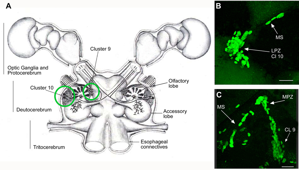
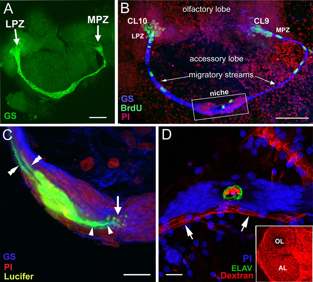
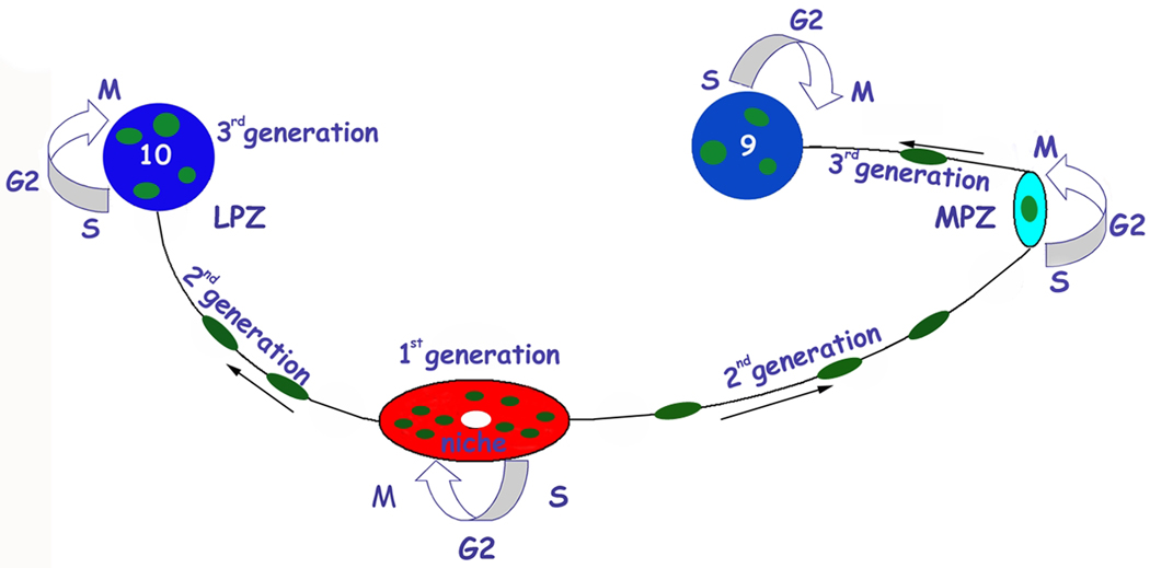
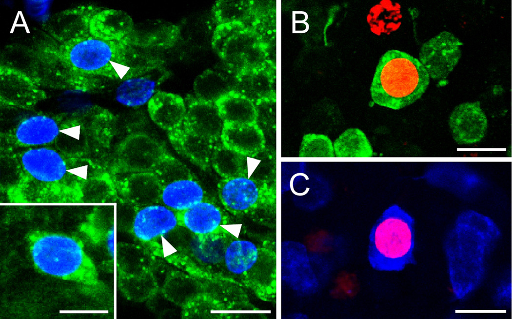
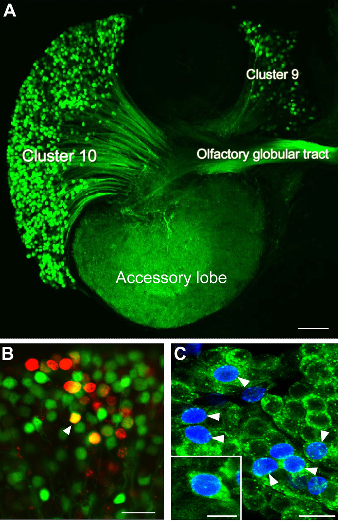
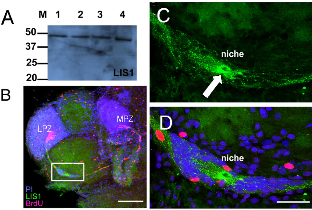
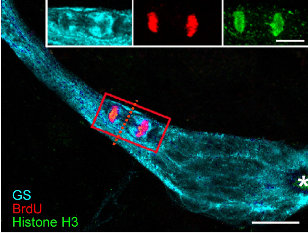
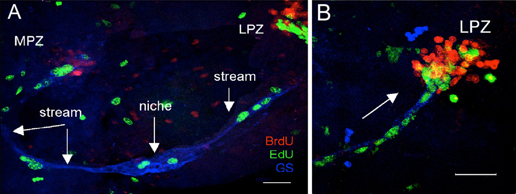
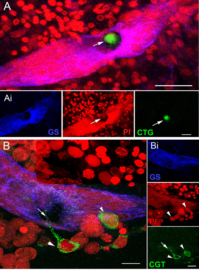
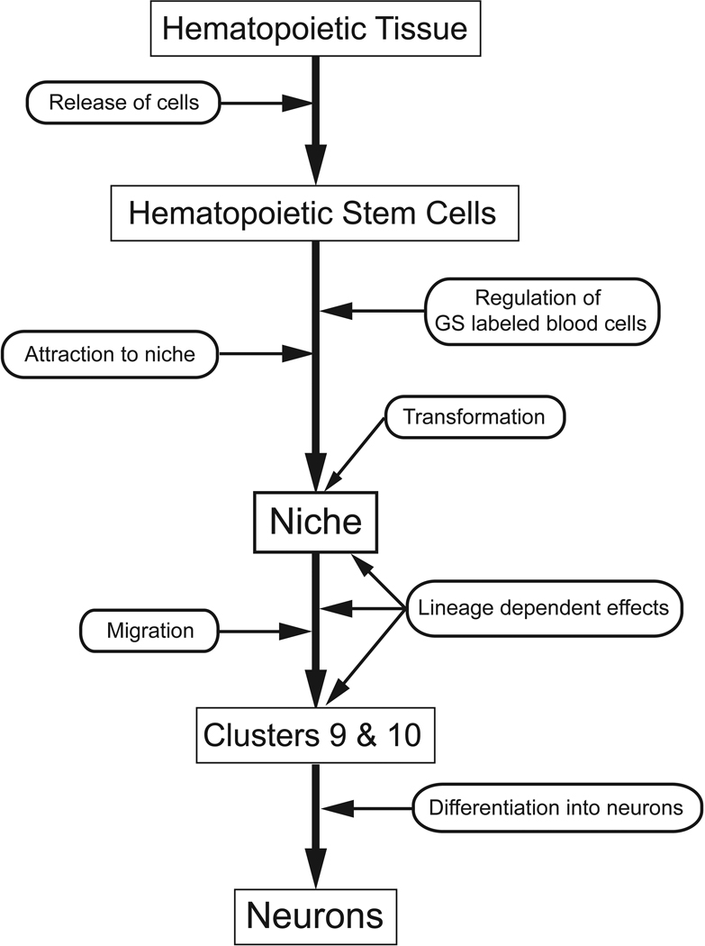
New neurons are produced and integrated into circuits in the adult brains of many organisms, including crustaceans. In some crustacean species, the first-generation neuronal precursors reside in a niche exhibiting characteristics analogous to mammalian neurogenic niches. However, unlike mammalian niches where several generations of neuronal precursors co-exist, the lineage of precursor cells in crayfish is spatially separated allowing the influence of environmental and endogenous regulators on specific generations in the neuronal precursor lineage to be defined. Experiments also demonstrate that the first-generation neuronal precursors in the crayfish Procambarus clarkii are not self-renewing. A source external to the neurogenic niche must therefore provide cells that replenish the first-generation precursor pool, because although these cells divide and produce a continuous efflux of second-generation cells from the niche, the population of first-generation niche precursors is not diminished with growth and aging. In vitro studies show that cells extracted from the hemolymph, but not other tissues, are attracted to and incorporated into the neurogenic niche, a phenomenon that appears to involve serotonergic mechanisms. We propose that, in crayfish, the hematopoietic system may be a source of cells that replenish the niche cell pool. These and other studies reviewed here establish decapod crustaceans as model systems in which the processes underlying adult neurogenesis, such as stem cell origins and transformation, can be readily explored. Studies in diverse species where adult neurogenesis occurs will result in a broader understanding of fundamental mechanisms and how evolutionary processes may have shaped the vertebrate/mammalian condition.


Figures

Figure 1
Figure 1
A. Diagram of the eureptantian (crayfish/lobster) brain including the optic ganglia, and showing the locations of the proto-, trito- and deutocerebral neuropils. The soma clusters 9 and 10 (circled), locations of neurogenesis in the adult brain, flank two prominent neuropil regions of the deutocerebrum, the olfactory (OL) and accessory (AL) lobes. (Names of brain areas are according to Sandeman et al., 1992). B. Bromodeoxyuridine (BrdU)-labeled nuclei in the migratory stream (MS), the lateral proliferation zone (LPZ) and the adjacent cluster 10 (CL10). C. BrdU-labeled cells in the migratory stream, the medial proliferation zone (MPZ) and cluster 9 (CL9) which is separated from the MPZ by a stream of migrating cells. Scale bars: B, C, 20µm. (Figure from Sullivan et al., 2007b).
Figure 2
Figure 2
The proliferative system maintaining adult neurogenesis in the crayfish (Procambarus clarkii) brainA. Both the LPZ and MPZ are contacted by the processes of a population of glial cells immunoreactive to glutamine synthetase (green). The somata of these cells form a cluster, the neurogenic niche, on the ventral surface of the brain. B. Left side of the brain of P. clarkii labeled immunocytochemically for the S-phase marker BrdU (green). Labeled cells are found in the lateral proliferation zone (LPZ) contiguous with cluster 10 and in the medial proliferation zone (MPZ) near cluster 9. The two zones are linked by a chain of labeled cells in a migratory stream that originates in the oval region labeled “niche”. Labeling for Drosophila synapsin (blue) and propidium iodide (red) is shown. C. Niche cells (green), labeled by intracellular injection of Lucifer yellow, have short processes (arrowheads) projecting to the vascular cavity (arrow) and longer fibers (double arrowheads) that fasciculate together to form the tracts projecting to the LPZ and MPZ. (blue: glutamine synthetase; red: propidium iodide). D. The vascular connection of the cavity in the centre of the glial soma cluster was demonstrated by injecting a dextran dye into the cerebral artery. The cavity, outlined in green by its reactivity to an antibody to Elav, contains the dextran dye (red) which is also contained within a larger blood vessel that runs along beneath the niche. Propidium iodide (blue) labeling of the glial cell nuclei is also shown. The inset shows the dextran-filled vasculature in the olfactory (OL) and accessory (AL) lobes on the left side of the brain. Scale bars: A, 100 µm; B, 75 µm; C, D, 20 µm. (Images A, C and D from Sullivan et al., 2007a; B from Benton et al., 2011).
Figure 3
Figure 3
Model summarizing a view of events leading to the production of new olfactory interneurons in adult crayfish (from Sullivan et al., 2007b). Neuronal precursor (1st generation) cells reside within a neurogenic niche where they undergo mitosis. Their daughters (2nd generation precursors) migrate along tracts created by the fibers of the niche cells, towards either the LPZ or the MPZ. At least one more division will occur in the LPZ and MPZ before the progeny (3rd and subsequent generations) differentiate into neurons. (Image from Zhang et al., 2011).
Figure 4
Figure 4
A. BrdU (blue) and SIFamide (green) immunolabeling in cluster 10 six months after the exposure of P. clarkii to BrdU. Double-labeled cells are indicated by the arrow- heads. The inset shows a higher magnification image of the soma of a double-labeled neuron. B. Double labeling of a soma for BrdU (red) and orcokinin (green) in cluster 9 of an animal exposed 6 months previously to BrdU. C. Double labeling of a cluster 9 soma for BrdU (red) and allatostatin-like peptide (blue). Scale bars: A, 100 µm; B, 20 µm; inset in B, 10 µm; C, D, 15 µm. (Images from Sullivan et al., 2007a).
Figure 5
Figure 5
A. The left side of a brain of the crayfish Cherax destructor in which dextran was applied to the accessory lobe. The dextran (green) enters neurons that have their terminals in the accessory lobe and labels the corresponding cell bodies and axons. From this it is clear that both projection neurons in cluster 10, and local interneurons in cluster 9, have their terminals in the accessory lobes and the axons from the projection neurons lie in the olfactory globular tract. B. Cluster 10 cell bodies from an animal that was exposed to BrdU for 12 days and then maintained in fresh pond water for 4 months. At this stage the animal was killed and dextran fluorescein 3000 MW was applied to the accessory lobe. Cells labeled red indicate that they passed through the cell cycle in the presence of BrdU. Cells labeled green indicate that they have terminals in the accessory lobe but did not pass through a cell cycle in the presence of BrdU. Double-labeled cells (orange) are cells that passed through a cell cycle in the presence of BrdU and have differentiated into neurons with their terminals in the accessory lobe. C. Cluster 10 cell bodies with BrdU (blue) and crustacean-SIFamide (green) label six months after being exposed to BrdU. Double-labeled cells, green and blue (arrowheads). Crustacean-SIFamide immunoreactivity is known to be expressed in olfactory interneurons in P. clarkii (Yasuda-Kamatani and Yasuda, 2006) and the presence of double labeling indicates that these cells were born in the adult animal and have differentiated into olfactory interneurons. Scale bars: A, 10µm; B, C, 20µm; C inset, 10µm. (Images from Sullivan and Beltz, 2005a).
Figure 6
Figure 6
Precursor cells in the niche express LIS1 protein. A. Western blot for LIS1 on protein preparations from the P. clarkii brain (lane 1, 35mm carapace length (CL); lane 2, 16mm CL; lane 3, 8mm CL and adult mouse brain (lane 4). B. Whole mount brains were double-labeled for BrdU (red) and LIS1 (green) and counterstained with propidium iodide (blue), and optically sectioned with the confocal microscope. The white box outlines the niche, which is magnified in C and D. C. LIS1 staining is found throughout the niche cells and their fibers that form the stream, but is particularly strong immediately around the vascular cavity (white arrow). D. The same magnification as in C, but combining all three fluorescence channels in a single image. Scare bars: B, 200 µm; C, D, 50 µm. (Images from Zhang et al., 2009).
Figure 7
Figure 7
This image shows a triple-labeled M-phase cell near the emergence of the stream from the niche, immunolabeled with glutamine synthetase (cyan), phosphohistone-H3 (green) and BrdU (red). Each fluorescent channel is shown separately in the inset. The cleavage planes of niche cells are always oriented perpendicular to the track of the stream. Cytoplasmic labeling of the dividing cell for GS confirms that the GS-labeled niche cells are the precursors of the neuronal lineage. The outlines of other niche cell nuclei are GS-labeled. The asterisk marks the vascular cavity. Scale bar 20 µm. (Images from Zhang et al., 2009).
Figure 8
Figure 8
A. Live crayfish were incubated in BrdU for 24 hours and then maintained in fresh pond water for 7 days. Just before sacrifice, they were treated with EdU (green) for 6 hours. Fixed brains also were labeled immunocytochemically for glutamine synthetase (blue) to reveal the niche and streams. S phase cells in the niche and streams are labeled only with EdU, demonstrating that migration is uni-directional (away from the niche) and that niche cell divisions are not self-renewing, as no BrdU-labeled cells remain in the niche. B. Higher magnification image of the LPZ. The BrdU-labeled cells which were labeled first in the sequence are found only in the proliferation zones (MPZ and LPZ) and not in the niche. Arrow indicates direction of migration. Scale bars: A, 100 µm; B, 50 µm. (Images from Benton et al., 2011).
Figure 9
Figure 9
CellTracker™-labeled cells (CTG-labeled cells) are found in the vascular cavity and in, and on, the neurogenic niche itself. A. Projections of confocal stacked images of the neurogenic niche. Position of a CTG-labeled cell in the vascular cavity with all fluorescence channels merged. Ai. (Left, center, right panels) Glutamine synthetase (GS, left) immunoreactivity in the niche; propidium iodide (PI, center) labeling all cell nuclei, with arrow pointing to the nucleus of the CTG-labeled cell; CTG-labeled cell (right). B. Other CTG-labeled cells have interesting morphological characteristics, two of which can be seen just outside the niche (arrowheads); one has a long process extending into the niche (arrow), and another CTG-labeled cell inserts on the outer margin of the niche. Bi. Top, middle, and bottom panels represent the separate channels: GS outlining the niche (top); PI revealing cell nuclei, arrowheads pointing to the respective CTG-labeled cells in B (middle); and CTG-labeled cells with arrow pointing to the fine process coming from the CTG-labeled cell (bottom). Scale bars: A, 50 µm; Ai, 20 µm; B, 20 µm; Bi, 10 µm. (Images from Benton et al., 2011).
Figure 10
Figure 10
Our current model of adult neurogenesis in the crayfish P. clarkii, beginning with hematopoietic tissue, the release of hematopoietic stem cells, their attraction to the niche and transformation into niche cells. These aspects of our model are hypothetical. The niche cells (1st generation neuronal precursors), which label for glutamine synthetase, divide symmetrically to produce daughters that migrate along processes of the niche cells towards cell cluster 9 or 10. These 2nd generation neuronal precursors divide at least once more in the proliferation zones in the cell clusters, before differentiating into neurons. These aspects of the model are supported by several types of data, reviewed in this paper.

References

    1. Ayub N, Benton JL, Zhang Y, Beltz BS. Environmental enrichment influences neuronal stem cells in the adult crayfish brain. Dev. Neurobiol. 2011 epub ahead of print, PMID 21192037. - PMC - PubMed
    1. Barnea A, Nottebohm F. Seasonal recruitment of hippocampal neurons in adult free-ranging black-capped chickadees. Proc. Natl. Acad. Sci. USA. 1994;91:11217–11221. - PMC - PubMed
    1. Bazin F. Étude comparée de l’organe deutocérébral des Macroures Reptantia et des Anomoures (Crustacés Décapodes) Arch. Zool. Exp. Gen. 1970a;111:245–264.
    1. Bazin F. Les organs deutocérébraux chez deux Crustacés Décapodes Macroures Reptantia: Panulirus regius de Brito Capello, Scyllarus arctus (L.) B. Soc. Zool. Fr. 1970b;96:87–92.
    1. Bazin F, Demeusy N. Existance d’organes intracérébraux énigmatiques chez le Crustacé Décapode Carcinus maenas (L.) C. R. Acad. Sci. 1968;267:356–358.

Publication types

Substances