. 2011 Dec;104(4):537-45.
doi: 10.1016/j.ymgme.2011.08.030. Epub 2011 Sep 3.

Biotinylation is a natural, albeit rare, modification of human histones

Affiliations

Biotinylation is a natural, albeit rare, modification of human histones

Toshinobu Kuroishi et al. Mol Genet Metab. 2011 Dec.

Abstract

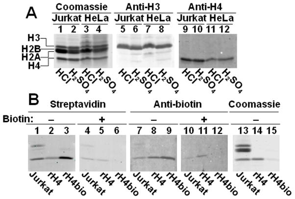
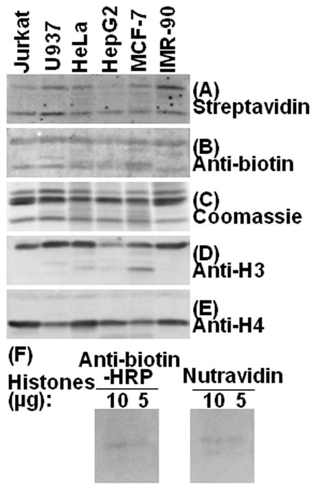
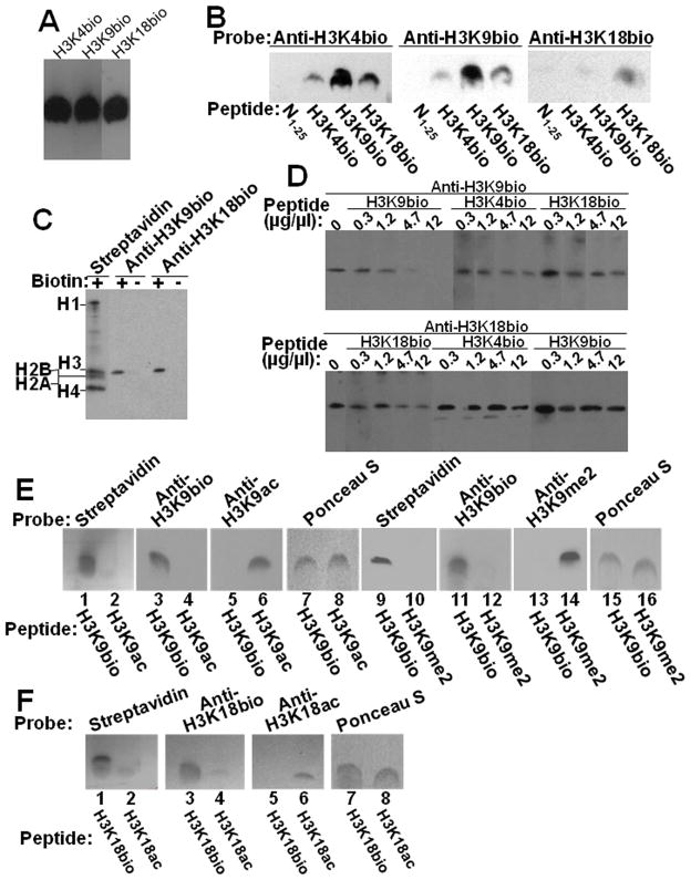
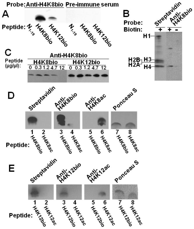
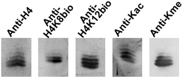
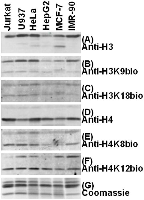
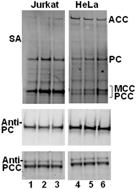
Previous studies suggest that histones H3 and H4 are posttranslationally modified by binding of the vitamin biotin, catalyzed by holocarboxylase synthetase (HCS). Albeit a rare epigenetic mark, biotinylated histones were repeatedly shown to be enriched in repeat regions and repressed loci, participating in the maintenance of genome stability and gene regulation. Recently, a team of investigators failed to detect biotinylated histones and proposed that biotinylation is not a natural modification of histones, but rather an assay artifact. Here, we describe the results of experiments, including the comparison of various analytical protocols, antibodies, cell lines, classes of histones, and radiotracers. These studies provide unambiguous evidence that biotinylation is a natural, albeit rare, histone modification. Less than 0.001% of human histones H3 and H4 are biotinylated, raising concerns that the abundance might too low to elicit biological effects in vivo. We integrated information from this study, previous studies, and ongoing research efforts to present a new working model in which biological effects are caused by a role of HCS in multiprotein complexes in chromatin. In this model, docking of HCS in chromatin causes the occasional binding of biotin to histones as a tracer for HCS binding sites.


Figures

Fig. 1
Fig. 1. Comparison of histone extraction protocols, and specificity testing of streptavidin and anti-biotin
Panel A: Nuclear histones were extracted from Jurkat cells (lanes 1, 5, and 9) or HeLa cells (lanes 3, 7, and 11) by using HCl; for comparison histones were extracted from Jurkat cells (lanes 2, 6, and 10) or HeLa cells (lanes 4, 8, and 12) by using H2SO4+TCA+acetone/HCl+acetone. Histones were probed with coomassie blue (lanes 1–4) and antibodies to the C-termini in histone H3 (lanes 5–8) and H4 (lanes 9–12). Panel B: HCl extracts of Jurkat cell histones (lanes 1, 4, 7, 10, and 13), recombinant human histone H4 (lanes 2, 5, 8, 11, and 14), and chemically biotinylated histone H4 (lanes 3, 6, 9, 12, and 15) were probed with streptavidin without biotin competitor (lanes 1–3) and with 5 mM free biotin (lanes 4–6), and with anti-biotin without biotin competitor (lanes 7–9) and with 5 mM free biotin (lanes 10–12), and with coomassie blue (lanes 13–15).
Fig. 2
Fig. 2. Biotinylation marks can be detected in bulk extracts from human cells, using streptavidin and anti-biotin as probes
Bulk extracts of histone extracts from various cell lineages were probed with streptavidin (panel A), anti-biotin (panel B), and the loading and transfer controls coomassie blue (panel C), anti-H3 (panel D), and anti-H4 (panel E). Panel F: Histones from HeLa cells were extracted with H2SO4+TCA+acetone/HCl+acetone. Ten or five microgram of histones were loaded per well. Blots were blocked with PBS containing 5% BSA. After probing with horseradish peroxidase-conjugated anti-biotin or Nutravidin, the blots were washed for 6 hrs, and exposed to autoradiography film for 1 min.
Fig. 3
Fig. 3. Specificity of antibodies to H3K4bio, H3K9bio and H3K18bio
Panel A: Confirmation of equal loading of peptides H3K4bio, H3K9bio, and H3K18bio with streptavidin. Panel B: Transblots of peptides N1–25 (non-biotinylated negative control), H3K4bio, H3K9bio, and H3K18bio were probed with anti-H3K4bio (left), anti-H3K9bio (middle), and anti-H3K18bio (right). Panel C: HCl extracts of histones from Jurkat cells were probed using streptavidin, anti-H3K9bio, and anti-H3K18bio; samples of biotin-free histones (“-”) were generated by using avidin agarose. Panel D: Bulk HCl extracts of histones from Jurkat cells were probed with anti-H3K9bio (top) and anti-H3K18bio (bottom) after pre-incubation of antibodies with increasing amounts of competing peptides H3K4bio, H3K9bio, and H3K18bio; controls (“C”) were prepared without peptide competitors. Note the difference in the order of peptide competitors in the two gels. For some gels, bands from the same analytical runs were electronically re-arranged to facilitate comparisons. Panel E: Peptides H3K9bio, H3K9ac, and H3K9me2 were probed with streptavidin (lanes 1, 2, 9 and 10), anti-H3K9bio (lanes 3, 4, 11 and 12), anti-H3K9ac (lanes 5 and 6), and anti-H3K9me2 (lanes 13 and 14); Ponceau S was used as loading control (lanes 7, 8, 15 and 16). Panel F: Peptides H3K18bio and H3K18ac were probed with streptavidin (lanes 1 and 2), anti-H3K18bio (lanes 3 and 4) and anti-H3K18ac (lanes 5 and 6); Ponceau S was used as loading control (lanes 7 and 8).
Fig. 4
Fig. 4. Specificity of antibodies to H4K8bio and H4K12bio
Panel A: Transblots of peptides N1–19 (non-biotinylated negative control), H4K8bio, and H4K12bio were probed with anti-H4K8bio; pre-immune serum was used as negative control. Panel B: HCl extracts of histones from Jurkat cells were probed using streptavidin and anti-H4K8bio; samples of biotin-free histones (“-”) were generated by using avidin agarose. Panel C: HCl extracts of histones from Jurkat cells were probed with anti-H4K8bio after pre-incubation of antibodies with increasing amounts of competing peptides H4K8bio and H4K12bio; controls (“C”) were prepared without peptide competitors. For some gels, bands from the same analytical runs were electronically rearranged to facilitate comparisons. Panel D: Peptides H4K8bio and H4K8ac were probed with streptavidin (lanes 1 and 2), anti-H4K8bio (lanes 3 and 4) and anti-H4K8ac (lanes 5 and 6); Ponceau S was used as loading control (lanes 7 and 8). Panel E: Peptides H4K12bio and H4K12ac were probed with streptavidin (lanes 1 and 2), anti-H4K12bio (lanes 3 and 4) and anti-H4K12ac (lanes 5 and 6); Ponceau S was used as loading control (lanes 7 and 8).
Fig. 5
Fig. 5. Detection of biotinylated histone H4 by TAU-PAGE
Histone H4 fraction from Jurkat cells was separated by TAU-PAGE. Transblots were probed with anti-H4, anti-H4K8bio, anti-H4K12bio, anti-acetyl Lysine (anti-Kac), and anti-pan methyl Lysine (anti-Kme).
Fig. 6
Fig. 6. Detection of biotinylated histones in human cells by immunoblots
Histones were extracted from Jurkat, U937, HeLa, HepG2, MCF-7 and IMR-90 cells by using HCl. Transblots were probed with anti-H3 (panel A), anti-H3K9bio (panel B), anti-H3K18bio (panel C), anti-H4 (panel D), anti-H4K8bio (panel E) and anti-H4K12bio (panel F). Sample integrity, and equal loading of histones were confirmed using coomassie blue staining (panel G).
Fig. 7
Fig. 7. Validation of the biotin depletion and repletion protocol
Panel A: Jurkat cells after a 2-wk depletion in biotin-deficient medium (0.025 nM, lane 1) compared with cells cultured in medium containing a physiological concentration of biotin (0.25 nM) (lane 2), and cells after a 1-wk repletion in medium containing a pharmacological concentration of biotin (10 nM) (lane 3). Biotinylated carboxylases were probed using streptavidin (SA). Equal expression, loading, and transfer of carboxylases was confirmed using anti-PC and anti-PCC. Panel B: As described for panel A, but HeLa cells (lanes 4–6) were substituted for Jurkat cells.
Fig. 8
Fig. 8. Binding of radiolabeled biotin to histones
Panel A: Whole cell extracts prepared by using our protocol (lanes 1, 4, 7 and 10), prepared according to Healy’s protocol (lanes 2, 5, 8 and 11), and acid histones extracts (lanes 3, 6, 9 and 12) from Jurkat cells were resolved using 4–12% Bis-Tris gels and probed with streptavidin (SA). Protein identities were verified by using antibodies to PC, histone H3, and histone H4. Panel B: As described for panel B, but HeLa cells were substituted for Jurkat cells. Lanes loaded with acid extracts were electronically combined with those loaded with whole cell extracts.

References

    1. Wolffe A. Chromatin. 3. Academic Press; San Diego, CA: 1998.
    1. Luger K, Mader AW, Richmond RK, Sargent DF, Richmond TJ. Crystal structure of the nucleosome core particle at 2.8 A resolution. Nature. 1997;389:251–260. - PubMed
    1. Kouzarides T, Berger SL. Chromatin modifications and their mechanism of action. In: Allis CD, Jenuwein T, Reinberg D, editors. Epigenetics. Cold Spring Harbor Press; Cold Spring Harbor, NY: 2007. pp. 191–209.
    1. Cheung WL, Ajiro K, Samejima K, Kloc M, Cheung P, Mizzen CA, Beeser A, Etkin LD, Chernoff J, Earnshaw WC, Allis CD. Apoptotic phosphorylation of histone H2B is mediated by mammalian sterile twenty kinase. Cell. 2003;113:507–517. - PubMed
    1. Hymes J, Fleischhauer K, Wolf B. Biotinylation of histones by human serum biotinidase: assessment of biotinyl-transferase activity in sera from normal individuals and children with biotinidase deficiency. Biochem Mol Med. 1995;56:76–83. - PubMed

Publication types

MeSH terms

LinkOut - more resources