. 2011 Nov 18;286(46):39945-57.
doi: 10.1074/jbc.M111.224451. Epub 2011 Sep 19.

Protein phosphatase 2A (PP2A)-specific ubiquitin ligase MID1 is a sequence-dependent regulator of translation efficiency controlling 3-phosphoinositide-dependent protein kinase-1 (PDPK-1)

Affiliations

Protein phosphatase 2A (PP2A)-specific ubiquitin ligase MID1 is a sequence-dependent regulator of translation efficiency controlling 3-phosphoinositide-dependent protein kinase-1 (PDPK-1)

Beatriz Aranda-Orgillés et al. J Biol Chem. .

Abstract

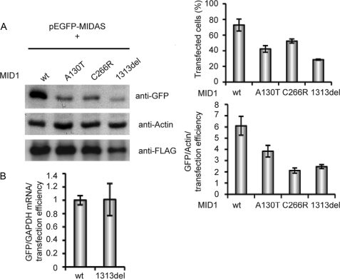
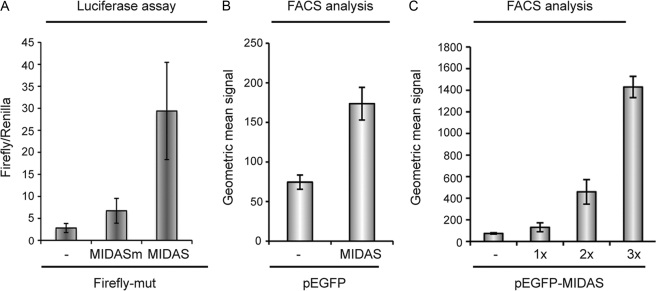
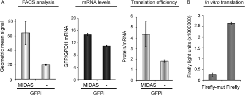
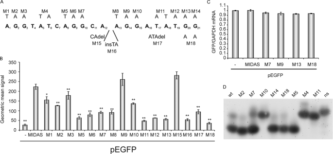
We have shown previously that the ubiquitin ligase MID1, mutations of which cause the midline malformation Opitz BBB/G syndrome (OS), serves as scaffold for a microtubule-associated protein complex that regulates protein phosphatase 2A (PP2A) activity in a ubiquitin-dependent manner. Here, we show that the MID1 protein complex associates with mRNAs via a purine-rich sequence motif called MIDAS (MID1 association sequence) and thereby increases stability and translational efficiency of these mRNAs. Strikingly, inclusion of multiple copies of the MIDAS motif into mammalian mRNAs increases production of the encoded proteins up to 20-fold. Mutated MID1, as found in OS patients, loses its influence on MIDAS-containing mRNAs, suggesting that the malformations in OS patients could be caused by failures in the regulation of cytoskeleton-bound protein translation. This is supported by the observation that the majority of mRNAs that carry MIDAS motifs is involved in developmental processes and/or energy homeostasis. Further analysis of one of the proteins encoded by a MIDAS-containing mRNA, namely PDPK-1 (3-phosphoinositide dependent protein kinase-1), which is an important regulator of mammalian target of rapamycin/PP2A signaling, showed that PDPK-1 protein synthesis is significantly reduced in cells from an OS patient compared with an age-matched control and can be rescued by functional MID1. Together, our data uncover a novel messenger ribonucleoprotein complex that regulates microtubule-associated protein translation. They suggest a novel mechanism underlying OS and point at an enormous potential of the MIDAS motif to increase the efficiency of biotechnological protein production in mammalian cells.


Figures

FIGURE 1.
FIGURE 1.
MIDAS motif and MID1 expression confer increased protein production from mRNAs that associate with the MID1-PP2A signaling complex. A, Firefly luciferase activity recorded from HeLa cells transfected with vectors either containing (Firefly-wt) or lacking (Firefly2) a functional MIDAS motif and co-transfected with empty pCMV vector (−), wild-type MID1 (wt), or mutated, inactive MID1 (A130T, C145S). Standard deviations were calculated from triplicates. B, sequence comparison of Firefly-wt and Firefly2 vectors. C, RNA-protein pulldown assay. Lysates from FLAG-MID1-overexpressing HeLa cells were incubated with biotinylated RNA transcribed in vitro from either Firefly-wt or Firefly2 vectors or from a Renilla vector without MIDAS motif (Renilla). Binding fractions and lysates were analyzed on a Western blot with an anti-FLAG antibody. D, consensus sequence that has been extracted from mRNAs containing G-quartet like sequence motifs. E, Firefly luciferase activity recorded from HeLa cells transfected solely with Firefly-wt vectors containing either a functional MIDAS motif (Firefly-wt) or a mutated MIDAS motif (Firefly-mut) within the open reading frame, and also co-expressing either empty pCMV vector (−), wild-type MID1 (wt), or mutated, dysfunctional MID1 (A130T, C145S). Standard deviations were calculated from triplicates. F, sequence comparison of the functional (Firefly-wt) and mutated (Firefly-mut) MIDAS motifs of the pGL3 vector. G, RNA-protein pulldown assay. Lysates from FLAG-MID1-overexpressing HeLa cells were incubated alone (No RNA) or with biotinylated RNA transcribed in vitro from a pGL3 vector containing either a functional MIDAS motif (Firefly-wt) or a mutated MIDAS motif (Firefly-mut) or from a Renilla vector lacking the MIDAS motif (Renilla). Binding fractions and lysate were analyzed on a Western blot with an anti-FLAG antibody. H, Firefly luciferase activity recorded from HeLa cells transfected with the Firefly-wt vector and either nonsilencing control siRNA oligonucleotides or α4 or MID1-specific oligonucleotides. Standard deviations were calculated from triplicates. I, real time PCR analysis of the Firefly mRNA relative to Renilla mRNA. All standard deviations were calculated from triplicates. J, knockdown efficiencies were controlled on a Western blot.
FIGURE 2.
FIGURE 2.
Mutant MID1 loses its influence on MIDAS-containing mRNAs. EGFP-MIDAS was co-transfected with wild-type MID1 (wt) or the A130T mutant as negative control or two other MID1 mutants (C266R and 1313del). A, left panel, lysates were analyzed on a Western blot using specific anti-GFP and anti-actin antibodies. Right upper panel, transfection efficiencies were assessed by FACS. Right lower panel, band intensities were measured, and GFP amounts were adjusted to actin and the number of transfected cells in each sample. Standard deviations were calculated from triplicates. B, real time PCR analysis of the GFP mRNA relative to GAPDH normalized to the transfection control. Standard deviations were calculated from triplicates.
FIGURE 3.
FIGURE 3.
MIDAS motif increases production of proteins. A, luciferase activity in CHO cells expressing a mutated pGL3 vector (Firefly-mut) carrying a functional (Firefly-mut-MIDAS) or a nonfunctional (Firefly-mut-MIDASm) MIDAS motif 3′ to the Firefly luciferase coding region. B, intensities of green fluorescence recorded from CHO cells transfected with GFP-encoding vectors (pEGFP) either containing (MIDAS) or lacking (−) a MIDAS motif 3′ to the coding region for the GFP protein, measured by FACS. C, production of GFP from pEGFP vectors with no (−), single (1×) double (2×) or triple (3×) functional MIDAS motif 3′ of the GFP open reading frame of a pEGFP vector in CHO cells. Fluorescence intensities were measured by FACS. All standard deviations were calculated from triplicates.
FIGURE 4.
FIGURE 4.
MIDAS motif mainly influences the translation efficiency and to a lesser extent the mRNA stability of MIDAS-containing mRNAs. A, amount of GFP (left panel) and GFP-mRNA (middle panel) produced from a GFP-IRES (GFPi) vector with (MIDAS) or without (−) MIDAS 3′ of the GFP open reading frame in CHO cells. Translation efficiencies are calculated as ratio of protein to mRNA amount (right panel). GFP amounts were measured by FACS, and mRNA amounts were measured by real time PCR. All standard deviations were calculated from triplicates. B, in vitro translation of a Firefly luciferase gene containing either a functional MIDAS (Firefly) or a mutated MIDAS (Firefly-mut) motif. Luciferase activities are shown. All standard deviations were calculated from triplicates.
FIGURE 5.
FIGURE 5.
MIDAS motif is a novel RNA protein-binding sequence with the ability to form hairpin structures. A, summary of MIDAS mutations inserted into pEFGP-MIDAS used. B, intensities of green fluorescence recorded from HeLa cells transfected with EGFP-MIDAS mutants, measured by FACS. Standard deviations are calculated from triplicates. t test (two-tailed, homoscedastic): *, p < 0.01; **, p < 0.001. C, quantification of GFP-mRNAs of selected pEGPF-MIDAS mutants after transfection into HeLa cells. GFP-mRNAs were quantified by real time PCR relative to GAPDH. Standard deviations were calculated from triplicates. D, native gel electrophoresis of RNA oligonucleotides carrying selected MIDAS mutations. Salt-free TBE buffer was used, and gels were stained with SYBR Gold.
FIGURE 6.
FIGURE 6.
mRNAs that are involved in ventral midline development, mTOR signaling, and energy homeostasis carry MIDAS motifs. A, extraction of a MIDAS consensus sequence, F and R are the forward and reverse nucleotides of the stem, respectively. B, binding of a selection of the identified mRNAs to the MID1 protein complex was tested in SHSY cells using FLAG-tagged MID1, RNA-protective immunoprecipitation, and RT-PCR. RT-PCR results were randomly confirmed using real time PCR. C, summary of mRNAs with known functions that were extracted from the data base using the MIDAS consensus sequence. Several mRNAs with related functions were identified.
FIGURE 7.
FIGURE 7.
PDPK-1 protein amount is significantly reduced in cells from an OS patient (OS) compared with age-matched control cells. A, schematic showing the predicted MIDAS motif within the PDPK-1 mRNA. B, representative Western blot showing the analysis of PDPK-1 and GAPDH and quantification of three independent experiments measuring PDPK-1 protein levels relative to GAPDH. C, RT-PCR analysis of the PDPK-1 mRNA relative to GAPDH. Standard deviations are calculated from triplicates. D, analysis of PDPK-1 protein stability after treatment with cycloheximide in a time course by Western blot and quantification of PDPK-1 protein levels relative to GAPDH. E, rescue of PDPK-1 protein levels by overexpression of wild-type MID1 in OS cells. Representative Western blot showing the analysis of MID1, PDPK-1, and GAPDH and quantification of three independent experiments measuring PDPK-1 protein levels relative to GAPDH (*, p = 0.035).

References

    1. Quaderi N. A., Schweiger S., Gaudenz K., Franco B., Rugarli E. I., Berger W., Feldman G. J., Volta M., Andolfi G., Gilgenkrantz S., Marion R. W., Hennekam R. C., Opitz J. M., Muenke M., Ropers H. H., Ballabio A. (1997) Nat. Genet. 17, 285–291 - PubMed
    1. Inui S., Sanjo H., Maeda K., Yamamoto H., Miyamoto E., Sakaguchi N. (1998) Blood 92, 539–546 - PubMed
    1. Trockenbacher A., Suckow V., Foerster J., Winter J., Krauss S., Ropers H. H., Schneider R., Schweiger S. (2001) Nat. Genet. 29, 287–294 - PubMed
    1. Jacinto E., Hall M. N. (2003) Nat. Rev. Mol. Cell Biol. 4, 117–126 - PubMed
    1. Short K. M., Hopwood B., Yi Z., Cox T. C. (2002) BMC Cell Biol. 3, 1. - PMC - PubMed

Publication types

MeSH terms

Supplementary concepts

LinkOut - more resources