. 2011 Dec;79(12):4902-11.
doi: 10.1128/IAI.05591-11. Epub 2011 Sep 19.

Candida albicans cell wall glycosylation may be indirectly required for activation of epithelial cell proinflammatory responses

Affiliations

Candida albicans cell wall glycosylation may be indirectly required for activation of epithelial cell proinflammatory responses

Celia Murciano et al. Infect Immun. 2011 Dec.

Abstract

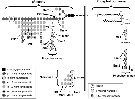
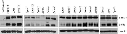
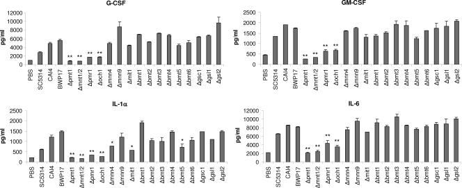
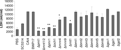
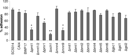
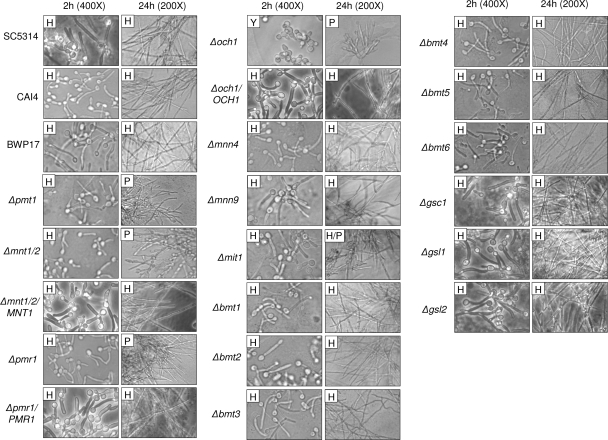
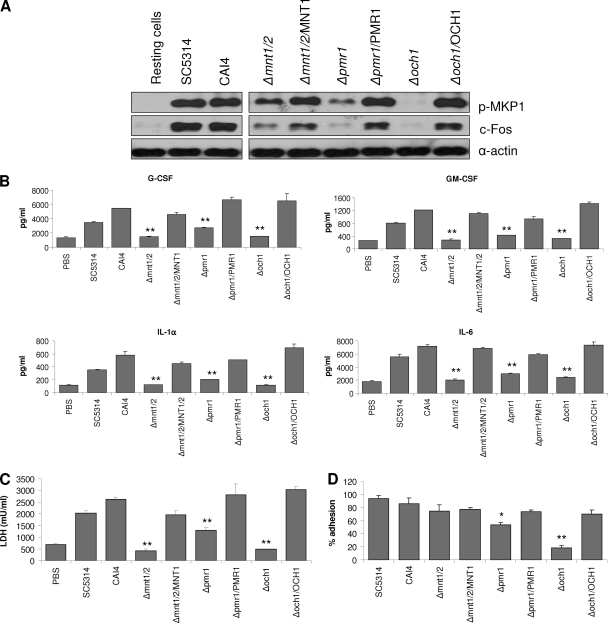
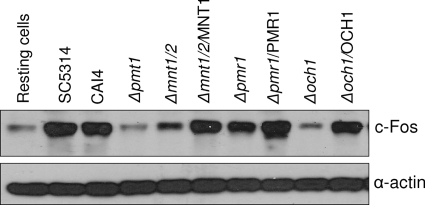
Oral epithelial cells discriminate between the yeast and hyphal forms of Candida albicans via the mitogen-activated protein kinase (MAPK) signaling pathway. This occurs through phosphorylation of the MAPK phosphatase MKP1 and activation of the c-Fos transcription factor by the hyphal form. Given that fungal cell wall polysaccharides are critical in host recognition and immune activation in myeloid cells, we sought to determine whether β-glucan and N- or O-glycosylation was important in activating the MAPK/MKP1/c-Fos hypha-mediated response mechanism and proinflammatory cytokines in oral epithelial cells. Using a series of β-glucan and N- and O-mannan mutants, we found that N-mannosylation (via Δoch1 and Δpmr1 mutants) and O-mannosylation (via Δpmt1 and Δmnt1 Δmnt2 mutants), but not phosphomannan (via a Δmnn4 mutant) or β-1,2 mannosylation (via Δbmt1 to Δbmt6 mutants), were required for MKP1/c-Fos activation, proinflammatory cytokine production, and cell damage induction. However, the N- and O-mannan mutants showed reduced adhesion or lack of initial hypha formation at 2 h, resulting in little MKP1/c-Fos activation, or restricted hypha formation/pseudohyphal formation at 24 h, resulting in minimal proinflammatory cytokine production and cell damage. Further, the α-1,6-mannose backbone of the N-linked outer chain (corresponding to a Δmnn9 mutant) may be required for epithelial adhesion, while the α-1,2-mannose component of phospholipomannan (corresponding to a Δmit1 mutant) may contribute to epithelial cell damage. β-Glucan appeared to play no role in adhesion, epithelial activation, or cell damage. In summary, N- and O-mannosylation defects affect the ability of C. albicans to induce proinflammatory cytokines and damage in oral epithelial cells, but this may be due to indirect effects on fungal pathogenicity rather than mannose residues being direct activators of the MAPK/MKP1/c-Fos hypha-mediated immune response.


Figures

Fig. 1.
Fig. 1.
Schematic diagram representing the locations of N- and O-mannosylation gene activity on cell wall glycosylation. For N-mannosylation, the first α-1,6-mannose residue is added to the triantennary core N-mannan structure by the α-1,6-mannosyltransferase Och1, followed by a second α-1,6-mannose via Mnn9. This creates an α-1,6-mannose backbone onto which α-1,2- and α-1,3-linked mannose residues are subsequently attached by a series of other Mnn protein family members to form the N-linked outer chain (not shown). Phosphomannan is attached to this outer chain via a phosphodiester bond, which requires Mnn4. β-1,2-Oligomannoses are also added onto the N-linked outer chain via Bmt1 and Bmt3 and onto phosphomannan via Bmt2, Bmt3, and Bmt4. Another major constituent of the cell wall is phospholipomannan, which has α-1,2-mannose attached via Mit1 followed by multiple additions of β-1,2-oligomannoses via Bmt5 and Bmt6. For O-mannosylation, addition of the initial α-mannan is performed by Pmt1, and the remaining α-1,2-mannoses are added by Mnt1 and Mnt2. Pmr1 is not a glycosyltransferase and does not add any mannans directly but exerts its effects on both N-mannosylation and O-mannosylation, as it is a Golgi P-type Ca2+/Mn2+ ATPase that floods the Golgi apparatus with manganese ions, which represent an essential cofactor for the Mnt, Mnn, Och1, Bmt, and Pmt enzymes. The thick arrows represent where truncations in N-mannosylation and O-mannosylation occur.
Fig. 2.
Fig. 2.
Activation of MAPK signaling by different C. albicans cell wall mutants. Different C. albicans cell wall mutants, the parent strains (CAI4 and BWP17), and wild-type strain (SC5314) were added to TR146 oral epithelial cells under standard culture conditions for 2 h. Total protein was isolated, and phosphorylation of MKP1 and induction of c-Fos were assessed. Bands are shown relative to an α-actin loading control. A fungal/epithelial cell MOI of 10:1 was used. Data are representative of three independent experiments.
Fig. 3.
Fig. 3.
Cytokine production by different C. albicans cell wall mutants. Different C. albicans cell wall mutants, the parent strains (CAI4 and BWP17), and a wild-type strain (SC5314) were added to TR146 oral epithelial cells under standard culture conditions for 24 h. Cell culture medium was collected and assessed for cytokine proteins by multiplex microbead assay (Luminex). A fungal/epithelial cell MOI of 0.01 was used. Data represent mean values ± standard errors of the means (SEM) and are representative of at least two independent experiments. *, P < 0.05; **, P < 0.01 (compared with the respective parent strain).
Fig. 4.
Fig. 4.
Induction of cell damage by different C. albicans cell wall mutants. Different C. albicans cell wall mutants, the parent strains (CAI4 and BWP17), and wild-type strain (SC5314) were added to TR146 oral epithelial cells under standard culture conditions for 24 h. Cell culture medium was collected and assessed for lactate dehydrogenase (LDH) release as a measure of epithelial cell damage. A fungal/epithelial cell MOI of 0.01 was used. Data represent mean values ± SEM and are representative of two independent experiments. *, P < 0.05; **, P < 0.01 (compared with the respective parent strain).
Fig. 5.
Fig. 5.
Adhesion of C. albicans cell wall mutants to oral epithelial cells. Yeast cells (100 CFU) of C. albicans cell wall mutants, the parent strains (CAI4 and BWP17), and wild-type strain (SC5314) were added to TR146 oral epithelial cells for 90 min. After extensive washing, molten (45°C) Sabouraud's dextrose agar was added and incubated at 37°C for 24 h for colony development of adhered yeasts. Results are expressed as the percent adhered yeast cells. Data represent mean values ± SEM and are representative of two independent experiments. *, P < 0.05; **, P < 0.01 (compared with the respective parent strain).
Fig. 6.
Fig. 6.
Morphologies of different C. albicans cell wall mutants. Different C. albicans cell wall mutants, the parent strains (CAI4 and BWP17), and wild-type strain (SC5314) were added to TR146 oral epithelial cells for 2 or 24 h and formalin fixed. Morphology was assessed by DIC microscopy at ×400 (2 h) or ×200 (24 h). H, hyphae; P, pseudohyphae; Y, yeast.
Fig. 7.
Fig. 7.
Infection of oral epithelial cells with C. albicans N- and O-glycosylation mutants and reintegrant strains. (A) Immunoblot of phosphorylated MKP1 and c-Fos induction after 2 h infection of TR146 cells with Δmnt1 Δmnt2, Δmnt1 Δmnt2/MNT1, Δpmr1, Δpmr1/PMR1, Δoch1, Δoch1/OCH1, parent (CAI4), and wild-type (SC5314) strains. Production of cytokines (B) and induction of cell damage assessed by LDH release (C) after 24 h infection with these strains. A MOI of 10 was used for panel A, and an MOI of 0.01 was used for panels B and C. (D) Percent adhesion to TR146 cells by these strains after 90 min. Data are representative of three (A) or two (B to D) independent experiments (±SEM). *, P < 0.05; **, P < 0.01 [compared with CAI4(CIp10) parent strain].
Fig. 8.
Fig. 8.
Late activation of c-Fos by different C. albicans N- and O-glycosylation mutants and reintegrant strains. Different C. albicans N- and O-glycosylation mutants, their reintegrated strains, the parent strain (CAI4), and a wild-type strain (SC5314) were added to TR146 oral epithelial cells under standard culture conditions for 24 h. Total protein was isolated and induction of c-Fos assessed. Bands are shown relative to an α-actin loading control. A fungal/epithelial cell MOI of 0.01 was used. Data are representative of two independent experiments.

References

    1. Bates S., et al. 2005. Candida albicans Pmr1p, a secretory pathway P-type Ca2+/Mn2+-ATPase, is required for glycosylation and virulence. J. Biol. Chem. 280:23408–23415 - PubMed
    1. Bates S., et al. 2006. Outer chain N-glycans are required for cell wall integrity and virulence of Candida albicans. J. Biol. Chem. 281:90–98 - PubMed
    1. Brown G. D. 2006. Dectin-1: a signalling non-TLR pattern-recognition receptor. Nat. Rev. Immunol. 6:33–43 - PubMed
    1. Buurman E. T., et al. 1998. Molecular analysis of CaMnt1p, a mannosyl transferase important for adhesion and virulence of Candida albicans. Proc. Natl. Acad. Sci. U. S. A. 95:7670–7675 - PMC - PubMed
    1. Cambi A., et al. 2003. The C-type lectin DC-SIGN (CD209) is an antigen-uptake receptor for Candida albicans on dendritic cells. Eur. J. Immunol. 33:532–538 - PubMed

Publication types

MeSH terms

LinkOut - more resources