. 2011 Sep 26;208(10):2005-16.
doi: 10.1084/jem.20101159. Epub 2011 Sep 19.

Host type I IFN signals are required for antitumor CD8+ T cell responses through CD8{alpha}+ dendritic cells

Affiliations

Host type I IFN signals are required for antitumor CD8+ T cell responses through CD8{alpha}+ dendritic cells

Mercedes B Fuertes et al. J Exp Med. .

Abstract

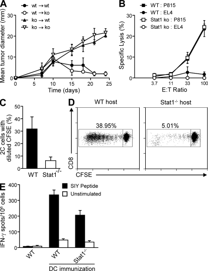
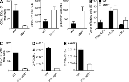
Despite lack of tumor control in many models, spontaneous T cell priming occurs frequently in response to a growing tumor. However, the innate immune mechanisms that promote natural antitumor T cell responses are undefined. In human metastatic melanoma, there was a correlation between a type I interferon (IFN) transcriptional profile and T cell markers in metastatic tumor tissue. In mice, IFN-β was produced by CD11c(+) cells after tumor implantation, and tumor-induced T cell priming was defective in mice lacking IFN-α/βR or Stat1. IFN signaling was required in the hematopoietic compartment at the level of host antigen-presenting cells, and selectively for intratumoral accumulation of CD8α(+) dendritic cells, which were demonstrated to be essential using Batf3(-/-) mice. Thus, host type I IFNs are critical for the innate immune recognition of a growing tumor through signaling on CD8α(+) DCs.


Figures

Figure 1.
Figure 1.
Human melanoma metastases show a positive correlation between T cell markers and IFN-induced transcripts. Tumor samples were obtained by core biopsy or excisional biopsy or obtained from material resected from patients as part of routine clinical management. Total RNA was extracted from tumor samples (n = 52) and gene levels were analyzed by Affymetrix. Arbitrary expression units according to Affymetrix gene levels are shown. (IRF1, R2 = 0.648; p30, R2 = 0.658).
Figure 2.
Figure 2.
Tumors induce CD8+ T cell priming, which is accompanied by IFN-β production by CD11c+ DCs in the tumor-draining lymph nodes. (A and B) C57BL/6 mice were inoculated or not with 5 × 10⁶ B16.SIY tumor cells (s.c.), splenocytes were harvested 6 d later, and restimulated for 16 h in the presence or absence of soluble SIY peptide (A). The frequency of tumor-specific IFN-γ–producing cells was assessed by ELISPOT. ***, P < 0.0001 versus No tumor. (B) Cells were gated on CD8+CD4B220, and the frequency of SIY-specific CD8+ T cells was assessed by FACS using specific tetramers. **, P = 0.0063 versus No tumor. (C and D) C57BL/6 mice were inoculated (s.c.) or not with 5 × 10⁶ B16.SIY tumor cells, and inguinal lymph nodes were recovered 4–6 d later. IFN-β mRNA expression was assessed by real-time RT-PCR analysis in total lymph nodes, and the results are expressed as 2−ΔCt using GAPDH as endogenous control. **, P = 0.0046 versus No tumor (C) or in CD11c+ and CD11c cells sorted from lymph nodes. ***, P = 0.0008 versus CD11c (D). (E) Wild-type C57BL/6 mice (expressing the congenic marker CD45.1+) were lethally irradiated and reconstituted with either wild-type (CD45.2+) or CD11c-DTR (CD45.2+) BM cells. Mice were allowed to reconstitute for 3 mo, and then were injected i.p. with diphtheria toxin (100 ng in 100 µl of DPBS) once a day for 8 d, starting 2 d before s.c challenge with 5 × 10⁶ B16.SIY tumor cells in the left flank (n = 5). Inguinal lymph nodes were recovered 6 d later, and IFN-β expression in total inguinal lymph nodes was measured by real-time PCR. The results are expressed as 2−ΔCt using 18s as endogenous control. (F and G) C57BL/6 mice were inoculated s.c. with the indicated tumor cell lines (5 × 10⁶; F), and splenocytes were harvested 6 d later and restimulated for 16 h in the presence or absence of soluble SIY peptide. Frequency of tumor-specific IFN-γ–producing cells was assessed by ELISPOT. (G) Real-time RT-PCR analysis of IFN-β mRNA expression in total inguinal lymph nodes. The results are expressed as 2−ΔCt using 18s as endogenous control. Data represent mean ± SEM (n = 5) and are representative of four independent experiments (A–C) or two independent experiments (D–G).
Figure 3.
Figure 3.
IFN-α/β, and not IFN-γ signaling, is critical for spontaneous CD8+ T cell priming to tumor-associated antigens. (A and B) Wild-type, IFN-α/βR−/−, or IFN-α/βR−/−/IFN-γR−/− mice were inoculated s.c. with 106 B16.SIY tumor cells. Splenocytes were harvested 17 d later and restimulated for 16 h in the presence or absence of or soluble SIY peptide (A). The frequency of tumor-specific IFN-γ–producing cells was assessed by ELISPOT. ***, P < 0.0001 versus WT. (B) cells were gated on CD8+CD4B220 and the frequency of SIY-specific CD8+ T cells was assessed by FACS using specific tetramers. ***, P < 0.0009; **, P < 0.0027 versus WT. (C) Wild-type and IFN-γR−/− mice were inoculated s.c. with 106 B16.SIY tumor cells, and splenocytes were harvested 17 d later and restimulated for 16 h in the presence or absence of soluble SIY peptide, and then the frequency of tumor-specific IFN-γ–producing cells was assessed by ELISPOT. P = 0.268 versus WT. (D and E) Wild-type and Stat1−/− mice were inoculated s.c. with 106 B16.SIY tumor cells, and splenocytes were harvested 17 d later and restimulated for 16 h in the presence or absence of soluble SIY peptide (D). The frequency of tumor-specific IFN-γ–producing cells was assessed by ELISPOT. ***, P < 0.0001 versus WT. (E) cells were gated on CD8+CD4B220, and the frequency of SIY-specific CD8+ T cells was assessed by FACS using specific tetramers. **, P = 0.0029 versus WT. Data represent mean ± SEM (n = 5), and are representative of three independent experiments.
Figure 4.
Figure 4.
IFN signaling is required in non–T cell BMDCs for tumor-specific T cell priming and for spontaneous rejection of immunogenic tumors in vivo. (A) Wild-type and Stat1−/− DBA/2 mice were lethally irradiated and reconstituted with either wild-type or Stat1−/− DBA/2 BM cells, and 3 mo later they were challenged s.c. with 106 P198 cells in the left flank (n = 5). Tumor size was measured at different time points. Results are shown as mean tumor diameter ± SEM. Data are representative of two independent experiments. (B) T cells from wild-type and Stat1−/− (H-2b) mice were stimulated with T cell–depleted irradiated splenocytes from DBA/2 (H-2d) mice for 5 d in an allogeneic MLR. Cytotoxic activity was measured by standard 51Cr-release assay against P815 (H-2d, cognate targets) and EL4 cells (H-2b, control targets). (C and D) CD8+ T cells were purified from the spleens of 2C Tg/RAG2−/− (Stat1-sufficient) mice, CFSE labeled, and transferred by retroorbital injection to wild-type or Stat1−/− mice. The next day, those mice were inoculated s.c. with 106 B16.SIY cells in the flank, and 7 d later splenocytes were harvested and CFSE dilution of 2C CD8+ T cells was assessed by FACS. (C) Percent of cells with decreased CFSE intensity within the DAPICD8+1B2+ gate. Data show mean ± SEM of individual mice in each group. Data are representative of two independent experiments. (D) Representative dot plots of CFSE dilution. Numbers indicate percent of cells in the indicated gate. (E) 106 SIY-pulsed wild-type BMDCs were s.c. inoculated in the flank of wild-type or Stat1−/− mice, and 20 d later splenocytes were harvested and restimulated in vitro for 16 h in the presence of culture medium or soluble SIY peptide. The frequency of tumor-specific IFN-γ–producing cells was assessed by ELISPOT. Data are representative of two independent experiments.
Figure 5.
Figure 5.
Analysis of DCs from WT, Stat1−/−, and IFN-α/βR−/− mice. (A) Wild-type and IFN-α/βR−/− mice were inoculated s.c. with 106 B16.SIY cells. 6 d later, surface expression of CD80, CD86, CD40, and class I and II MHC was assessed by FACS in tumor-draining lymph node cells gated on CD11c+ cells. Filled histograms correspond to an isotype control (IC), continuous line corresponds to wild-type, and dashed line corresponds to IFN-α/βR−/− mice. (B) Adherent splenocytes from wild-type and Stat1−/− mice were loaded with SIY peptide or left untreated and used to stimulate 2C CD8+ T cells. IL-2 production was assessed by ELISA. (C and D) Wild-type Rag1−/− mice (top) or Rag1−/−Stat1−/− mice (bottom) were inoculated with 106 MC57 or MC57.SIY tumor cells. 14 d later SIY/Kb expression was assessed by FACS using high-affinity 2C TCR tetramers gated on the tumor-infiltrating CD11c+ population (C) and CD11b+ population (D). Filled histograms correspond to staining with streptavidin-phycoerythrin alone. Data are representative of two independent experiments (n = 4).
Figure 6.
Figure 6.
Endogenous type I IFN signaling is required for intratumoral accumulation of CD8α+ DCs. (A and B) Wild-type and Stat1−/− mice were inoculated s.c. with 106 B16.SIY cells, and 15 d later tumors were harvested and frequency (A) and percentages (B) of CD8α+ DCs, mDCs, and pDCs infiltrating tumors were analyzed by FACS. GFP+DAPI+CD3+ cells were gated out, and the different DCs subpopulations were identified as follows: mDCs, CD11C+B220CD8αCD11b+; CD8α+DCs, CD11C+B220CD8α+CD11b; and pDCs, CD11CintB220+PDCA+. Results are shown as mean ± SEM of 3 independent experiments (n = 4). (C–E) Wild-type and IFN-α/βR−/− mice were inoculated s.c. with 106 B16.SIY cells, and 15 d later tumors were harvested and frequency of intratumoral CD8α+ DCs was assessed by FACS (C). (D and E) XCR1 mRNA expression (D) and Batf3 mRNA expression (E) were assessed by real-time RT-PCR analysis on tumor homogenates. The results are expressed as 2−ΔCt using 18s as endogenous control. Results are shown as mean ± SEM of 2 independent experiments (n = 5).
Figure 7.
Figure 7.
CD8α+ DCs are critical for antitumor CD8+ T cell priming. Wild-type and Batf3−/− mice were inoculated s.c. with 106 B16.SIY cells. 6 d later, splenocytes were harvested and restimulated for 16 h in the presence of culture medium or soluble SIY peptide (A). The frequency of tumor-specific IFN-γ–producing cells was assessed by ELISPOT. ***, P < 0.0001 versus WT. (B) the frequency of SIY-specific CD8+ T cells was assessed by FACS using specific anti–Kb-SIY tetramers, cells were gated on the CD8+CD4B220 population. ***, P < 0.001 versus WT. (C) IFN-β mRNA expression was assessed by real-time RT-PCR analysis in total lymph nodes. The results are expressed as 2−ΔCt using 18s as endogenous control. Results are shown as mean ± SEM (n = 5) and are representative of at least two experiments.
Figure 8.
Figure 8.
Type I IFN signaling must occur on the CD8α+ DC lineage for antitumor CD8+ T cell priming to occur. (A) Wild-type mice were lethally irradiated and reconstituted with wild-type, Batf3−/−, or IFN-α/βR−/− BM cells, or a mix of wild-type and IFN-α/βR−/− or Batf3−/− and IFN-α/βR−/− in a 50/50 proportion. Mice were allowed to reconstitute for 3 mo, and were then inoculated s.c. with 106 B16.SIY cells. (A) splenocytes were harvested 6 d later, and the frequency of SIY-specific CD8+ T cells was assessed by FACS using specific tetramers. Cells were gated on CD8+CD4B220. (B) Tumor size was measured at the end of the experiment. *, P < 0.05 versus IFN-α/βR−/− + WT. Results are shown as mean ± SEM of 2 independent experiments (n = 4 each).

Comment in

References

    1. Anderson M.J., Shafer-Weaver K., Greenberg N.M., Hurwitz A.A. 2007. Tolerization of tumor-specific T cells despite efficient initial priming in a primary murine model of prostate cancer. J. Immunol. 178:1268–1276 - PubMed
    1. Anichini A., Molla A., Mortarini R., Tragni G., Bersani I., Di Nicola M., Gianni A.M., Pilotti S., Dunbar R., Cerundolo V., Parmiani G. 1999. An expanded peripheral T cell population to a cytotoxic T lymphocyte (CTL)–defined, melanocyte-specific antigen in metastatic melanoma patients impacts on generation of peptide-specific CTLs but does not overcome tumor escape from immune surveillance in metastatic lesions. J. Exp. Med. 190:651–667 10.1084/jem.190.5.651 - DOI - PMC - PubMed
    1. Apetoh L., Ghiringhelli F., Tesniere A., Obeid M., Ortiz C., Criollo A., Mignot G., Maiuri M.C., Ullrich E., Saulnier P., et al. 2007. Toll-like receptor 4-dependent contribution of the immune system to anticancer chemotherapy and radiotherapy. Nat. Med. 13:1050–1059 10.1038/nm1622 - DOI - PubMed
    1. Basu S., Binder R.J., Suto R., Anderson K.M., Srivastava P.K. 2000. Necrotic but not apoptotic cell death releases heat shock proteins, which deliver a partial maturation signal to dendritic cells and activate the NF-kappa B pathway. Int. Immunol. 12:1539–1546 10.1093/intimm/12.11.1539 - DOI - PubMed
    1. Belz G.T., Smith C.M., Eichner D., Shortman K., Karupiah G., Carbone F.R., Heath W.R. 2004. Cutting edge: conventional CD8 alpha+ dendritic cells are generally involved in priming CTL immunity to viruses. J. Immunol. 172:1996–2000 - PubMed

Publication types

MeSH terms

Substances