. 2011 Nov;31(22):4405-16.
doi: 10.1128/MCB.06163-11. Epub 2011 Sep 19.

Stress-stimulated mitogen-activated protein kinases control the stability and activity of the Cdt1 DNA replication licensing factor

Affiliations

Stress-stimulated mitogen-activated protein kinases control the stability and activity of the Cdt1 DNA replication licensing factor

Srikripa Chandrasekaran et al. Mol Cell Biol. 2011 Nov.

Abstract

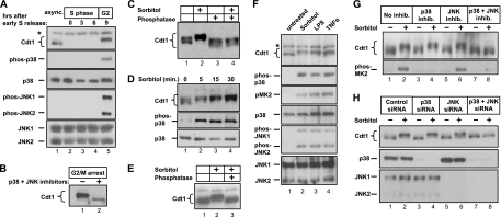
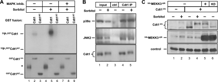
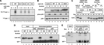
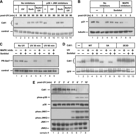
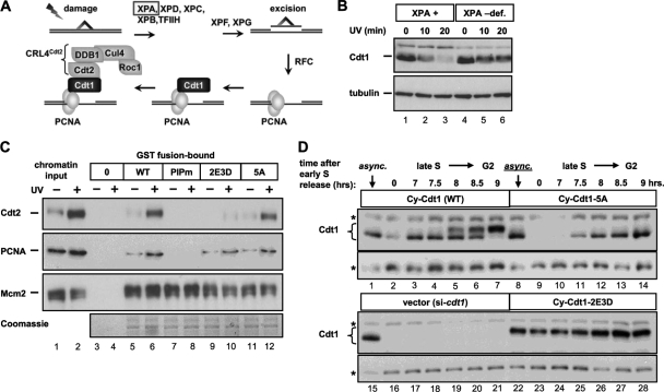
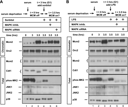
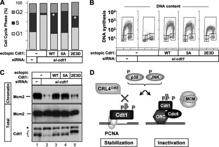
DNA replication is tightly coordinated both with cell cycle cues and with responses to extracellular signals to maintain genome stability. We discovered that human Cdt1, an essential origin licensing protein whose activity must be restricted to G(1) phase, is a substrate of the stress-activated mitogen-activated protein (MAP) kinases p38 and c-Jun N-terminal kinase (JNK). These MAP kinases phosphorylate Cdt1 both during unperturbed G(2) phase and during an acute stress response. Phosphorylation renders Cdt1 resistant to ubiquitin-mediated degradation during S phase and after DNA damage by blocking Cdt1 binding to the Cul4 adaptor, Cdt2. Mutations that block normal cell cycle-regulated MAP kinase-mediated phosphorylation interfere with rapid Cdt1 reaccumulation at the end of S phase. Phosphomimetic mutations recapitulate the stabilizing effects of Cdt1 phosphorylation but also reduce the ability of Cdt1 to support origin licensing. Two other CRL4(Cdt2) targets, the cyclin-dependent kinase (CDK) inhibitor p21 and the methyltransferase PR-Set7/Set8, are similarly stabilized by MAP kinase activity. These findings support a model in which MAP kinase activity in G(2) promotes reaccumulation of a low-activity Cdt1 isoform after replication is complete.


Figures

Fig. 1.
Fig. 1.
Stress triggers MAPK-dependent phosphorylation of Cdt1. (A) Lysates from HeLa cells synchronized by double thymidine block and release were probed for the indicated endogenous antigens (e.g., phos-p38, phosphorylated p38). The position of a nonspecific band is indicated by the asterisk to the left of the gel. async., asynchronous; hrs, hours. (B) HeLa cells were arrested in prometaphase by thymidine-nocodazole synchronization and then treated with a combination of p38 and c-Jun N-terminal kinase (JNK) inhibitors (+) or mock treated (−) for 1 h prior to harvest and detection of endogenous Cdt1. (C) Asynchronous HeLa cells were treated with 500 mM sorbitol for 60 min. Cell lysates were resolved by SDS-PAGE, and endogenous Cdt1 was detected by immunoblotting. For lanes 3 and 4, lysates were treated with a mixture of calf intestinal phosphatase and lambda phosphatase (in the absence of phosphatase inhibitors) prior to electrophoresis. The gel was prepared and electrophoresed using conditions optimized to detect phosphorylation-dependent mobility changes as described in Materials and Methods. (D) Lysates from HeLa cells treated with sorbitol for the indicated times were immunoblotted to detect endogenous Cdt1, phosphorylated p38 (phospho-p38), and total p38. (E) Primary human diploid fibroblasts were treated with sorbitol, and lysates were incubated with protein phosphatase as indicated prior to analysis of endogenous Cdt1. (F) HeLa cells were treated with sorbitol for 30 min, 100 ng/ml lipopolysaccharide (LPS) for 60 min, or 100 μg/ml tumor necrosis factor alpha (TNFα) for 60 min. Lysates were probed for the indicated proteins by immunoblotting. (G) HeLa cells were treated with MAPK inhibitors (inhib.) for 15 min prior to sorbitol treatment, and lysates were probed with anti-Cdt1 antibody or phosphorylated MAPK-activated protein kinase 2 (phos-MK2) antibody. (The p38 inhibitor was SB203580, and the JNK inhibitor was SP600125.) (H) HeLa cells were transfected with control small interfering RNA (siRNA) targeting green fluorescent protein (GFP) (lanes 1 and 2), siRNA molecules targeting p38α and p38β (lanes 3 and 4), siRNA targeting JNK1 and JNK2 (lanes 5 and 6), or siRNA simultaneously targeting all four stress-activated MAPK isoforms (lanes 7 and 8) for 60 h and then mock treated or treated with sorbitol as indicated. Endogenous Cdt1, p38α, p38β, JNK1, and JNK2 were detected by immunoblotting. The p38α and p38β isoforms are recognized by the same antibody and migrate together by SDS-PAGE.
Fig. 2.
Fig. 2.
Cdt1 associates with stress-inducible MAP kinases. (A) Cells were subjected to sorbitol treatment with or without prior treatment with the p38 and JNK MAPK inhibitors as indicated. Cell lysates were incubated with recombinant GST fusion protein GST-Cdt1 (amino acids 1 to 546) or GST-Cdt1ΔC (amino acids 1 to 320) immobilized on glutathione-Sepharose beads. Bound complexes were then incubated in kinase buffer with [γ-32P]ATP, and phosphorylated substrates were detected by autoradiography. Total protein in the reaction mixture was detected by Coomassie blue staining. (The position of a nonspecific band in the control GST preparation is indicated by the asterisk to the left of the gel [lanes 1, 4, and 7]. The absence of an obvious phosphorylation-dependent mobility shift reflects the different electrophoresis conditions needed in this experiment.) (B) Endogenous Cdt1 was isolated from lysates of sorbitol-treated or mock-treated cells using anti-Cdt1 antibody or normal rabbit serum as a control (ctrl). Proteins coimmunoprecipitating with Cdt1 were analyzed by immunoblotting with antibodies to detect endogenous p38 and JNK. IP, immunoprecipitation. (C) Cells were transfected with plasmids encoding HA-MEKK3ΔN (hemagglutinin [HA]-tagged MEK kinase 3 lacking its autoinhibitory domain, constitutively active) or a kinase dead (KD) version of MEKK3ΔN and cotransfected with plasmid encoding V5-tagged Cdt1 (Cdt1-V5). Cells were treated with sorbitol as indicated and probed with V5 and HA antibodies to detect the ectopic proteins. The loading control is a nonspecific background band.
Fig. 3.
Fig. 3.
Five sites in Cdt1 are phosphorylated by stress-inducible MAP kinases. (A) Recombinant full-length N-terminal GST-Cdt1 fusion proteins were incubated with active p38 in the presence of [γ-32P]ATP followed by autoradiography. Total Cdt1 was detected by Coomassie blue staining (note the mobility shift in the phosphorylated substrates). The mutated residues in the different GST-Cdt1 constructs are as follows: S372A T402A T406A for 3A-1, T402A T406A S411A for 3A-2, S372A S491A for 2A, and S391A T402A T406A S411A S491A for 5A (in subsequent analysis, S372 was not important for Cdt1 phosphorylation in vitro and thus was not included in the 5A mutant). The substrate for the reaction in lane 1 is GST alone. WT, wild type. (B) HCT-116 cells stably expressing WT Cdt1, Cdt1-5A, or Cdt1-2E3D (S391D T402E T406E S411D S491D) were subjected to sorbitol treatment as indicated. Ectopic Cdt1 or endogenous phospho-p38 and p38 were detected by immunoblotting. (C) HeLa cells stably expressing (untagged) WT Cdt1, Cdt1-5A, or Cdt1-2E3D were synchronized in early S phase by double thymidine block and then released into transfection medium containing cdt1 siRNA or control siRNA plus nocodazole (Noc.) for 10 h. The ectopic Cdt1 expression constructs harbor synonymous mutations at the siRNA binding site. Cells were then released into G1 for 2 h prior to analysis by immunoblotting with anti-Cdt1 antibody. We confirmed that the cells had completed S phase at the G2 time points (data not shown). The position of a nonspecific band is indicated by the asterisk to the left of the gel. (D) HeLa cells stably expressing V5-tagged WT Cdt1 or Cdt1-5A were treated with 0.5 M sorbitol (+) or treated with PBS (−) as a control for 30 min. Lysates were then immunoprecipitated with crude antisera raised against phosphorylated Cdt1 peptides (phosphorylated threonines at positions 402, 206, and 411 [pT402/T406/T411] and phosphorylated serine at position 491 [p491]) and then probed for the V5 tag. The control immunoprecipitate with normal rabbit serum (NS) is indicated. The antibody heavy chain is labeled IgG. (E) HeLa cells were synchronized in G2 by sequential thymidine and nocodazole treatments and then released into G1 for 2 h. Lysates were immunoprecipitated with phospho-specific Cdt1 antisera and then probed for endogenous Cdt1. The control immunoprecipitate with normal rabbit serum (NS) is indicated. The positions of the antibody heavy chain (IgG) and a nonspecific band (asterisk) are indicated to the left of the gel.
Fig. 4.
Fig. 4.
MAPK-mediated phosphorylation prevents UV-mediated degradation of three substrates of the CRL4Cdt2 ubiquitin ligase. (A) Asynchronous HeLa cells were subjected to UV irradiation, sorbitol treatment, and combined treatments in the presence and absence of MAPK inhibitors as indicated. Sorbitol (Sorb) and/or inhibitors were added 15 min prior to UV irradiation. The cells were harvested at the indicated times (in minutes), and the lysates were analyzed by immunoblotting with anti-Cdt1 antibody. A nonspecific band serves as a loading control. (B) Lysates of cells irradiated and/or treated with p38 and JNK inhibitors as described above for panel A for the indicated times (in hours) were analyzed for endogenous p21 and tubulin proteins by immunoblotting. (C) Lysates of cells irradiated and/or treated with p38 and JNK inhibitors as in panel A for the indicated times were analyzed for endogenous PR-Set7/Set8 by immunoblotting. A nonspecific band serves as a loading control. (D) HeLa cells transfected with plasmids encoding WT Cdt1, Cdt1-5A (unphosphorylatable) or Cdt1-2E3D (phosphomimetic) mutants were treated with UV and sorbitol as indicated and analyzed by immunoblotting with anti-Cdt1 antibody. (E) HeLa cells were UV irradiated, and whole-cell lysates from time points from 0 to 10 min postirradiation were probed for the indicated endogenous antigens.
Fig. 5.
Fig. 5.
Phosphorylation blocks Cdt1 association with Cdt2 and controls Cdt1 stability during S phase. (A) In the case of UV-induced damage, a collection of nucleotide excision repair factors that includes the Xeroderma pigmentosum group A (XPA) protein remove the damaged region, creating a substrate for PCNA loading, Cdt1 interaction, and recruitment of the CRL4Cdt2 ubiquitin E3 ligase via the Cdt2 adaptor. TFIIH, transcription factor IIH; RFC, replication factor complex. (B) UV-induced Cdt1 degradation was monitored by immunoblotting whole-cell lysates of cells derived from patients with a profound XPA deficiency (XPA –def.) that are hence also PCNA loading deficient (3). Cdt1 degradation was compared to degradation in derivatives of those cells reconstituted with the normal XPA cDNA (XPA +). (C) C-terminal GST fusions of wild-type and mutant Cdt1 were partially purified from E. coli and incubated with sonicated chromatin fragments from untreated and UV-irradiated cells. Cdt1PIPm is Cdt1 with a three-amino acid substitution in the PCNA-interacting motif (30). Association of endogenous PCNA, Cdt2, and Mcm2 with GST alone (lanes 3 and 4) or GST-Cdt1 fusion proteins was monitored by immunoblotting, and the isolated GST-Cdt1 proteins were detected by Coomassie blue staining. (D) Stable derivatives of HeLa cells were created with a cyclin-dependent kinase (CDK)-resistant form of Cdt1 that lacks the cyclin/CDK binding site at amino acids 68 to 70 (Cy motif). These Cdt1 constructs also bear mutations in MAP kinase phosphorylation sites as indicated. These cell lines were then synchronized in early S phase by double thymidine block (time zero). Cells were released from the early S block into transfection medium to deplete endogenous Cdt1, and samples were collected from 7 h until 9 h after release to monitor Cdt1 phosphorylation and accumulation during late S and G2. Whole-cell lysates were probed to detect Cdt1 by immunoblotting. The positions of nonspecific bands that serve as loading controls are indicated by asterisks to the left of the gel.
Fig. 6.
Fig. 6.
Stress MAP kinases inhibit MCM loading. (A) HCT-116 cells were synchronized in G0/G1 by serum deprivation and then restimulated with serum for the indicated number of hours. Where indicated, sorbitol was added to a concentration of 0.35 M 3 h after stimulation with serum. A mixture of p38 and JNK inhibitors was added 15 min before sorbitol addition to the cells in lane 5. The cells in lane 6 were transfected with a mixture of four siRNAs targeting p38α and p38β plus JNK1 and JNK2 24 h prior to serum stimulation. Whole-cell lysates (total) and chromatin fractions were analyzed for the indicated endogenous proteins by immunoblotting. (B) As in panel A except that LPS was added to 100 ng/ml 3 h after serum stimulation instead of sorbitol.
Fig. 7.
Fig. 7.
The phosphomimetic Cdt1 mutant is hypomorphic for MCM loading and DNA replication. (A) HCT-116 cells stably expressing WT Cdt1, Cdt1-5A (unphosphorylatable), or Cdt1-2E3D (phosphomimetic mutant) were treated with siRNA targeting endogenous Cdt1 or GST as a control for 80 h and labeled with bromodeoxyuridine (BrdU) for the final hour. DNA content and DNA synthesis were evaluated by flow cytometry using anti-BrdU and propidium iodide. The average proportion of cells in each cell cycle phase was quantified from gated populations of three independent experiments. The standard errors of the means for the cell populations in the G1, S, and G2 cell cycle phases, respectively, are the following: 3.5, 5.1, and 3.0 for control siRNA; 6.5, 2.9, and 6.1 for empty vector plus siRNA targeting cdt1 (si-cdt1); 3.8, 0.6, and 3.9 for WT Cdt1 plus si-cdt1; 0.8, 1.8, and 1.2 for Cdt1-5A plus si-cdt1; and 5.9, 8.4, and 3.0 for Cdt1-2E3D plus si-cdt1. Asterisks indicate statistical significance (P < 0.05) determined by paired Student's t test. (B) Two-dimensional histograms of cell lines transfected with control siRNA or cdt1 siRNA expressing siRNA-resistant WT Cdt1, Cdt1-5A, Cdt1-2E3D, or empty vector. The y axis shows DNA synthesis as measured by fluorescein isothiocyanate (FITC)-conjugated anti-BrdU signal on a logarithmic scale, and the linear x axis shows DNA content measured by propidium iodide staining. (C) The cell lines in panel A were depleted of endogenous Cdt1 with siRNA for 80 h. Chromatin fractions and whole-cell lysates (total) were analyzed for Mcm2 and Cdt1. (D) Model of MAPK-dependent phosphorylation of Cdt1. MAPK-dependent phosphorylation of Cdt1 has two biological consequences: protection from CRL4Cdt2-mediated degradation and functional inhibition. P, phosphate; ORC, origin recognition complex.

Similar articles

Cited by

References

    1. Abbas T., et al. 2010. CRL4(Cdt2) regulates cell proliferation and histone gene expression by targeting PR-Set7/Set8 for degradation. Mol. Cell 40:9–21 - PMC - PubMed
    1. Abbas T., et al. 2008. PCNA-dependent regulation of p21 ubiquitylation and degradation via the CRL4Cdt2 ubiquitin ligase complex. Genes Dev. 22:2496–2506 - PMC - PubMed
    1. Aboussekhra A., et al. 1995. Mammalian DNA nucleotide excision repair reconstituted with purified protein components. Cell 80:859–868 - PubMed
    1. Alvarez E., et al. 1991. Pro-Leu-Ser/Thr-Pro is a consensus primary sequence for substrate protein phosphorylation. Characterization of the phosphorylation of c-myc and c-jun proteins by an epidermal growth factor receptor threonine 669 protein kinase. J. Biol. Chem. 266:15277–15285 - PubMed
    1. Arentson E., et al. 2002. Oncogenic potential of the DNA replication licensing protein CDT1. Oncogene 21:1150–1158 - PubMed

Publication types

MeSH terms

LinkOut - more resources