. 2011 Nov;31(22):4593-608.
doi: 10.1128/MCB.05798-11. Epub 2011 Sep 19.

Distinct functions of Sox2 control self-renewal and differentiation in the osteoblast lineage

Affiliations

Distinct functions of Sox2 control self-renewal and differentiation in the osteoblast lineage

Eunjeong Seo et al. Mol Cell Biol. 2011 Nov.

Abstract

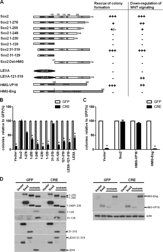
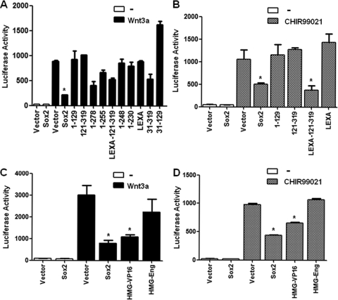
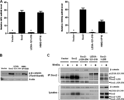
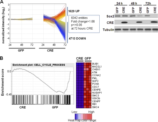
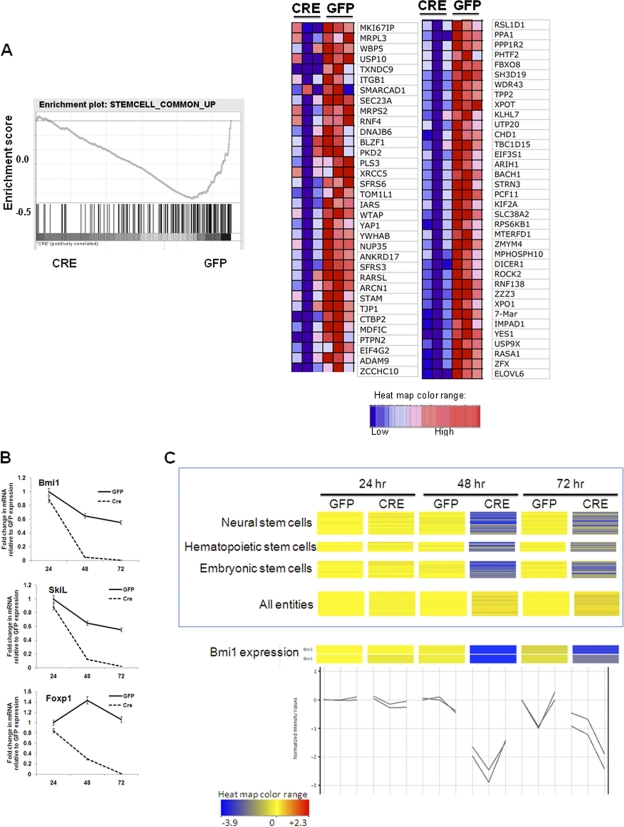
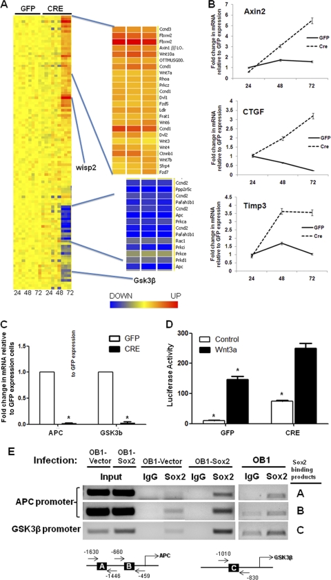
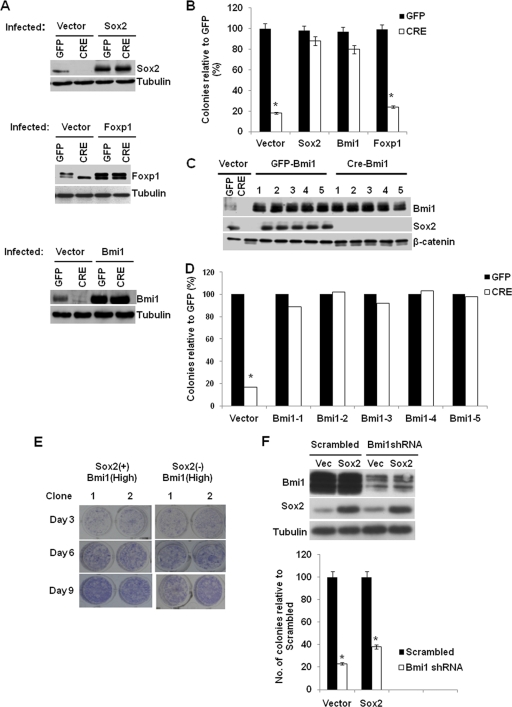
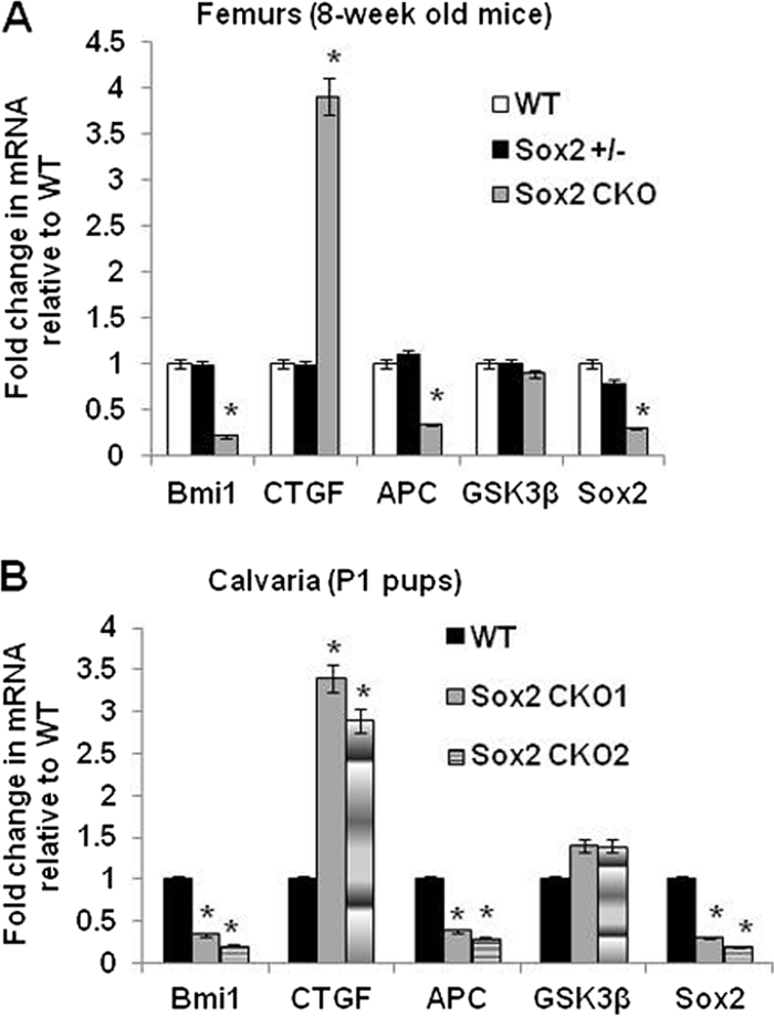
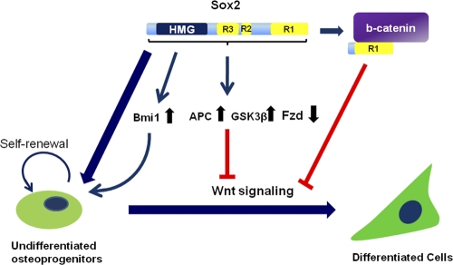
The transcription factor Sox2 is a key player in the maintenance of pluripotency and "stemness." We have previously shown that Sox2 maintains self-renewal in the osteoblast lineage while inhibiting differentiation (U. Basu-Roy et al., Cell Death Differ. 17:1345-1353, 2010; A. Mansukhani, D. Ambrosetti, G. Holmes, L. Cornivelli, and C. Basilico, J. Cell Biol. 168:1065-1076, 2005). Sox2 also interferes with Wnt signaling by binding β-catenin, a central mediator of the Wnt pathway. Here we show that these multiple functions of Sox2 are encoded in distinct domains. The self-renewal function of Sox2 is dependent on its transcriptional activity and requires both its DNA-binding and C-terminal activation regions, while only the third C-terminal transactivation (TA) region is required for binding β-catenin and interfering with Wnt-induced transcription. The results of gene expression analysis upon Sox2 deletion strongly support the notion that Sox2 maintains stemness. We show also that Sox2 suppresses differentiation by attenuating Wnt signaling by posttranscriptional and transcriptional mechanisms and that adenomatous polyposis coli (APC) and GSK3β, which are negative regulators of the Wnt pathway, are direct Sox2 targets in osteoblasts. Several genes, such as the FoxP1 and BMI-1 genes, that are associated with stemness are downregulated upon Sox2 inactivation. Constitutive expression of the Polycomb complex member BMI-1 can bypass the Sox2 requirement for self-renewal but does not affect differentiation. Our results establish a connection between Sox2 and BMI-1 in maintaining self-renewal and identify BMI-1 as a key mediator of Sox2 function.


Figures

Fig. 1.
Fig. 1.
Identification of Sox2 domains required for self-renewal and the inhibition of Wnt signaling. (A) Schematic representation of Sox2 constructs and summary of their activity in rescue of colony formation and Wnt inhibition. +++, complete effect; ++ and +, partial effect; +/−, negligible effect; −, no effect; HMG, DNA binding domain of Sox2; LEXA, DNA binding domain of LexA; RI, R2, and R3, transactivation domains of Sox2; VP16, activation domain of VP16; Eng, repressor domain of Engrailed. The results of a colony assay and a Wnt reporter assay are presented in panel B and Fig. 2A. (B) Colony assay with Sox2 deletion mutants. Sox2-floxed (Sox2flox/flox) osteoblasts were transduced with vector, Sox2, or the indicated mutants for 36 h and then reinfected with either GFP or CRE lentivirus for 72 h. A total of 1,000 cells were plated in triplicate in six-well plates. Colonies were stained and counted after 10 days. Numbers of colonies obtained in the CRE infection are plotted as percentages of the number of colonies obtained in the corresponding GFP infection. *, P < 0.05. (C) Colony assay with Sox2 chimeric proteins. Sox2flox/flox osteoblasts were transduced with vector, Sox2, HMG-VP16, or HMG-Eng, and a colony assay was performed as described for panel B. *, P < 0.05. (D) Western analysis of Sox2 mutants and chimeric proteins. Sox2flox/flox osteoblasts were infected with the indicated constructs as described above. Expression of viral constructs was verified using antibodies against the C-terminal or HMG domain of Sox2 or LexA. Error bars, standard deviations (SD).
Fig. 2.
Fig. 2.
Independent domains of Sox2 inhibit the Wnt-β-catenin pathway. (A and B) Wnt-responsive luciferase activity in osteoblasts expressing Sox2 mutants. OB1-TOP cells (osteoprogenitor cells containing a stably integrated Wnt-responsive luciferase reporter construct) were infected with empty vector or the indicated constructs for 36 h and plated in triplicate. The next day, cells were treated with Wnt3A (100 ng/ml) for 10 h (A) or with CHIR 99021 (4.5 μM) for 18 h (B), and luciferase activity was measured. *, P < 0.05. (C and D) Wnt-responsive luciferase activity in osteoblasts expressing Sox2-chimeric fusion proteins. OB1-TOP cells were infected with empty vector, Sox2, HMG-VP16, or HMG-Eng and treated with Wnt3A (100 ng/ml) for 10 h (C) or with CHIR 99021 (4.5 μM) for 18 h (D), and luciferase activity was measured as described in Materials and Methods. *, P < 0.05. Error bars, SD.
Fig. 3.
Fig. 3.
Sox2 inhibits Wnt signaling by induction of APC and GSK3β mRNA and by binding to β-catenin. (A) Gene expression analysis of APC and GSK3β in osteoblasts expressing Sox2 and chimeric proteins. OB1 cells were infected with vector, Sox2, LexA-121-319, or HMG-VP16 for 48 h. Expression levels of APC and GSK3β were analyzed using qRT-PCR and specific primers. All values are normalized to actin and are expressed as severalfold increases compared to vector-infected control results. *, P < 0.05. (B) Western analysis of phosphorylated β-catenin. Empty vector, Sox2, LexA 121-319, and HMG-VP16 viral vectors were expressed in OB1 cells, and phospho-β-catenin (serine 33 and threonine 45) was detected after 48 h. (C) Interaction of Sox2 (R1) with β-catenin. OB1 cells were infected with empty vector, Sox2, Sox2-Δ129-254 (containing R1 only), LexA 121-319, or Sox2 1-255 for 36 h. Cells were treated with control media or Wnt3A-conditioned media for 24 h. Immunoprecipitation (IP) was performed with anti-Sox2 antibody against the C terminus of Sox2 or against the HMG domain, and blot analysis was performed with anti-β-catenin antibody, anti-Sox2 HMG antibody, or anti-Sox2 C-terminal antibody. The lower panel shows expression levels of proteins in the whole-cell lysate used in IP. Error bars, SD.
Fig. 4.
Fig. 4.
Gene expression changes following Sox2 deletion. (A) Expression profiles of GFP- and CRE-infected Sox2flox/flox cells, showing entities upregulated >1.66-fold at 72 h. Normalized expression values plotted on a log2 scale were determined relative to expression in GFP-infected samples. Expression changes upregulated more than 2-fold are shown in red, and those downregulated over 2-fold are shown in blue. The right panel shows Sox2 and CRE protein expression as determined by Western analysis of GFP- and CRE-infected Sox2flox/flox cells at the indicated time points. Tubulin was used as a loading control. (B) Cell cycle genes are downregulated upon Sox2 deletion. A GSEA enrichment plot showing expression enrichment of a set of cell cycle-related genes is presented. A negative enrichment score indicates that expression of the majority of these genes in the GFP-infected cells was enriched. Bars represent individual genes in a ranked data set list. A heat map of the genes in the leading edge (most highly ranked) with the greatest significant differences revealed in experiments performed using triplicate samples is shown.
Fig. 5.
Fig. 5.
Stem cell genes are downregulated upon Sox2 deletion. (A) A GSEA enrichment plot showing expression enrichment of a set of common stem cell-related genes at 48 h is presented. Bars represent individual genes in a ranked data set list. Expression of the majority of these genes was enriched in the GFP-infected cells. A heat map of the genes in the leading edge showing the strongest downregulation in CRE-infected cells is present. (B) Gene expression analysis of stemness genes in osteoblasts following Sox2 deletion. Sox2flox/flox cells were transduced with either GFP or CRE lentivirus for 72 h. Expression levels of indicated genes were analyzed using qRT-PCR and specific primers. All values are normalized to actin and are expressed as fold changes in comparison to vector-infected control results. (C) Heat map of the indicated stem cell gene sets in GFP- or CRE-infected Sox2flox/− cells at 24, 48, and 72 h after infection. The expression level of all 22,690 entities is shown as a control. Significant downregulation upon Sox2 deletion is indicated in blue. Expression of BMI-1 from two independent BMI-1 probes in the same samples is shown in the bottom panel. Error bars, SD.
Fig. 6.
Fig. 6.
Deregulation of the Wnt signaling pathway in Sox2-depleted cells. (A) Heat map showing expression of Wnt pathway genes in triplicate samples of GFP- and CRE-infected Sox2flox/flox cells at 24, 48, and 72 h. The most strongly upregulated (orange) and downregulated (blue) gene clusters are shown in an expanded view on the right. (B) Gene expression analysis of Wnt target genes in osteoblasts following Sox2 deletion. Sox2flox/flox cells were transduced with either GFP- or CRE-expressing lentivirus for 72 h. Expression levels of the indicated genes were analyzed using qRT-PCR and specific primers. All values are normalized to actin and are expressed as fold changes in comparison to vector-infected control results. (C) APC and GSK3β are downregulated in Sox2-depleted osteoblasts. Sox2flox/flox cells were transduced with either GFP- or CRE-expressing lentivirus for 72 h. Expression levels of APC and GSK3β were analyzed using qRT-PCR and specific primers. All values are normalized to actin and are expressed as fold changes in comparison to vector-infected control results. *, P < 0.05. (D) Wnt reporter activity in Sox2-depleted osteoblasts. Sox2flox/− cells containing stably transfected pTOPflash vector were infected with either GFP- or CRE-expressing lentivirus for 48 h and treated with control or Wnt3A-conditioned media. Luciferase activity was measured as described in Materials and Methods. *, P < 0.05. (E) APC and GSK3β are direct targets of Sox2 in osteoblasts. Cells were infected with empty vector or Sox2 and used for ChIP analysis using anti-Sox2 antibody or IgG as a negative control. PCR was performed using immunoprecipitated chromatin and primers specific for APC or GSK3β promoter regions (see Fig. S4 at http://www.med.nyu.edu/sites/all/files/Seo_et_al_MCB_Supplementary_figures.pdf).
Fig. 7.
Fig. 7.
BMI-1 is a critical regulator of Sox2-dependent self-renewal. (A) Western analysis of Sox2, Foxp1, and BMI-1 expression. Sox2flox/− cells were infected with empty vector or Sox2-, Foxp1-, or BMI-1-expressing lentivirus, and then endogenous Sox2 was deleted using CRE-expressing virus-based excision as described in Materials and Methods. Protein expression was analyzed by immunoblotting with the indicated antibodies. (B) Colony assay of Sox2-depleted cells expressing transgenic Sox2, Foxp1, or BMI-1. Cells were infected as described for panel A, and 1,000 cells were plated in triplicate. Colonies were stained and counted after 10 days. Numbers of colonies obtained after CRE infection are plotted as percentages of the numbers of colonies in the corresponding GFP infection. Each experiment was repeated at least twice. Results from a representative experiment are shown. *, P < 0.05. (C and D) High-BMI-1, Sox2-null cells can be propagated in culture. Colonies obtained from BMI-overexpressing and Sox2-deleted osteoblasts were analyzed by Western blotting (C) and colony assay (D) to determine the levels of the indicated proteins. The colony assay was performed as described above. (E) Differentiation of Sox2-positive, high-Bmi-1 osteoblasts and Sox2-negative, high-Bmi-1 osteoblasts. Sox2-positive, high-Bmi-1 cells and Sox2-negative, high-Bmi-1 cells were differentiated in the presence of BMP2 (100 ng/ml) and stained for alkaline phosphatase activity at the indicated times. (F) Sox2 overexpression does not restore colony formation by Bmi-1-depleted osteoblasts. OB1 cells were transduced with either an empty vector or Sox2-expressing virus and then infected with either scrambled or Bmi-1-specific shRNA. Western blot analysis confirmed expression of Sox2 and a decrease in Bmi-1 protein levels. For the colony assay, 1,000 cells were plated in triplicate. Numbers of colonies were counted after 10 days and are plotted as a percentage of control numbers. Each experiment was repeated at least twice. Results from a representative experiment are shown. *, P < 0.05. Error bars, SD.
Fig. 8.
Fig. 8.
Expression of Sox2-regulated genes in the bones of mice with osteoblast-specific conditional knockout of Sox2 (6). Total RNA was extracted from femurs of 8-week old mice (A) and calvaria of P1 pups (B). Expression of indicated genes was analyzed by qRT-PCR using specific primers. All values are normalized to 18S rRNA and are expressed as fold change compared to wild-type (WT) results. Sox2 CKO, Sox2 conditional knockout. *, P < 0.05. Error bars, SD.
Fig. 9.
Fig. 9.
Model for the multiple mechanisms by which Sox2 regulates the osteoblast lineage. Sox2 maintains stemness and self-renewal by transcriptional mechanisms, fostering expression of genes such as Bmi-1 that are necessary for the self-renewal of osteoprogenitor cells. Sox2 also regulates transcription of genes in the Wnt pathway (e.g., APC, GSK3β, and Fzd receptor genes) whose upregulation or downregulation would inhibit Wnt signaling. Additionally, Sox2 downregulates canonical Wnt signaling by interacting through its C-terminal activation domain (R1) with β-catenin, thus inhibiting the prodifferentiation Wnt pathway.

References

    1. Ambrosetti D., Holmes G., Mansukhani A., Basilico C. 2008. Fibroblast growth factor signaling uses multiple mechanisms to inhibit Wnt-induced transcription in osteoblasts. Mol. Cell. Biol. 28:4759–4771 - PMC - PubMed
    1. Ambrosetti D. C., Basilico C., Dailey L. 1997. Synergistic activation of the fibroblast growth factor 4 enhancer by Sox2 and Oct-3 depends on protein-protein interactions facilitated by a specific spatial arrangement of factor binding sites. Mol. Cell. Biol. 17:6321–6329 - PMC - PubMed
    1. Ambrosetti D. C., Scholer H. R., Dailey L., Basilico C. 2000. Modulation of the activity of multiple transcriptional activation domains by the DNA binding domains mediates the synergistic action of Sox2 and Oct-3 on the fibroblast growth factor-4 enhancer. J. Biol. Chem. 275:23387–23397 - PubMed
    1. Aubin J., Triffit J. 2002. Mesenchymal cells and osteoblast differentiation, p. 59–82 In Bilezikian J., Raisz L. G., Rodan G. A.(ed.), Principles of bone biology, vol. 1 Academic Press, New York, NY
    1. Baron R., Rawadi G. 2007. Targeting the Wnt/beta-catenin pathway to regulate bone formation in the adult skeleton. Endocrinology 148:2635–2643 - PubMed

Publication types

MeSH terms

LinkOut - more resources