Comparative Study
. 2011 Nov;31(22):4633-45.
doi: 10.1128/MCB.05940-11. Epub 2011 Sep 19.

Differential role of Nkx2-5 in activation of the atrial natriuretic factor gene in the developing versus failing heart

Affiliations
Comparative Study

Differential role of Nkx2-5 in activation of the atrial natriuretic factor gene in the developing versus failing heart

Sonisha A Warren et al. Mol Cell Biol. 2011 Nov.

Abstract

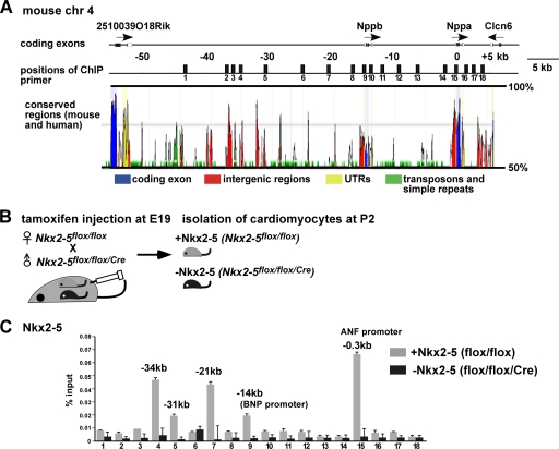
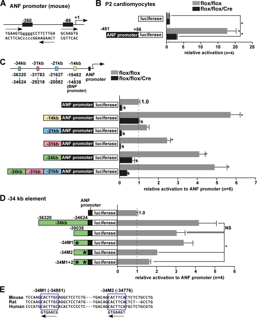
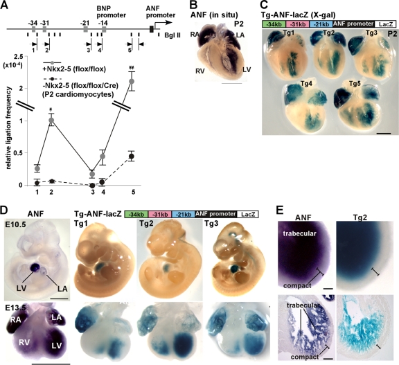
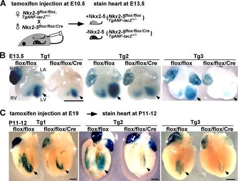
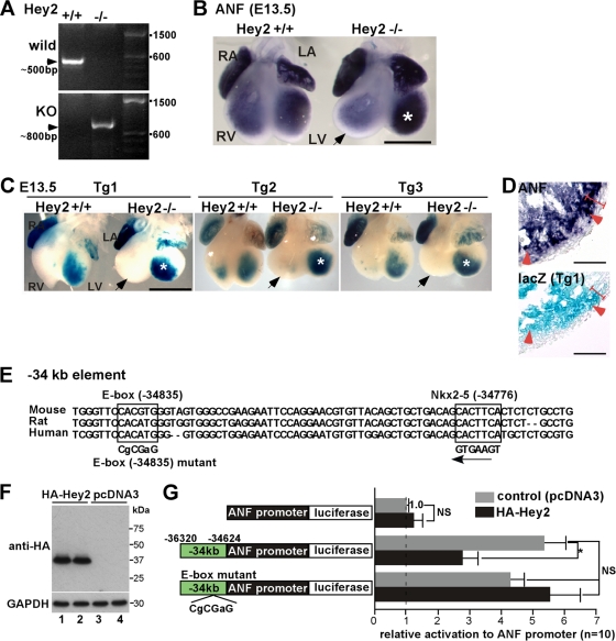
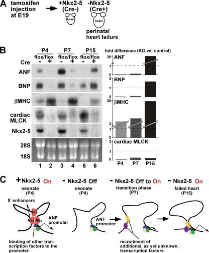
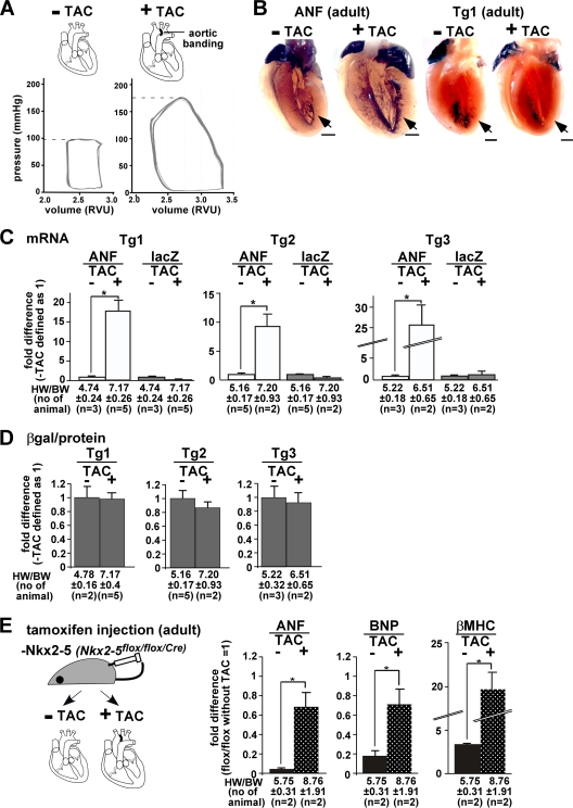
Atrial natriuretic factor (ANF) is abundantly expressed in atrial cardiomyocytes throughout ontogeny and in ventricular cardiomyocytes in the developing heart. However, during cardiac failure and hypertrophy, ANF expression can reappear in adult ventricular cardiomyocytes. The transcription factor Nkx2-5 is one of the major transactivators of the ANF gene in the developing heart. We identified Nkx2-5 binding at three 5' regulatory elements (kb -34, -31, and -21) and at the proximal ANF promoter by ChIP assay using neonatal mouse cardiomyocytes. 3C analysis revealed close proximity between the distal elements and the promoter region. A 5.8-kb fragment consisting of these elements transactivated a reporter gene in vivo recapitulating endogenous ANF expression, which was markedly reduced in tamoxifen-inducible Nkx2-5 gene knockout mice. However, expression of a reporter gene was increased and expanded toward the outer compact layer in the absence of the transcription repressor Hey2, similar to endogenous ANF expression. Functional Nkx2-5 and Hey2 binding sites separated by 59 bp were identified in the -34 kb element in neonatal cardiomyocytes. In adult hearts, this fragment did not respond to pressure overload, and ANF was induced in the absence of Nkx2-5. These results demonstrate that Nkx2-5 and its responsive cis-regulatory DNA elements are essential for ANF expression selectively in the developing heart.


Figures

Fig. 1.
Fig. 1.
Profile of Nkx2-5 occupancy at a genomic locus, including the ANF and BNP genes. (A) Organization of the genomic locus, including the ANF (Nppa) and BNP (Nppb) genes. Numbers indicate distances, in kilobases (kb), from the ANF transcription start site. Eighteen positions of ChIP-PCR primer-probe sets are indicated. (B) Diagram of experiment demonstrating tamoxifen injection at E19 into pregnant female (Nkx2-5flox/flox) mice mated with male (Nkx2-5flox/flox/Cre) mice, followed by isolation of cardiomyocytes at P2. (C) ChIP analysis of Nkx2-5 in cardiomyocytes expressing Nkx2-5 isolated from flox/flox hearts or lacking Nkx2-5 isolated from flox/flox/Cre hearts. Results are shown as percentage of input DNA recovered (means plus standard errors [SE]) from two independent experiments with PCR performed in duplicate. ANF, atrial natriuretic factor; BNP, brain natriuretic peptide.
Fig. 2.
Fig. 2.
Functional characterization of the Nkx2-5-responsive sites in the proximal promoter and enhancer elements for ANF gene expression. (A) Schematics of two Nkx2-5 consensus binding sites in the mouse ANF proximal promoter located at bp −260 and −86 from the ANF transcription start site. The bp −260 site corresponds to the rat bp −242 site, including palindromic consensus binding sites, and the bp −86 site corresponds to the rat bp −87 site (18). (B) Relative luciferase reporter activities of ANF proximal promoter (−451 to +56) normalized to β-galactosidase activity, with the value of the promoterless luciferase reporter defined as 1 (values are means ± SE). *, P < 0.0001 (ANOVA). (C) Analysis of enhancer activities of the four 5′ upstream Nkx2-5-binding elements located around kb −14 (BNP promoter), −21, −31, and −34 relative to the ANF transcriptional start site in the presence of Nkx2-5 (flox/flox). The corresponding induction of luciferase reporter activities was normalized to β-galactosidase activity, with the value for the ANF(−451 to +56) luciferase reporter defined as 1 (values are means ± SE). *, P < 0.05 in comparison to the ANF promoter; #, P < 0.05 for flox/flox versus flox/flox/Cre mice (ANOVA). (D) Analysis of deletion and point mutations in the −34-ANF promoter luciferase construct demonstrating that the M2 mutation eliminates enhancer activity. (E) Sequence of two consensus Nkx2-5 binding sites located at bp −34851 and −34776 relative to the ANF transcription start site.
Fig. 3.
Fig. 3.
Proximity between upstream enhancers and the ANF proximal promoter. (A) Schematic of the ANF locus showing BglII sites and the positions of PCR primers (arrows). 3C analysis of the ANF gene locus was done with cardiomyocytes expressing (gray circles) or lacking (black circles) Nkx2-5. Data were normalized to amplification of BglII-digested and religated BAC clones containing the entire ANF locus (means ± SE) from two independent experiments with PCR performed in duplicate. * and **, P < 0.0001 and P < 0.0018 for flox/flox versus flox/flox/Cre mice (ANOVA). (B and C) Whole-mount in situ hybridization demonstrating endogenous ANF mRNA expression (B) and X-Gal staining of −343121-ANF-lacZ in 5 transgenic lines (C) at P2. (D) Endogenous ANF mRNA expression in comparison to X-Gal staining in developing hearts (E10.5 and E13.5) of −343121-ANF-lacZ transgenic mice (Tg1, Tg2, and Tg3). (E) Enlarged images of left ventricular expression of ANF mRNA and X-Gal staining of −343121-ANF-lacZ, which is positive in the trabecular layer and negative in the compact layer. Bars = 1 mm (B and D) and 100 μm (C). LA, left atrium; LV, left ventricle; RA, right atrium; RV, right ventricle.
Fig. 4.
Fig. 4.
Reduction of X-Gal staining of −343121-ANF-lacZ transgenic mice in hearts lacking Nkx2-5. (A) Diagram of experimental system indicating tamoxifen injection at E10.5 and X-Gal staining at E13.5. (B) Representative images of reduced X-Gal intensity in hearts lacking Nkx2-5 in comparison to the flox/flox litters at E13.5 with tamoxifen injection at E10.5. Arrows and arrowheads indicate left ventricular X-Gal staining. Numbers of mice examined were as follows: Tg1, flox/flox = 8 and flox/flox/Cre = 5; Tg2, flox/flox = 5 and flox/flox/Cre = 3; Tg3, flox/flox = 6 and flox/flox/Cre = 3. (C) Representative images of P11-P12 flox/flox and flox/flox/Cre hearts after perinatal tamoxifen injection. Arrows and arrowheads indicate left ventricular X-Gal staining. Number of mice examined: Tg1, flox/flox = 5 and flox/flox/Cre = 2; Tg2, flox/flox = 4 and flox/flox/Cre = 3; Tg3, flox/flox = 3 and flox/flox/Cre = 4. Bars = 1 mm. LA, left atrium; LV, left ventricle; RA, right atrium; RV, right ventricle.
Fig. 5.
Fig. 5.
Enhanced X-Gal staining of −343121-ANF-lacZ transgenic mice in the left ventricle of transcription repressor hearts lacking Hey2. (A) PCR genotyping of Hey2+/+ (lane 1) and germ line Hey2/ mice (lane 2). (B) Representative images of in situ hybridization of ANF in Hey2+/+ versus Hey2/ litters. ANF expression in the left ventricle of Hey2−/− is marked with an asterisk. The right ventricle was rounder and larger in Hey2 knockout mice, in which ANF mRNA was slightly reduced (arrow). (C) Increased X-Gal intensity in the left ventricle of hearts lacking Hey2 (asterisk) in comparison to the control litters at E13.5 in Tg1 to Tg3. The right ventricles of Hey2/ mice are marked with arrows. Representative images from a total of 9 Hey2+/+ and 8 Hey2/ heterozygous −343121-ANF-lacZ-positive embryos are shown. (D) Enlarged images of tissue sections demonstrate ectopic expression of ANF and lacZ in the compact layer (arrowheads). The compact layer is defined morphologically as the appearance of a compact band of uniform tissue, while the endocardial noncompacted trabecular layer consists of trabecular meshwork with deep endomyocardial spaces (16). (E) An E-box sequence is located at −34835 bp, close to the Nkx2-5-binding sequence at −34776 bp. The mutated E-box sequence used in the −34-ANF luciferase reporter construct is indicated. (F) Western blotting demonstrates HA-tagged Hey2 protein expression in the transfected cells (lanes 1 and 2) in comparison to control cells transfected with pcDNA3. (G) Effects of Hey2 on ANF(−451 to +56), −34-ANF promoter, and the E-box mutant of −34-ANF luciferase constructs. Induction of luciferase reporter activities was normalized to β-galactosidase activity, with the value in ANF(−451 to +56) luciferase reporter cotransfected with empty pcDNA3 plasmid defined as 1 (values are means ± SE). *, P < 0.05 (ANOVA). Bars = 1 mm (B and C) and 50 μm (D). LA, left atrium; LV, left ventricle; RA, right atrium; RV, right ventricle.
Fig. 6.
Fig. 6.
Distinct role of Nkx2-5 in the regulation of the ANF genes under physiologically normal and pathological conditions. (A) Diagram of the experimental system indicating tamoxifen injection at E19. Nkx2-5 deletion causes progressive heart failure within a few weeks (3). (B) Northern blotting shows time-dependent changes in ANF, BNP, and βMHC mRNA in comparison to decreased cardiac MLCK and Nkx2-5 mRNA levels at P4 (lanes 1 and 2), P7 (lanes 3 and 4), and P15 (lanes 5 and 6) in control (flox/flox) versus Nkx2-5 knockout (flox/flox/Cre) hearts. RNA was purified from two or three pooled hearts. (Lanes 1 and 2 are reproduced from reference with permission of the publisher.) Differences in the expression in Nkx2-5 knockout versus control hearts normalized to 28S RNA are shown. (C) Model for transcription of the ANF gene in the heart with (+) or without (−) Nkx2-5 under normal physiological conditions at P4 and at early (P7) and later stages (P15) of failing hearts. Other transcription factors potentially binding at the promoter region (blue, green, and pink) may be GATA4/5/6, serum-responsive factor, TBX5, MEF2C, Baf60C, glucocorticoid receptor, adrenergic signaling through AP1 and SP1, endothelin 1, and Zac1 (14, 23, 25, 36, 49, 55). cis elements and responsive transcription factors (yellow and purple) that mediate reactivation of the ANF gene at P7 and P15 remain to be identified. P, postnatal day; MHC, myosin heavy chain; MLCK, myosin light chain kinase.
Fig. 7.
Fig. 7.
Nkx2-5-independent ANF transcription in pressure-overloaded adult hearts. (A) Diagram of experiments, demonstrating pressure overload (transverse aortic constriction [TAC]), and a representative LV pressure-volume curve with and without TAC, demonstrating increased LV systolic pressure 2 weeks after TAC. Volume is shown in relative volume units (RVU) without additional calibration. (B) Endogenous ANF mRNA expression compared to X-Gal staining of −343121-ANF-lacZ transgenic mice in adult hearts with or without pressure overload (TAC). Bars = 1 mm. (C) Difference, as determined by TaqMan real-time RT-PCR, between ANF and lacZ mRNA expression, with expression without TAC defined as 1. Increase of ANF but not lacZ mRNA expression in pressure-overloaded hearts is demonstrated in three transgenic lines. (D) Difference of lacZ enzymatic activities normalized to the amount of proteins with or without TAC in three transgenic lines. lacZ expression without TAC was defined as 1. The heart weight-to-body weight ratios (HW/BW) (means ± SE) and the number of mice examined are indicated. (E) Diagram of experiments utilizing mice lacking Nkx2-5 with or without TAC in adult mice. The difference in ANF, BNP, and βMHC mRNA expression in Nkx2-5 knockout hearts with and without TAC relative to the control flox/flox heart without TAC (defined as 1) is shown. Increase in ANF, BNP, and βMHC mRNA expression in the pressure-overloaded hearts is demonstrated in Nkx2-5 knockout hearts. The HW/BW ratios (means ± SE) and the number of mice examined are indicated.

References

    1. Bar H., Kreuzer J., Cojoc A., Jahn L. 2003. Upregulation of embryonic transcription factors in right ventricular hypertrophy. Basic Res. Cardiol. 98:285–294 - PubMed
    1. Beltrami A. P., et al. 2003. Adult cardiac stem cells are multipotent and support myocardial regeneration. Cell 114:763–776 - PubMed
    1. Briggs L. E., et al. 2008. Perinatal loss of Nkx2-5 results in rapid conduction and contraction defects. Circ. Res. 103:580–590 - PMC - PubMed
    1. Bruneau B. G., et al. 2001. A murine model of Holt-Oram syndrome defines roles of the T-box transcription factor Tbx5 in cardiogenesis and disease. Cell 106:709–721 - PubMed
    1. Chan J. Y., et al. 2008. Identification of cardiac-specific myosin light chain kinase. Circ. Res. 102:571–580 - PMC - PubMed

Publication types

MeSH terms

LinkOut - more resources