. 2011 Oct 15;187(8):4347-59.
doi: 10.4049/jimmunol.1003404. Epub 2011 Sep 19.

Nucleosides from Phlebotomus papatasi salivary gland ameliorate murine collagen-induced arthritis by impairing dendritic cell functions

Affiliations

Nucleosides from Phlebotomus papatasi salivary gland ameliorate murine collagen-induced arthritis by impairing dendritic cell functions

Vanessa Carregaro et al. J Immunol. .

Abstract

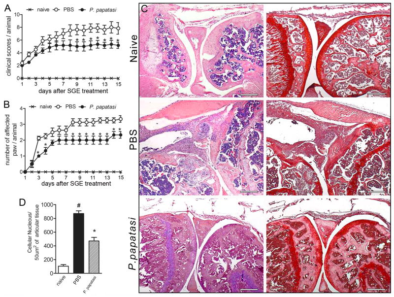
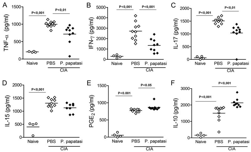
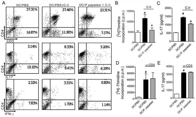
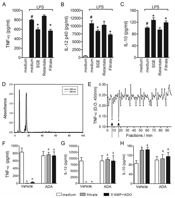
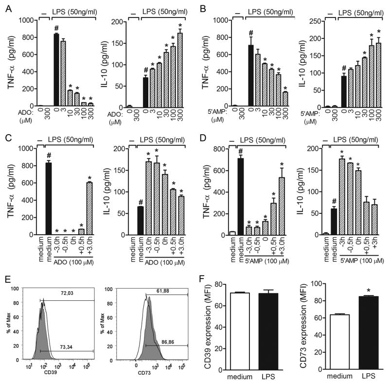
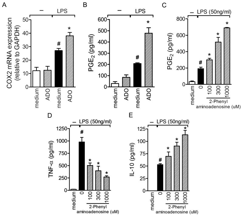
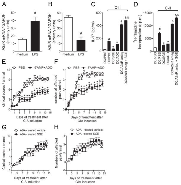
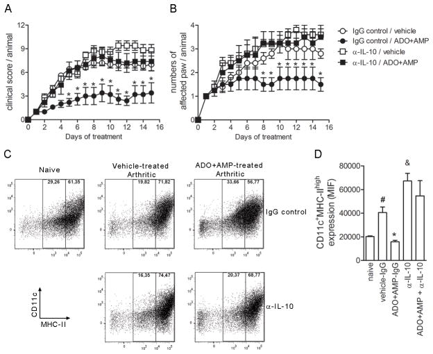
Among several pharmacological compounds, Phlebotomine saliva contains substances with anti-inflammatory properties. In this article, we demonstrated the therapeutic activity of salivary gland extract (SGE) of Phlebotomus papatasi in an experimental model of arthritis (collagen-induced arthritis [CIA]) and identified the constituents responsible for such activity. Daily administration of SGE, initiated at disease onset, attenuated the severity of CIA, reducing the joint lesion and proinflammatory cytokine release. In vitro incubation of dendritic cells (DCs) with SGE limited specific CD4(+) Th17 cell response. We identified adenosine (ADO) and 5'AMP as the major salivary molecules responsible for anti-inflammatory activities. Pharmacologic inhibition of ADO A2(A) receptor or enzymatic catabolism of salivary nucleosides reversed the SGE-induced immunosuppressive effect. Importantly, CD73 (ecto-5'-nucleotidase enzyme) is expressed on DC surface during stage of activation, suggesting that ADO is also generated by 5'AMP metabolism. Moreover, both nucleosides mimicked SGE-induced anti-inflammatory activity upon DC function in vitro and attenuated establishment of CIA in vivo. We reveal that ADO and 5'AMP are present in pharmacological amounts in P. papatasi saliva and act preferentially on DC function, consequently reducing Th17 subset activation and suppressing the autoimmune response. Thus, it is plausible that these constituents might be promising therapeutic molecules to target immune inflammatory diseases.


Conflict of interest statement

DISCLOSURES: The authors have no conflicting financial interests

Figures

Figure 1
Figure 1. SGE from P. papatasi attenuated collagen-induced arthritis
Naïve (×) or collagen-immunized and challenged DBA/1 mice were injected i.v. daily with PBS (○) or P. papatasi SGE (1 gland/animal) (●) for 14 d. Mice were monitored for disease progression as indicated by clinical scores (A)and number of affected paws(B). On d15 of SGE treatment, mice were euthanized, the articular joints were harvested, and histopathologic analysis was performed. Knee joint sections were stained by H&E (left row) or with safranin-O (right row), a proteoglycan red marker showing profound cartilage damage in PBS-treated mice (absence of proteoglycan) and preservation of cartilage in SGE-treated mice(C). Quantification of cellular infiltrate was performed by ImageJ software (NIH, USA)in 40 fields with 400× magnification for each animal/group (D). Morphometric histologic examination shows markedly less cellular infiltration in the SGE-treated than in the PBS-treated group (black bar vs. striped bar). Results show the mean±SEM, N =4; *, P<0.05 compared with PBS-treated group.
Figure 2
Figure 2. Decreased inflammatory cytokinesin articular jointsfrom SGE-treated arthritic mice
Ankle joints from naïve (○)orPBS-(◇) or SGE-(◆) treated arthritic mice were collected for determination of TNF-α (A), IFN-γ (B), IL-17(C), IL-15 (D), and IL-10 (F) levels by ELISA and PGE2 (E) by RIA in the homogenate supernatants. Results are expressed as mean±SEM, N =4 (naïve) and 9–10 (arthritic groups).
Figure 3
Figure 3. SGE treatment inhibits antigen-specific CD4+ T cell activation
BMDCs (106 cells/ml) from DBA/1 naïve mice were incubated overnight with P. papatasi SGE (8 glands/ml) or PBS. Non-adherent cells from LNs and spleen cells from CIA mice on d35 of disease were added to BMDC culture, followed by stimulation with or without collagen-II (5 μg/ml) for 96 h. Percentages of CD4+CD3+, CD4+IL-17+, and CD4+IFN-γ+ cells were determined by flow cytometry (A). Purified CD4+T cells (107 cells/ml) from arthritic mice were added to BMDC culture, and lymphoproliferation induced by C-II (5μg/ml) (B) or plate-bound α-CD3 (5μg/ml) (D) for 96h was assessed by [3H]thymidine incorporation. Culture supernatant was harvested to measure IL-17 levels from C-II-(C) or α-CD3-(E) stimulated cultures. The results are expressed as the mean±SEM obtained from two or three independent experiments made in triplicate (N =3 per group). #, P<0.05 when compared with medium; *, P<0.05 when compared with stimuli; *, P <0.05 compared with saliva vehicle (PBS).
Figure 4
Figure 4. ADO and 5′AMP are the major suppressive molecules in P. papatasi SGE
BMDCs were preincubated overnight with SGE (16 μg/ml) or SGE microcon YM-3 fractions: retentate (>3 kDa) and filtrate (<3 kDa) and then stimulated with LPS (50 ng/mL) for 24 h. TNF-α (A), IL-12 p40 (B), and IL-10 (C) production were measured in the culture supernatants. To characterize DC modulator(s), the filtrate fraction was chromatographed by reverse-phase HPLC as described in Materials and Methods (D). The activity of each eluted fractions was evaluated in LPS-induced TNF-α production by BMDCs (E). Filtrate or ADO+5′AMP commercial standards were previously incubated with vehicle (DMSO) or ADA (4.3U) for 3 h before LPS stimulation, and cytokine production (F–H) was measured in the culture supernatant. #, P<0.05 compared with medium; &, P<0.05 when compared with vehicle treatment.
Figure 5
Figure 5. Effects of ADO and 5′AMP on TNF-α and IL-10 production by LPS-stimulated DCs
BMDCs were incubated without (open bars) or with (shaded bars) 50 ng/ml LPS for 24 h after overnight pre-incubation with increasing concentrations of ADO (A) or 5′AMP (B) (μM). Cytokine production was measured in the culture supernatants by ELISA. Time-course inhibition of cytokine production by 100μM of ADO (C) or 5′AMP (D) in LPS-stimulated BMDCs. The results are expressed as the mean±SEM obtained from one of three independent experiments made in triplicate (N =3 per group). *, P <0.05 compared with LPS stimuli. Surface molecules were labeled with FITC or PE conjugated with anti-CD11c, anti-CD39, or anti-CD73; representative mean fluorescence intensity (MFI) of BMDC staining from medium or LPS cultures is shown in each box (E). The filled histograms represent cells labeled with the specific mAb; the empty histograms represent the same cell suspension labeled with isotypic control mAb. In (F), the bars display the relative MFI obtained from one of four independent experiments made in quadruplicate (N =4). *, P<0.05 compared with medium.
Figure 6
Figure 6. ADO potentiates COX2 mRNA expression and PGE2 production induced by LPS stimulation
COX2 mRNA quantification (A) and PGE2 production (B) were quantified in BMDCs pre-incubated with ADO or medium with or without LPS-stimulation for 24h. The indicated concentration of adenosinesynthetic analogous (2-phenylaminoadenosine) wasadded into BMDC culture 3h before LPS stimuli. PGE2 (C), TNF-α (D), and IL-10 (E) levels were detected 24 h later. PGE2 was measured by RIA and cytokines by ELISA assay. The results are expressed as the mean±SEM obtained from one of three independent experiments made in triplicate (N =3 per group). #, P<0.05 compared with medium; *, P<0.05 compared with the control (LPS).
Figure 7
Figure 7. BlockingA2AR prevents SGE immunosuppressive effect on DC function and 5′AMP+ADO treatment-attenuated CIA
BMDCs (106 cells/mL) were incubated ± with ADO (100 μM). Cells were harvested 24h after LPS stimulation for A2AR (A) and A2BR (B) mRNA expression quantification. Purified CD4+T cells (107 cells/ml) from arthritic mice were added into BMDC culture overnight, pretreated with PBS, SGE (8 glands/ml), or ADO (100μM). Specific A2AR-antagonist was added into some wells 2h before nucleoside or saliva treatment. IL-17 production (C) was measured by ELISAand lymphocyte proliferation by [3H]-thymidine incorporation (D) was determined 96h after C-II stimulation (5μg/ml). #, P<0.05 when compared with medium; *, P<0.05 compared with stimuli; &, P<0.05 compared with salivary nucleosides; %P<0.05 compared with A2AR antagonist. Collagen-immunized DBA/1 mice were injected i.v. daily with salivary equimolar concentration of 5′AMP+ADO (20 uM each ones) (●) or PBS (○) for 14d after start of disease. Mice were monitored for disease progression as indicated by clinical scores (E) and numbers of affected paws (F) *, P<0.05 compared with PBS-treated group. Results show the mean±SEM; N =10. In some groups, salivary gland extract or vehicle (PBS) were previously incubated with ADA (4.3 U) for 3 h. Afterwards, collagen-immunized and challenged DBA/1 mice were injected i.v. daily with ADA-treated vehicle (○)or ADA-treated P. papatasi SGE (1gland/animal)(●) for 14 d. Mice were monitored for disease progression as indicated by clinical scores (G)and number of affected paws (H). Results show the mean±SEM, N =5.
Figure 8
Figure 8. Suppressive effect of salivary nucleosides in vivo is dependent of IL-10
At the onset of arthritis symptoms, naïve or collagen-immunized and challenged DBA/1 mice injected i.v. daily vehicle (PBS) or ADO+AMP were concomitantly treated (250ug/mice; i.p. route) with normal rat IgG (IgG control) or rat anti-IL-10 (αIL-10) antibody three times per week. Mice were monitored for disease progression as indicated by clinical scores (A) and number of affected paws (B). Results show the mean±SEM, N =5; *, P<0.05 compared with vehicle/IgG control. On day 15 of treatment, mice were euthanized, the draining lymph nodes were harvested, and DC surface molecules were labeled with anti-CD11c or anti-MHC class II monoclonal antibodies conjugated with FITC and PE. Representative dot plot of DC staining from naïve, vehicle or ADO+5′AMP treated mice injected with αIL-10 or rat IgG control is shown in each box (C). In (D), the bars display the relative MFI and results show the mean±SEM, N =5;#, P<0.05 compared with naïve mice, *, P<0.05 compared with vehicle/IgG control.&, P<0.05 compared with vehicle/IgG control.

References

    1. Ribeiro JM. Role of saliva in blood-feeding by arthropods. Annual review of entomology. 1987;32:463–478. - PubMed
    1. Ribeiro JM, I, Francischetti M. Role of arthropod saliva in blood feeding: sialome and post-sialome perspectives. Annual review of entomology. 2003;48:73–88. - PubMed
    1. Ribeiro JM, Weis JJ, Telford SR., 3rd Saliva of the tick Ixodes dammini inhibits neutrophil function. Experimental parasitology. 1990;70:382–388. - PubMed
    1. Warburg A, Schlein Y. The effect of post-bloodmeal nutrition of Phlebotomus papatasi on the transmission of Leishmania major. The American journal of tropical medicine and hygiene. 1986;35:926–930. - PubMed
    1. Donnelly KB, Lima HC, Titus RG. Histologic characterization of experimental cutaneous leishmaniasis in mice infected with Leishmania braziliensis in the presence or absence of sand fly vector salivary gland lysate. The Journal of parasitology. 1998;84:97–103. - PubMed

Publication types

MeSH terms