. 2011 Oct 28;52(11):8381-92.
doi: 10.1167/iovs.11-7973.

Histopathology and functional correlations in a patient with a mutation in RPE65, the gene for retinol isomerase

Affiliations

Histopathology and functional correlations in a patient with a mutation in RPE65, the gene for retinol isomerase

Vera L Bonilha et al. Invest Ophthalmol Vis Sci. .

Abstract

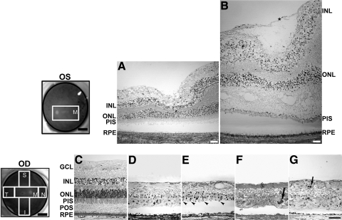
Purpose: Here the authors describe the structural features of the retina and retinal pigment epithelium (RPE) in postmortem donor eyes of a 56-year-old patient with a homozygous missense RPE65 mutation (Ala132Thr) and correlate the pathology with the patient's visual function last measured at age 51.

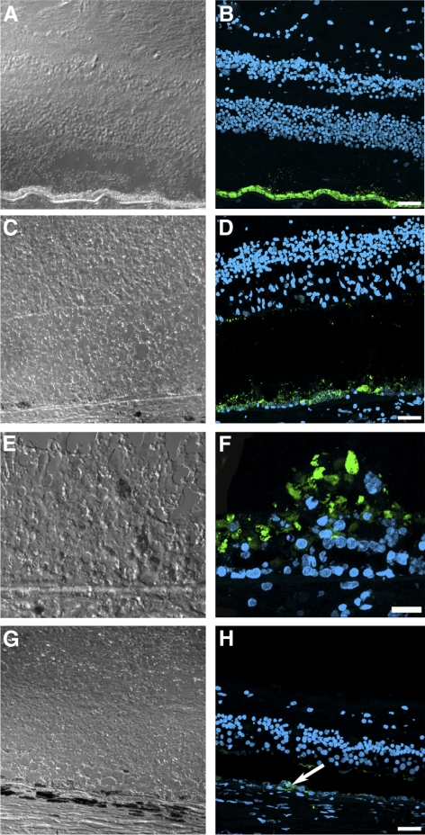
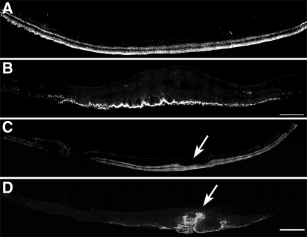
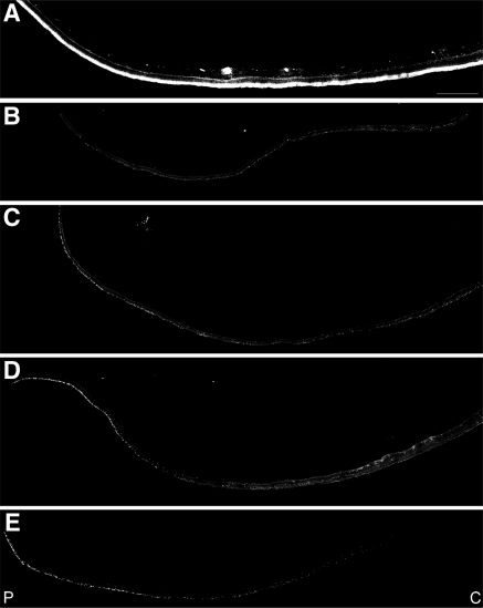
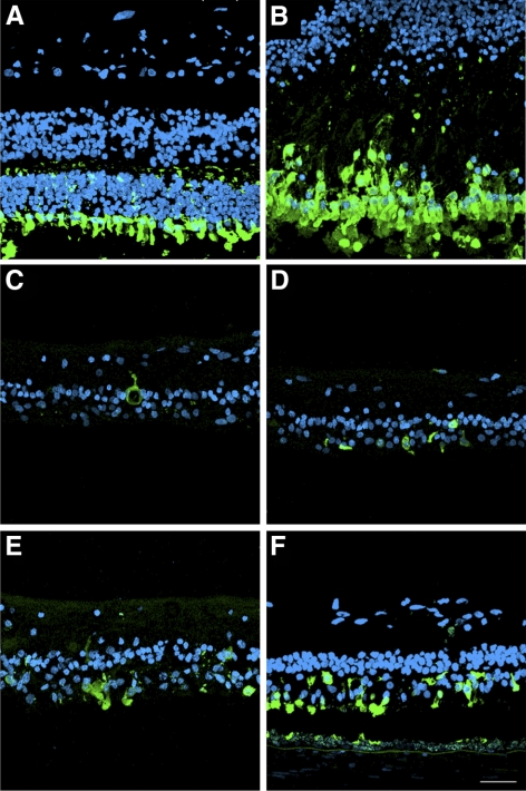
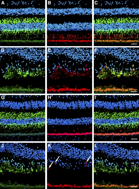
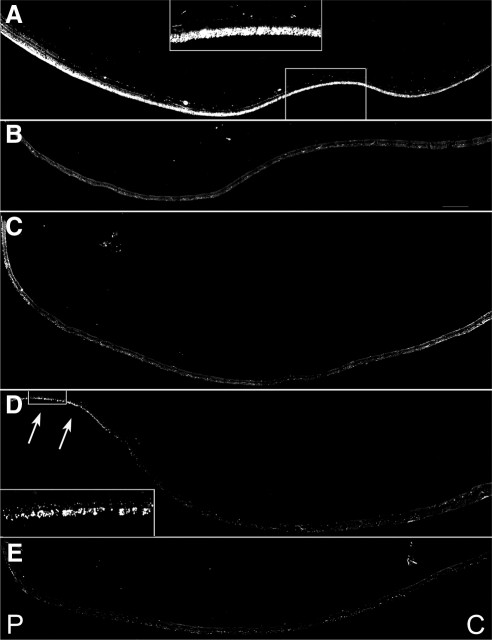
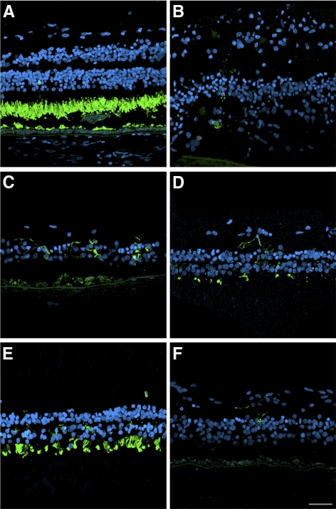
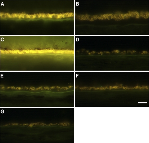
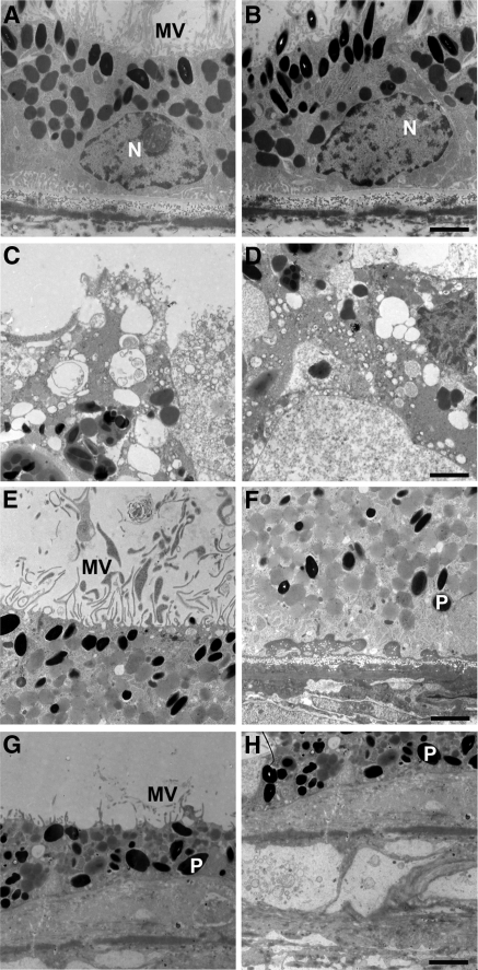
Methods: Eyes were enucleated within 13.5 hours after death. Representative areas from the macula and periphery were processed for light and electron microscopy. Immunofluorescence was used to localize the distribution of RPE65, rhodopsin, and cone arrestin. The autofluorescence in the RPE was compared with that of two normal eyes from age-similar donors.

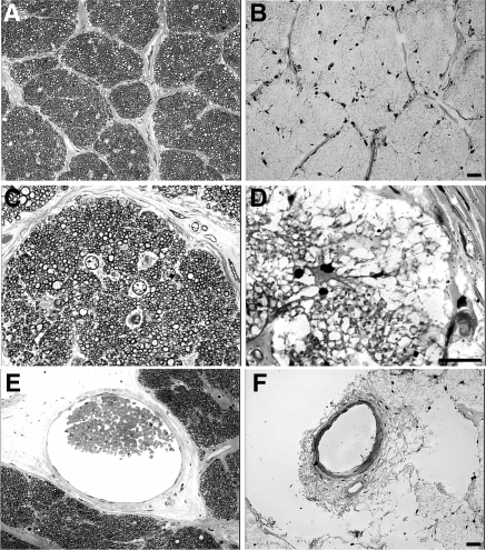
Results: Histologic examination revealed the loss of rods and cones across most areas of the retina, attenuated retinal vessels, and RPE thinning in both eyes. A small number of highly disorganized cones were present in the macula that showed simultaneous labeling with cone arrestin and red/green or blue opsin. RPE65 immunoreactivity and RPE autofluorescence were reduced compared with control eyes in all areas studied. Rhodopsin labeling was observed in rods in the far periphery. The optic nerve showed a reduced number of axons.

Conclusions: The clinical findings of reduced visual acuity, constricted fields, and reduced electroretinograms (ERGs) 5 years before death correlated with the small number of cones present in the macula and the extensive loss of photoreceptors in the periphery. The absence of autofluorescence in the RPE suggests that photoreceptor cells were probably missing across the retina for extended periods of time. Possible mechanisms that could lead to photoreceptor cell death are discussed.


Figures

Figure 1.
Figure 1.
Findings in patient with RPE65 gene mutation. (A, B) Goldmann visual fields to a V-4e white test light and I-4e white test light at age 22. (C, D) Goldmann visual fields to a V-4e white test light at age 51. (E, F) Fundus photographs OD and OS at age 51.
Figure 2.
Figure 2.
Degeneration in the retina of an RPE65 donor. Fundus images of both eyes with disc and macula delineated; schematic drawing of the regions cut and processed for cryosectioning (far left, top and bottom). One-micrometer plastic sections of both matched control (A, C) and RPE65 donor (B, DG) postmortem retinas stained with toluidine blue. The macula of the left eye of the RPE65 donor (B) displayed edema. It contained a prominent preretina (epiretinal) membrane composed of fibroblastlike cells that were vitread to a connective tissue lamina (*). In the periphery, the retina of the RPE65 donor (DG) displayed different degrees of retinal degeneration in all quadrants observed. Sparse inner and outer nuclear layers with stunted photoreceptor inner and outer segments are evident in the superior quadrant (E, arrowheads). A few pigmented cells are seen invading the degenerating retina of the temporal (F) and nasal quadrant (G, arrows). Finally, a thin, continuous area of pigmented RPE cells was present. Quadrants: M, macula; I, inferior; S, superior; T, temporal; N, nasal. RPE, retinal pigment epithelium; POS, photoreceptor outer segment; PIS, photoreceptor inner segment; ONL, outer nuclear layer; INL, inner nuclear layer; GCL, ganglion cell layer. Scale bars: 0.5 cm (fundus images upper and lower left); 400 μm (A, B); 200 μm (C–G).
Figure 3.
Figure 3.
Degeneration in the optic nerve of the RPE65 donor. Light microscopy of 1-μm plastic sections of optic nerve from both a matched control (A, C, E) and the RPE65 donor (B, D, F) stained with toluidine blue. Analysis of the optic nerve of the RPE65 donor in both low (B) and high (D) magnification showed the absence of myelinated axons when compared with the control (A, C). Only a few nuclei (astrocytes?) are evident (D). There is a paucity of axons consistent with the loss of ganglion cells in the retina. Finally, the RPE65 donor displayed a severely reduced central retinal vessel diameter (F). Scale bars, 200 μm.
Figure 4.
Figure 4.
Presence of remaining RPE cells expressing RPE65 in the postmortem eyes from an RPE65 donor. A continuous layer of cells was observed expressing RPE65 in the control (B) and in the macula of the RPE65 donor (D); however, the RPE65 donor expression was substantially lower than that observed in the control tissue. In a few areas in the macula of the RPE65 donor (F), several layers of RPE65-positive cells were observed in the RPE65 donor retina. In the periphery of the RPE65 donor retina, very few cells were detected expressing RPE65 (H, arrow). A, C, E, and G are differential interference contrast microscopy images of the same field shown in B, D, F, and H. Scale bars, 40 μm (B, D, H); 20 μm (F).
Figure 5.
Figure 5.
Presence of disorganized cones in the macular region of both postmortem eyes from an RPE65 donor. Montages of photomicrographs of the macular region of both the right (B) and the left (D) postmortem eyes from the RPE65 donor and control eyes (A, C) were analyzed using a cone arrestin antibody (7G6). Observations showed that in the RPE65 postmortem donor eyes, cones were still present in the macula but were highly disorganized and synapses were absent. (arrows) Macula pit. Scale bar, 500 μm.
Figure 6.
Figure 6.
Significant reduction in the cones in the periphery of an RPE65 postmortem donor eye. Montages of photomicrographs of the periphery tissue from the RPE65 donor (BE) and control eyes (A) were analyzed using a cone arrestin antibody (7G6). Comparison of the samples showed that cones were mostly absent in the affected retina in inferior (B), superior (C), temporal (D), and nasal (E) regions. P, periphery; C, central. Scale bar, 500 μm.
Figure 7.
Figure 7.
Disorganized morphology of the cones remaining in the retina of an RPE65 postmortem donor eye. High-magnification comparison of the control and RPE65 donor retina showed that cone arrestin was distributed along the entire plasma membrane of this cone type, from the tip of the outer segment to the synaptic base in the control retina (A). Cones were present in the macula of the RPE65 donor, but synapses were not visualized (B). On the other hand, cones were mostly absent in the periphery of the RPE65 mutant retina in the inferior (C), superior (D), temporal (E), and nasal (F) quadrants. Scale bar, 40 μm.
Figure 8.
Figure 8.
Disorganized expression of red/green and blue opsins in the cones in the macula of an RPE65 postmortem donor eye. The distribution of cones was also analyzed in control and RPE65 eyes labeled with the cone arrestin (7G6), red/green opsin (AB5405), and blue opsin (AB5407) antibodies. Control retinas displayed cone arrestin distributed along the entire cone cell body (A, G), however the RPE65 donor retina displayed disorganized cones (D, J). In the control eyes, red/green (B) and blue (H) opsins were restricted to the cone outer segments. In the RPE65 eyes, the red/green opsin displayed a more diffuse staining (E) that overlapped with cone arrestin. However, blue opsin localization was mostly to the cone cell boundaries (K, arrows). Overlaid images are shown in C, F, I, L. Scale bar, 40 μm.
Figure 9.
Figure 9.
Absence of rhodopsin in the rods in the periphery of an RPE65 postmortem donor eye. Montages of photomicrographs of the periphery tissue from the RPE65 donor (BE) and control eyes (A) were analyzed using a rhodopsin antibody (B630N). Comparison of the samples showed that rhodopsin was mostly absent in the affected retina in the inferior (B), superior (C), and nasal (E) regions. A few rods were still present in the far periphery of the temporal region of the RPE65 mutant retina (D, inset and arrows). P, periphery; C, central. Scale bar, 500 μm.
Figure 10.
Figure 10.
Disorganized morphology of the rods remaining in the retina of an RPE65 postmortem donor eye. High-magnification comparison of the control and RPE65 donor retina showed that rhodopsin was restricted to the rod outer segments in the control retina (A). In the RPE65 donor, rods were significantly decreased in the affected retina in the macula (B) and periphery in the inferior (C), superior (D), temporal (E), and nasal (F) quadrants, with the remaining rods expanding horizontally into the RPE65 mutant retina. Scale bar, 40 μm.
Figure 11.
Figure 11.
Significant decrease in the accumulation of autofluorescent material in the RPE of an RPE65 postmortem donor eye. Human cryosections of both a matched control (A, C) and an affected RPE65 donor (B, DG) were observed on epifluorescence in the green channel (FITC filter, excitation 495 nm/emission 519 nm). RPE from the RPE65 mutant retina displayed significantly decreased autofluorescent granules in the macula (B) and periphery in the inferior (D), superior (E), temporal (F), and nasal (G) quadrants when compared with the control RPE (A). Scale bar, 200 μm.
Figure 12.
Figure 12.
RPE degeneration in an RPE65 postmortem donor eye. Electron micrographs of the RPE from the control (A, B) and the RPE65 donor (CH). The control RPE displayed apical microvilli (A) and basal infoldings (B). The ultrastructure of the RPE65 donor RPE showed degenerating changes in the different quadrants studied. In the macula, apical microvilli were absent, and pleomorphic inclusions were common (C). The basal surface was characterized by the absence of infoldings and pleomorphic inclusions (D). In the nasal quadrant, apical microvilli were present (E). However, the basal surface was characterized by the presence of electron-dense material beneath the RPE cells (F). In the inferior quadrant, the RPE was discontinuous, and in some areas short apical microvilli were still present above the RPE (G) and a debris zone was present below it (H). N, nucleus; P, pigment; MV, microvilli. Scale bars, 2 μm.

Similar articles

Cited by

References

    1. Cremers FP, van den Hurk JA, den Hollander AI. Molecular genetics of Leber congenital amaurosis. Hum Mol Genet. 2002;11:1169–1176 - PubMed
    1. den Hollander AI, Roepman R, Koenekoop RK, Cremers FP. Leber congenital amaurosis: genes, proteins and disease mechanisms. Prog Retin Eye Res. 2008;27:391–419 - PubMed
    1. Jin M, Li S, Moghrabi WN, Sun H, Travis GH. Rpe65 is the retinoid isomerase in bovine retinal pigment epithelium. Cell. 2005;122:449–459 - PMC - PubMed
    1. Moiseyev G, Chen Y, Takahashi Y, Wu BX, Ma JX. RPE65 is the isomerohydrolase in the retinoid visual cycle. Proc Natl Acad Sci U S A. 2005;102:12413–12418 - PMC - PubMed
    1. Redmond TM, Poliakov E, Yu S, Tsai JY, Lu Z, Gentleman S. Mutation of key residues of RPE65 abolishes its enzymatic role as isomerohydrolase in the visual cycle. Proc Natl Acad Sci U S A. 2005;102:13658–13663 - PMC - PubMed

Publication types

MeSH terms