

A novel mechanism of sulfur transfer catalyzed by O-acetylhomoserine sulfhydrylase in the methionine-biosynthetic pathway of Wolinella succinogenes
- PMID: 21931214
- PMCID: PMC3176619
- DOI: 10.1107/S0907444911028010
A novel mechanism of sulfur transfer catalyzed by O-acetylhomoserine sulfhydrylase in the methionine-biosynthetic pathway of Wolinella succinogenes
Abstract
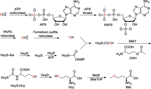
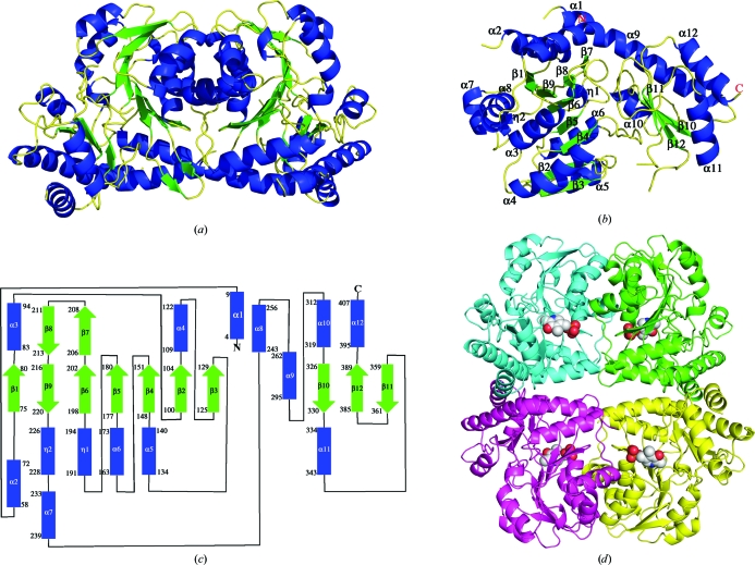
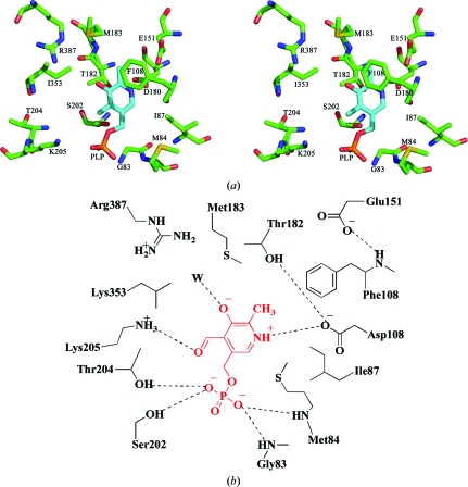
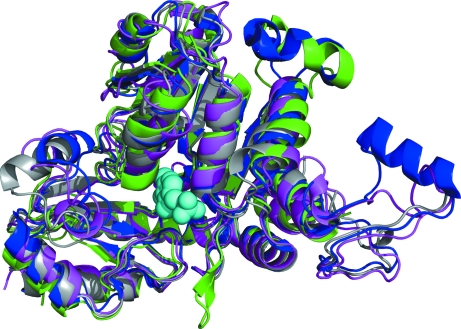
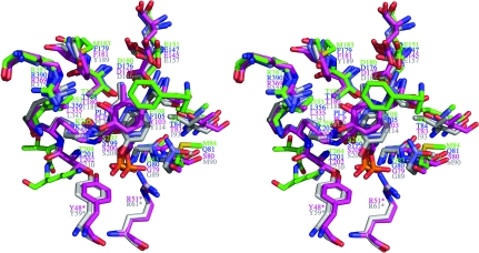
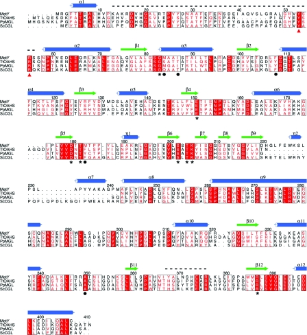
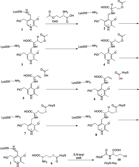
O-Acetylhomoserine sulfhydrylase (OAHS) is a pyridoxal 5'-phosphate (PLP) dependent sulfide-utilizing enzyme in the L-cysteine and L-methionine biosynthetic pathways of various enteric bacteria and fungi. OAHS catalyzes the conversion of O-acetylhomoserine to homocysteine using sulfide in a process known as direct sulfhydrylation. However, the source of the sulfur has not been identified and no structures of OAHS have been reported in the literature. Here, the crystal structure of Wolinella succinogenes OAHS (MetY) determined at 2.2 Å resolution is reported. MetY crystallized in space group C2 with two monomers in the asymmetric unit. Size-exclusion chromatography, dynamic light scattering and crystal packing indicate that the biological unit is a tetramer in solution. This is further supported by the crystal structure, in which a tetramer is formed using a combination of noncrystallographic and crystallographic twofold axes. A search for structurally homologous proteins revealed that MetY has the same fold as cystathionine γ-lyase and methionine γ-lyase. The active sites of these enzymes, which are also PLP-dependent, share a high degree of structural similarity, suggesting that MetY belongs to the γ-elimination subclass of the Cys/Met metabolism PLP-dependent family of enzymes. The structure of MetY, together with biochemical data, provides insight into the mechanism of sulfur transfer to a small molecule via a protein thiocarboxylate intermediate.
Figures
Similar articles
-
Protein thiocarboxylate-dependent methionine biosynthesis in Wolinella succinogenes.J Am Chem Soc. 2011 Jan 19;133(2):379-86. doi: 10.1021/ja107424t. Epub 2010 Dec 16. J Am Chem Soc. 2011. PMID: 21162571 Free PMC article.
-
Occurrence of transsulfuration in synthesis of L-homocysteine in an extremely thermophilic bacterium, Thermus thermophilus HB8.J Bacteriol. 2001 Mar;183(6):2086-92. doi: 10.1128/JB.183.6.2086-2092.2001. J Bacteriol. 2001. PMID: 11222609 Free PMC article.
-
Corynebacterium glutamicum utilizes both transsulfuration and direct sulfhydrylation pathways for methionine biosynthesis.J Bacteriol. 2002 Mar;184(5):1277-86. doi: 10.1128/JB.184.5.1277-1286.2002. J Bacteriol. 2002. PMID: 11844756 Free PMC article.
-
The enzymes of the transsulfuration pathways: active-site characterizations.Biochim Biophys Acta. 2011 Nov;1814(11):1511-7. doi: 10.1016/j.bbapap.2011.03.006. Epub 2011 Mar 22. Biochim Biophys Acta. 2011. PMID: 21435402 Review.
-
Interactions with sulfur acceptors modulate the reactivity of cysteine desulfurases and define their physiological functions.Biochim Biophys Acta Mol Cell Res. 2024 Oct;1871(7):119794. doi: 10.1016/j.bbamcr.2024.119794. Epub 2024 Jul 19. Biochim Biophys Acta Mol Cell Res. 2024. PMID: 39033933 Review.
Cited by
-
Thiamin biosynthesis: still yielding fascinating biological chemistry.Biochem Soc Trans. 2012 Jun 1;40(3):555-60. doi: 10.1042/BST20120084. Biochem Soc Trans. 2012. PMID: 22616866 Free PMC article. Review.
-
Structures and kinetics of Thermotoga maritima MetY reveal new insights into the predominant sulfurylation enzyme of bacterial methionine biosynthesis.J Biol Chem. 2021 Jan-Jun;296:100797. doi: 10.1016/j.jbc.2021.100797. Epub 2021 May 18. J Biol Chem. 2021. PMID: 34019879 Free PMC article.
-
Fungal L-Methionine Biosynthesis Pathway Enzymes and Their Applications in Various Scientific and Commercial Fields.Biomolecules. 2024 Oct 17;14(10):1315. doi: 10.3390/biom14101315. Biomolecules. 2024. PMID: 39456248 Free PMC article. Review.
-
pH-dependent regulation of an acidophilic O-acetylhomoserine sulfhydrylase from Lactobacillus plantarum.Appl Environ Microbiol. 2024 May 21;90(5):e0011824. doi: 10.1128/aem.00118-24. Epub 2024 Apr 3. Appl Environ Microbiol. 2024. PMID: 38568076 Free PMC article.
-
Conversion of methionine biosynthesis in Escherichia coli from trans- to direct-sulfurylation enhances extracellular methionine levels.Microb Cell Fact. 2023 Aug 11;22(1):151. doi: 10.1186/s12934-023-02150-x. Microb Cell Fact. 2023. PMID: 37568230 Free PMC article.
References
-
- Barton, G. J. (1993). Protein Eng. 6, 37–40. - PubMed
-
- Brünger, A. T., Adams, P. D., Clore, G. M., DeLano, W. L., Gros, P., Grosse-Kunstleve, R. W., Jiang, J.-S., Kuszewski, J., Nilges, M., Pannu, N. S., Read, R. J., Rice, L. M., Simonson, T. & Warren, G. L. (1998). Acta Cryst. D54, 905–921. - PubMed
Publication types
MeSH terms
Substances
Associated data
- Actions
Grants and funding
LinkOut - more resources
Full Text Sources
Other Literature Sources
Research Materials
Miscellaneous
