

Two-photon excited UV fluorescence for protein crystal detection
- PMID: 21931215
- PMCID: PMC3176620
- DOI: 10.1107/S0907444911028253
Two-photon excited UV fluorescence for protein crystal detection
Abstract
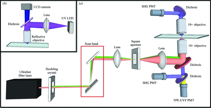
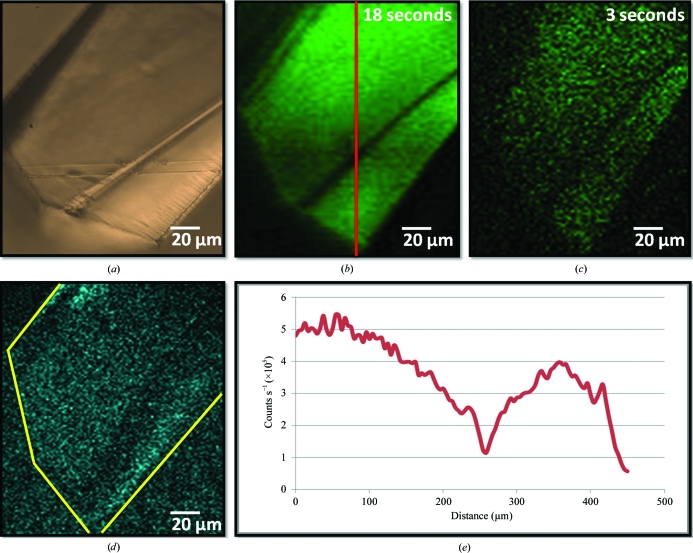
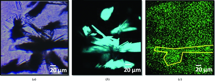
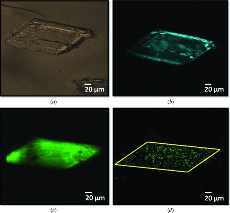
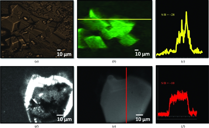
Two-photon excited ultraviolet fluorescence (TPE-UVF) microscopy is explored for sensitive protein-crystal detection as a complement to second-order nonlinear optical imaging of chiral crystals (SONICC). Like conventional ultraviolet fluorescence (UVF), TPE-UVF generates image contrast based on the intrinsic fluorescence of aromatic residues, generally producing higher fluorescence emission within crystals than the mother liquor by nature of the higher local protein concentration. However, TPE-UVF has several advantages over conventional UVF, including (i) insensitivity to optical scattering, allowing imaging in turbid matrices, (ii) direct compatibility with conventional optical plates and windows by using visible light for excitation, (iii) elimination of potentially damaging out-of-plane UV excitation, (iv) improved signal to noise through background reduction from out-of-plane excitation and (v) relatively simple integration into instrumentation developed for SONICC.
Figures
Similar articles
-
Selective imaging of active pharmaceutical ingredients in powdered blends with common excipients utilizing two-photon excited ultraviolet-fluorescence and ultraviolet-second order nonlinear optical imaging of chiral crystals.Anal Chem. 2012 Jul 17;84(14):5869-75. doi: 10.1021/ac300917t. Epub 2012 Jun 27. Anal Chem. 2012. PMID: 22816778 Free PMC article.
-
Label-free detection of single protein molecules using deep UV fluorescence lifetime microscopy.Anal Chem. 2006 Apr 15;78(8):2732-7. doi: 10.1021/ac052166u. Anal Chem. 2006. PMID: 16615786
-
Imaging of protein crystals with two-photon microscopy.Biochemistry. 2012 Feb 28;51(8):1625-37. doi: 10.1021/bi201682q. Epub 2012 Feb 16. Biochemistry. 2012. PMID: 22324807 Free PMC article.
-
Two-photon excitation of fluorescence for three-dimensional optical imaging of biological structures.J Photochem Photobiol B. 2000 Mar;55(1):1-8. doi: 10.1016/s1011-1344(00)00028-2. J Photochem Photobiol B. 2000. PMID: 10877060 Review.
-
Screening of protein crystallization trials by second order nonlinear optical imaging of chiral crystals (SONICC).Methods. 2011 Dec;55(4):379-86. doi: 10.1016/j.ymeth.2011.11.003. Epub 2011 Nov 17. Methods. 2011. PMID: 22101350 Free PMC article. Review.
Cited by
-
A low-cost method for visible fluorescence imaging.Acta Crystallogr F Struct Biol Commun. 2017 Dec 1;73(Pt 12):657-663. doi: 10.1107/S2053230X17015941. Epub 2017 Nov 10. Acta Crystallogr F Struct Biol Commun. 2017. PMID: 29199986 Free PMC article.
-
Crystallization of G protein-coupled receptors.Methods Cell Biol. 2013;117:451-68. doi: 10.1016/B978-0-12-408143-7.00024-4. Methods Cell Biol. 2013. PMID: 24143992 Free PMC article.
-
20 years of crystal hits: progress and promise in ultrahigh-throughput crystallization screening.Acta Crystallogr D Struct Biol. 2023 Mar 1;79(Pt 3):198-205. doi: 10.1107/S2059798323001274. Epub 2023 Feb 27. Acta Crystallogr D Struct Biol. 2023. PMID: 36876429 Free PMC article.
-
Non-invasive nanoscale imaging of protein micro- and nanocrystals for screening crystallization conditions.J Appl Crystallogr. 2024 Nov 22;57(Pt 6):1907-1912. doi: 10.1107/S1600576724010124. eCollection 2024 Dec 1. J Appl Crystallogr. 2024. PMID: 39628883 Free PMC article.
-
Protein-crystal detection with a compact multimodal multiphoton microscope.Commun Biol. 2020 Oct 13;3(1):569. doi: 10.1038/s42003-020-01275-8. Commun Biol. 2020. PMID: 33051587 Free PMC article.
References
-
- Andrey, P., Lavault, B., Cipriani, F. & Maurin, Y. (2004). J. Appl. Cryst. 37, 265–269.
-
- Beaurepaire, E., Oheim, M. & Mertz, J. (2001). Opt. Commun. 188, 25–29.
-
- Beechem, J. M. & Brand, L. (1985). Annu. Rev. Biochem. 54, 43–71. - PubMed
-
- Bern, M., Goldberg, D., Stevens, R. C. & Kuhn, P. (2004). J. Appl. Cryst. 37, 279–287.
Publication types
MeSH terms
Substances
Grants and funding
LinkOut - more resources
Full Text Sources
