. 2012 Apr;26(4):736-45.
doi: 10.1038/leu.2011.252. Epub 2011 Sep 20.

The bone marrow-expressed antimicrobial cationic peptide LL-37 enhances the responsiveness of hematopoietic stem progenitor cells to an SDF-1 gradient and accelerates their engraftment after transplantation

Affiliations

The bone marrow-expressed antimicrobial cationic peptide LL-37 enhances the responsiveness of hematopoietic stem progenitor cells to an SDF-1 gradient and accelerates their engraftment after transplantation

W Wu et al. Leukemia. 2012 Apr.

Abstract

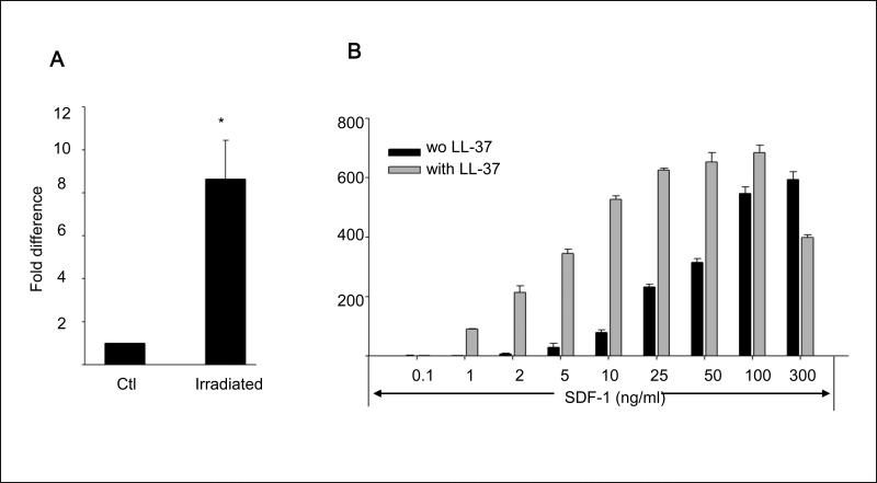
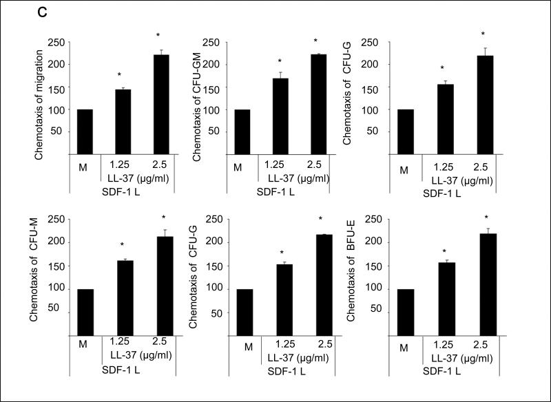
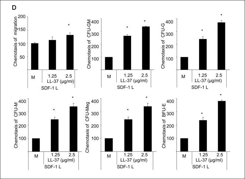
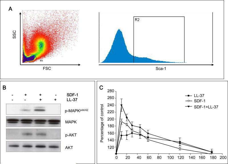
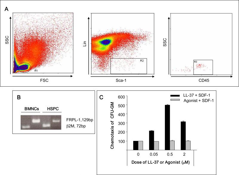
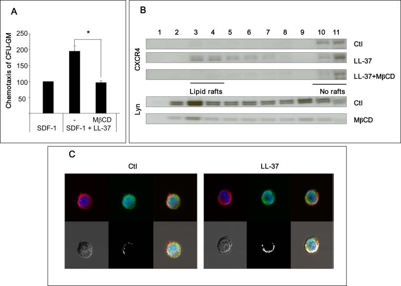
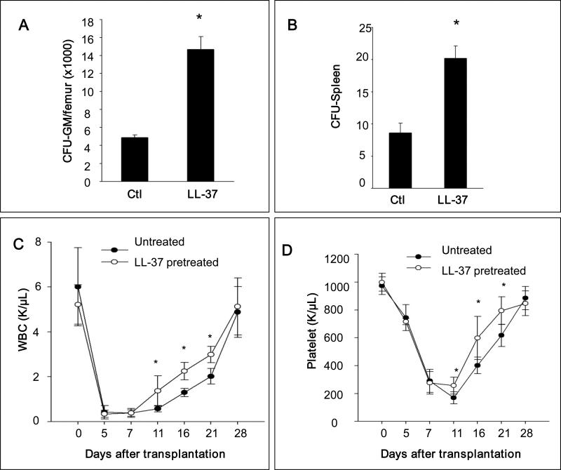
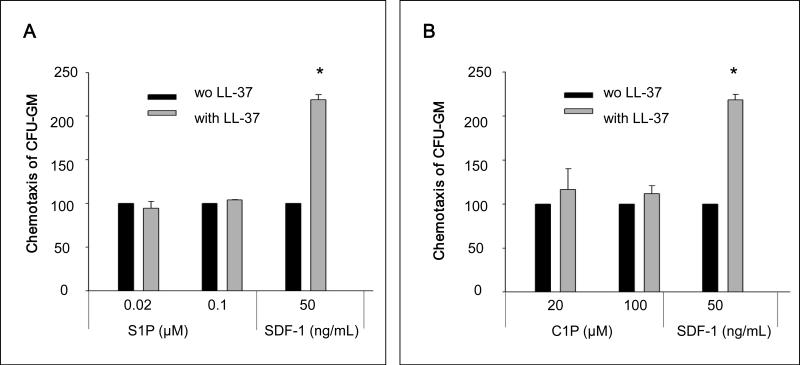
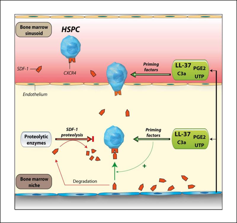
We report that the bone marrow (BM) stroma-released LL-37, a member of the cathelicidin family of antimicrobial peptides, primes/increases the responsiveness of murine and human hematopoietic stem/progenitor cells (HSPCs) to an α-chemokine stromal-derived factor-1 (SDF-1) gradient. Accordingly, LL-37 is upregulated in irradiated BM cells and enhances the chemotactic responsiveness of hematopoietic progenitors from all lineages to a low physiological SDF-1 gradient as well as increasing their (i) adhesiveness, (ii) SDF-1-mediated actin polymerization and (iii) MAPK(p42/44) phosphorylation. Mice transplanted with BM cells ex vivo primed by LL-37 showed accelerated recovery of platelet and neutrophil counts by ∼3-5 days compared with mice transplanted with unprimed control cells. These priming effects were not mediated by LL-37 binding to its receptor and depended instead on the incorporation of the CXCR4 receptor into membrane lipid rafts. We propose that LL-37, which has primarily antimicrobial functions and is harmless to mammalian cells, could be clinically applied to accelerate engraftment as an ex vivo priming agent for transplanted human HSPCs. This novel approach would be particularly important in cord blood transplantations, where the number of HSCs available is usually limited.


Figures

Figure 1
Figure 1. LL-37 enhances responsiveness of mBM- and hCB-derived MNCs and HSPCs to an SDF-1 gradient
Panel A – Cathelicidin mRNA is upregulated in irradiated BM cells. Whole mBM cells were harvested 24 h after lethal irradiation (myeloablative conditioning for transplantation) and cathelicidin mRNA expression was investigated by RQ-PCR in BM cells from irradiated and control non-irradiated mice. Values are the fold change compared to non-irradiated cells. Data were combined from three independent experiments. *p<0.001. Panel B – Chemotaxis of mBM CFU-GM in response to different concentrations of SDF-1, with and without LL-37. Values are the fold increase of the number of migrated cells compared to the number of migrated cells in medium alone. Gray bars indicate the presence of LL-37 (2.5ug/ml) in the lower Transwell chambers and black bars indicate its absence. The data represent the combined results from three independent experiments performed in duplicate per group (n = 6). Panel C – The priming effect of LL-37 on a low (50ng/ml) SDF-1 gradient in murine BMNCs and clonogenic progenitors. Panel D – The priming effect of LL-37 on a low (50ng/ml) SDF-1 gradient in human BMNCs and clonogenic progenitors. Values are the percentage increase in the number of migrated cells compared with the number of migrated cells in response to SDF-1 alone (M, without LL-37). The data represent the combined results from three independent experiments performed in duplicate per group (n = 6). * p<0.05
Figure 2
Figure 2. LL-37 enhances SDF-1-dependent intracellular signaling and actin polymerization
Panel A – Sca-1+ BMNCs were sorted from the lymph gate. Panel B – In purified Sca-1+ BMNC, LL-37 increases SDF-1-mediated phosphorylation of MAPKp42/44, but not AKT. Panel C – LL-37 enhances SDF-1-mediated F-actin polymerization in purified Sca-1+ BMNCs. In both experiments, SDF-1 was employed at a dose of 50ng/ml and LL-37 at 2.5μg/ml. Representative data from three independent experiments is shown.
Figure 3
Figure 3. The LL-37-induced priming effect is not mediated by FRPL-1
Panel A – Sorting of Sca-1+CD45+Lin BMNCs. Cells from lymph gate (R1) were subjected to lineage-negative, Sca-1-positive (R2) and CD45-positive (R3) selections. Panel B – RTPCR analysis of FPRL-1 mRNA expression in murine BMNCs and BM-purified Sca-1+CD45+Lin cells. Representative data from two independent experiments is shown. Panel C – Low-dose SDF-1(10ng/ml) chemotaxis priming assay in the presence of different concentrations (0, 0.05, 0.5, 2.0 μM) of LL-37 or the FPRL-1 agonist WKYMVM. LL-37, but not WKYMVM, enhances the responsiveness of clonogenic progenitors to SDF-1 gradients. Values are shown as the percentage increase in the number of migrated cells compared to the number of migrated cells in response to SDF-1 alone. The data represent the combined results from three independent experiments performed in duplicate per group (n = 6).
Figure 4
Figure 4. The priming effect of LL-37 is dependent on enhanced incorporation of CXCR4 into lipid rafts
Panel A – LL-37-mediated enhancement of migration of murine CFU-GM in response to an SDF-1 gradient was significantly inhibited by 1 h pretreatment of cells with MβCD (2.5mM). Values are the percentage increase in the number of migrated cells compared to the number of migrated cells in response to SDF-1 alone. SDF-1 was employed at a dose of 50ng/ml and LL-37 at a dose of 2.5μg/ml. The data represent the combined results from three independent experiments performed in duplicate per group (n = 6). *P<0.01. Panel B – Western blot analysis of the localization of CXCR4 and lipid rafts in different fractions of cell membrane lipids. Membranes were enriched in lipid rafts (fractions 3–4) or depleted of lipid rafts (fractions 10–11). The human monocytic cell line, THP-1, pretreated with or without MβCD (2.5mM), was stimulated with or without LL-37 (2.5μg/ml). CXCR4 was detected in membrane fractions by western blot along with Lyn, a marker of lipid rafts. Representative results are shown from three independent experiments. Panel C – Confocal microscopy analysis of the localization of CXCR4 in lipid rafts. THP-1 cells were cultured for 6 h in serum-free medium and than stimulated without (control) or with LL-37 (2.5 μg/ml). Lipid rafts were stained with cholera toxin B subunit conjugated with fluorescein isothiocyanate (FITC, green) and CXCR4 was stained with mouse monoclonal anti-hCXCR4 IgG and Alexa Fluor 568 goat anti-mouse IgG (red) antibodies. Stained cells were examined and images were generated by using a FLUOVIEW FV1000 laser-scanning confocal microscope. White areas indicate co-localization of lipid rafts with CXCR4. The experiment was repeated three times on different cell preparations and a representative image is shown.
Figure 5
Figure 5. Pre-incubation of BMNCs with LL-37 before transplantation enhances homing and engraftment of HSPCs
Panel A – Lethally irradiated mice (6 mice per group) were transplanted with 5 × 10⁵ mBMNCs, pretreated without (control) or with LL-37 (2.5ug/ml for 30 minutes), and 11 days after transplantation femoral BMNCs were harvested and cells isolated for counting CFU-GM. Panel B – In similar experiments mice were lethally irradiated and transplanted with 1 × 10⁵ mBMNCs. Eleven days after transplantation, spleens were harvested and fixed, and CFU-S on the spleen surface was counted. No colonies were formed in lethally irradiated and non-transplanted mice (irradiation control). The data in panel A and B represent the combined results from three independent experiments (n = 9). * p<0.01. Panel C – Lethally irradiated mice were transplanted with 5 × 10⁵ mBMNCs, pretreated without (control) or with LL-37 (2.5μg/ml for 30 minutes). White blood cells (WBC) and platelets were counted at intervals (0, 5, 7, 11, 16, and 21 days after transplantation). Results are combined from two independent experiments (5 mice per group, n = 10). Black circles indicate the un-pretreated mice, and white circles indicate the mice pretreated with LL-37. * p<0.05.
Figure 6
Figure 6. The LL-37-induced priming effect is specific to the SDF-1–CXCR axis
Chemotaxis assay of murine CFU-GM in response to different concentrations of S1P (Panel A) and C1P (Panel B) with and without LL-37. Values are the percentage increase in the number of migrated cells in the presence of LL-37 compared to the number migrated cell in the absence of LL-37. Gray bars indicate the presence of LL-37 (2.5μg/ml), while black bars indicate the absence of LL-37 in the lower Transwell chambers. The data represent the combined results from three independent experiments performed in duplicate per group (n = 6). * p<0.001
Figure 7
Figure 7. The involvement of LL-37 in homing and engraftment of HSPCs
Conditioning for transplantation by radio-chemotherapy induces a proteolytic microenvironment in BM and several proteolytic enzymes are released that decrease the SDF-1 level in BM. We report here that LL-37 released from BM stroma cells and/or granulocytes present in leucophoresis products enhance/sensitize responsiveness of HSPCs to decreasing SDF-1 gradient. It has been also reported that the complement cascade cleavage fragment anaphylatoxin C3a , uridine triphosphate (UTP) , and prostaglandin E2 (PGE2) enhance the responsiveness of HSPCs to an SDF-1 gradient. These observations collectively suggest that the homing responsiveness of HSPCs is positively modulated by several factors related to inflammation or tissue injury.

Similar articles

Cited by

References

    1. Delaney C, Ratajczak MZ, Laughlin MJ. Strategies to enhance umbilical cord blood stem cell engraftment in adult patients. Expert Review of Hematology. 2010;3:273–283. 2010/06/01. - PMC - PubMed
    1. Janowska-Wieczorek A, Majka M, Kijowski J, Baj-Krzyworzeka M, Reca R, Turner AR, et al. Platelet-derived microparticles bind to hematopoietic stem/progenitor cells and enhance their engraftment. Blood. 2001 Nov 15;98:3143–3149. 2001. - PubMed
    1. Reca R, Mastellos D, Majka M, Marquez L, Ratajczak J, Franchini S, et al. Functional receptor for C3a anaphylatoxin is expressed by normal hematopoietic stem/progenitor cells, and C3a enhances their homing-related responses to SDF-1. Blood. 2003 May 15;101:3784–3793. 2003. - PubMed
    1. Shirvaikar N, Marquez-Curtis LA, Ratajczak MZ, Janowska-Wieczorek A. Hyaluronic Acid and Thrombin Upregulate MT1-MMP Through PI3K and Rac-1 Signaling and Prime the Homing-Related Responses of Cord Blood Hematopoietic Stem/Progenitor Cells. Stem Cells and Development. 2011;20:19–30. - PMC - PubMed
    1. Kimura T, Boehmler AM, Seitz G, Kuçi S, Wiesner T, Brinkmann V, et al. The sphingosine 1-phosphate receptor agonist FTY720 supports CXCR4-dependent migration and bone marrow homing of human CD34+ progenitor cells. Blood. 2004 Jun 15;103:4478–4486. 2004. - PubMed

Publication types