. 2011 Sep;9(9):e1001149.
doi: 10.1371/journal.pbio.1001149. Epub 2011 Sep 13.

Binding of superantigen toxins into the CD28 homodimer interface is essential for induction of cytokine genes that mediate lethal shock

Affiliations

Binding of superantigen toxins into the CD28 homodimer interface is essential for induction of cytokine genes that mediate lethal shock

Gila Arad et al. PLoS Biol. 2011 Sep.

Erratum in

Abstract

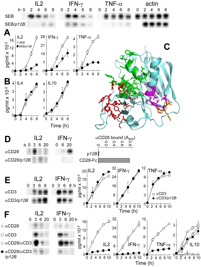
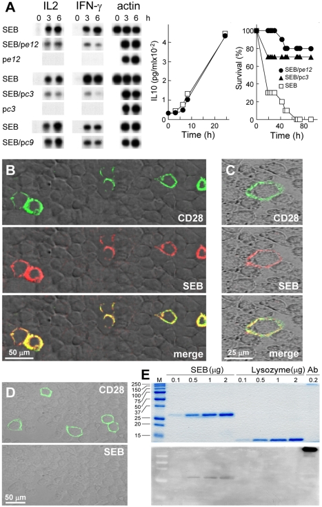
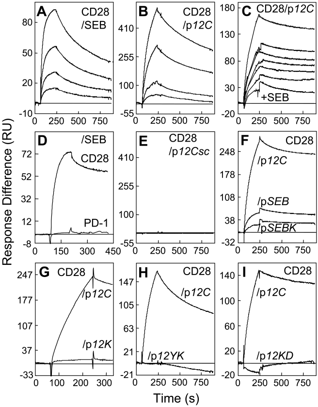
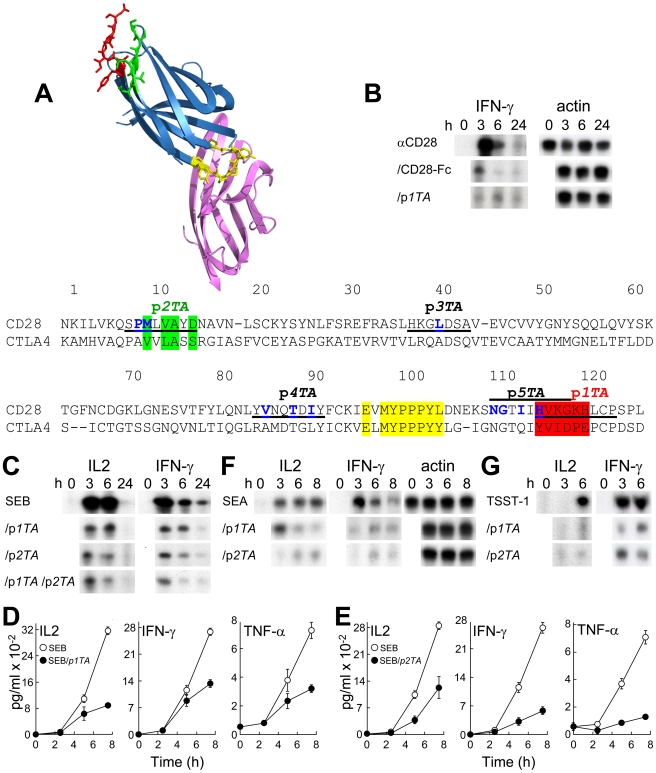
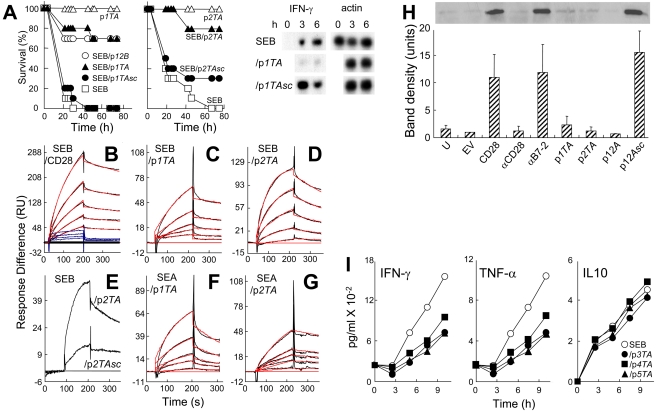
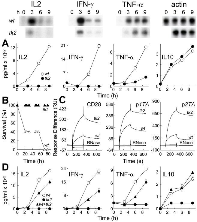
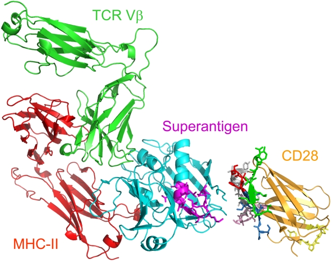
Bacterial superantigens, a diverse family of toxins, induce an inflammatory cytokine storm that can lead to lethal shock. CD28 is a homodimer expressed on T cells that functions as the principal costimulatory ligand in the immune response through an interaction with its B7 coligands, yet we show here that to elicit inflammatory cytokine gene expression and toxicity, superantigens must bind directly into the dimer interface of CD28. Preventing access of the superantigen to CD28 suffices to block its lethality. Mice were protected from lethal superantigen challenge by short peptide mimetics of the CD28 dimer interface and by peptides selected to compete with the superantigen for its binding site in CD28. Superantigens use a conserved β-strand/hinge/α-helix domain of hitherto unknown function to engage CD28. Mutation of this superantigen domain abolished inflammatory cytokine gene induction and lethality. Structural analysis showed that when a superantigen binds to the T cell receptor on the T cell and major histocompatibility class II molecule on the antigen-presenting cell, CD28 can be accommodated readily as third superantigen receptor in the quaternary complex, with the CD28 dimer interface oriented towards the β-strand/hinge/α-helix domain in the superantigen. Our findings identify the CD28 homodimer interface as a critical receptor target for superantigens. The novel role of CD28 as receptor for a class of microbial pathogens, the superantigen toxins, broadens the scope of pathogen recognition mechanisms.


Conflict of interest statement

The authors have declared that no competing interests exist.

Figures

Figure 1
Figure 1. Superantigen mimetic peptide blocks SEB action and CD28 signaling.
(A, B) p12B (10 µg/ml) inhibits SEB-mediated induction in human PBMC of IL2, IFN-γ, and TNF-α mRNA (autoradiograms) and of protein (graphs; data are shown as means ± SEM (n = 3 experiments)) (A) but not of IL4 and IL10 (B). mRNA was quantitated by RNase protection analysis; β-actin mRNA indicates equal loading of RNA. (C) In SEB structure (pdb3seb.ent), amino acid residues in the 150–161 β-strand(8)/hinge/α-helix(4) domain are shown in purple, with solvent-accessible residues N151, K153, and K154 in orange. Residues contacting MHC-II are colored red; those contacting the TCR are colored green . (D) p12B inhibits induction of IL2 and IFN-γ mRNA in PBMC by αCD28 mAb. PBMC were induced by αCD28 (2.5 µg/ml) alone or with 10 µg/ml p12B. To show that αCD28 does not bind p12B, p12B and CD28-Fc were immobilized and binding of mouse αCD28 was assayed. (E) p12B fails to inhibit induction of IL2, IFN-γ, and TNF-α genes by αCD3. PBMC were incubated with αCD3 (0.1 µg/ml) alone or with 10 µg/ml p12B. mRNA and secreted cytokines were quantitated. (F) p12B inhibits αCD28/αCD3-mediated induction of IL2, IFN-γ, and TNF-α genes but not of IL10. PBMC were incubated with αCD3, αCD28, or both, with or without 10 µg/ml p12B. mRNA and secreted cytokines were determined.
Figure 2
Figure 2. SEB binds to CD28.
(A) Phage display peptides selected by affinity for the SEB binding site in CD28 are SEB antagonists that protect mice from killing by SEB. PBMC were induced with SEB alone or with 0.1 µg/ml pc3, pe12, or pc9, a negative control. IL2 and IFN-γ mRNA are shown; β-actin mRNA indicates equal loading of RNA. For pe12, IL10 was determined (data are shown as means ± SEM (n = 3 experiments)). Mice (n = 10 per group) were challenged with 6 µg SEB alone or with 0.2 µg pe12 or 0.5 µg pc3; p for survival, 10−4. (B–D) Binding of SEB to cell surface CD28. Representative fields of confocal microscopy are shown. In (B), HEK293-T cells were transfected to express CD28-GFP fusion protein (green) and after 48 h incubated for 1 h with Alexa-Fluor-633-labeled SEB (red). In (C), BHK-21 hamster cells were transfected with CD28 cDNA vector and after 48 h incubated successively for 30 min with labeled SEB (red), goat polyclonal αCD28, and Cy2-labeled donkey anti-goat IgG (green). In (D), BHK-21 cells were transfected to express CD28 and after 48 h incubated first with goat polyclonal αCD28 and Cy2-labeled donkey anti-goat IgG (top) and only then with labeled SEB (bottom). (E) Binding of SEB to CD28. SEB, lysozyme, and polyclonal αCD28 (Ab) were separated on duplicate SDS-PAGE gels. Coomassie blue staining (top); far-western blot with CD28-Fc (bottom); M, size marker.
Figure 3
Figure 3. SEB binds CD28 through its β-strand(8)/hinge/α-helix(4) domain.
(A and B) Binding of CD28-Fc in 2-fold increments from 0.25 µM to immobilized SEB (A) and from 0.125 µM to p12C (B). (C) Binding of CD28-Fc (2 µM) to p12C without SEB (top curve) or with SEB in 2-fold increments from 0.78 µM (6 lower curves, top to bottom). (D) Binding of 2 µM PD1-Fc and CD28-Fc to immobilized SEB. (E) Binding of CD28-Fc in 2-fold increments from 0.125 µM to scrambled p12Csc (EKAKYTQLVKDN). (F–I) Mutations in SEB β-strand(8)/hinge/α-helix(4) domain peptides reduce binding of 2 µM CD28-Fc. SPR responses are compared for immobilized p12C and pSEB (TNKKKVTAQELD), pSEBK (TNAKKVTAQELD) (F), p12K (YNAKKATVQELD) (G), p12YK (ANAKKATVQELD) (H), and p12KD (YNAKKATVQELA) (I). Representative SPR responses are shown.
Figure 4
Figure 4. Peptide mimetics of the CD28 dimer interface are superantigen antagonists.
(A) CTLA-4/B7-2 complex (1I85.pdb; [1]) and the dimer interface in CTLA-4. B7-2 is shown in purple and CTLA-4 in blue, with MYPPPY in yellow, YVIDPE (HVKGKH in CD28) in red, and VVLASS (MLVAYD in CD28) in green, as in the sequence alignment of the extracellular domains of human CD28 and CTLA-4. Amino acids at the crystallographic dimer interface of CD28 are shown in blue. Positions of peptides are underlined. (B) p1TA antagonizes induction of IFN-γ mRNA by αCD28. PBMC were induced by αCD28 (2.5 µg/ml) alone or with 10 µg/ml of CD28-Fc or p1TA. IFN-γ and β-actin mRNA was determined. (C–E) CD28 dimer interface peptides p1TA and p2TA (0.1 µg/ml) inhibit induction by SEB of IL2 and IFN-γ mRNA (C) and IL2, IFN-γ, and TNF-α (D and E) in PBMC (data are shown as means ± SEM (n = 3 experiments)). (F and G) Induction of IL2 and IFN-γ mRNA by SEA (F) and TSST-1 (G) alone or with 0.1 (F) or 10 µg/ml peptide (G); β-actin mRNA indicates equal loading of RNA.
Figure 5
Figure 5. CD28 dimer interface mimetic peptides bind and antagonize superantigens and protect mice from lethal challenge.
(A) Mice (n = 10 per group) were challenged with 6 µg SEB alone or with 1 µg p1TA, 5 µg p12B, or 1 µg scrambled peptide p1TAsc (CHGHLVPKK), or with 0.2 µg p2TA or p2TAsc (ASMDYPVL); controls without SEB received 25 µg peptide; p for survival, 2×10⁻⁸ (p1TA), 0.38 (p1TAsc), 3×10⁻⁶ (p2TA), and 0.19 (p2TAsc). PBMC were induced with SEB alone or with 1 µg/ml p1TA or p1TAsc; IFN-γ and β-actin mRNA was determined. (B–G) Representative SPR responses for binding of SEB to immobilized CD28-Fc, p1TA, and p2TA (B–D), of SEB to p2TAsc or p2TA (E) and of SEA to p1TA and p2TA (F and G). Graphical fits to the binding curves are presented in red color; kinetic parameters show the specificity of these interactions (Table S1). Analyte concentrations increased in 2-fold increments from 0.156 (C and F), 0.312 (G), 1.56 (B), and 3.12 µM (D); in (E), binding of p2TA and p2TAsc is shown for the highest of 5 concentrations tested, 10 µM. In (B), curves in blue color show binding of ribonuclease A in four 2-fold increments from 3.12 µM to immobilized CD28-Fc. (H) Peptide mimetics of the CD28 dimer interface and of the β-strand(8)/hinge/α-helix(4) domain in SEB inhibit binding of SEB to cell-surface CD28. 293-T cells were transfected to express CD28 and after 36 h incubated for 1 h without addition (CD28) or with 0.1 µg/ml αCD28, anti-B7-2 or 10 µg/ml p1TA, p2TA, p12A, or its scrambled form p12Asc (EKAKYTQLVKDN), before further incubation for 1 h with 15 µg/ml wt SEB. Cells were washed 3 times with cold phosphate-buffered saline before lysis. Western blot of equal amounts of total cell protein (Bradford assay) with αSEB mAb and horseradish peroxidase-conjugated goat anti-mouse IgG shown is from a representative experiment. Bar graph shows intensity of bands for three independent experiments ± SD. U, untransfected; EV, empty vector. (I) IFN-γ, TNF-α, and IL10 (data are shown as means ± SEM (n = 3)) in medium from PBMC induced with SEB alone or with 0.1 µg/ml p3TA, p4TA, or p5TA.
Figure 6
Figure 6. Mutation of SEB β-strand(8)/hinge/α-helix(4) domain affects binding to CD28, Th1 cytokine gene induction and lethality.
(A) Induction of cytokine genes by wt and tk2 mutant SEB. PBMC were induced with 1 ng/ml wt or tk2 SEB; mRNA (autoradiograms) and secreted cytokines (graphs; data are shown as means ± SEM (n = 3)) are depicted. (B) Lack of lethality of tk2 SEB. Mice (n = 5 per group) were challenged with 10 µg wt or tk2 SEB; p for survival, 10−5. (C) Affinity of tk2 SEB for CD28. Representative SPR responses for binding of wt and tk2 SEB to immobilized CD28-Fc, p1TA, and p2TA are shown for 5 µM wt and tk2 SEB or RNase A to facilitate comparison; kinetic data were collected for five 2-fold increments in protein concentration from 1.25 µM (Table S2). (D) Dominant-negative phenotype of tk2 SEB. PBMC were induced with 1 ng/ml wt SEB, 0.1 ng/ml tk2 SEB, or both; secreted cytokines were determined as shown (data are shown as means ± SEM (n = 3)).
Figure 7
Figure 7. Model for superantigen action through binding to CD28.
Structural model for Th1 cell activation by superantigens through simultaneous binding to CD28, MHC-II, and TCR. SEC3 (cyan), a close relative of SEB, in complex with TCR Vβ (green) and MHC-II (red) extracellular domains (1jck.pdb; [38]). The SEC3 β-strand/hinge/α-helix domain in magenta is freely accessible to the N-terminal 118-residue portion of the CD28 extracellular domain (1yjd.pdb; [21]). CD28 homodimer interface peptides (Figure 4A) are shown: p1TA (HVK resolved in 1yjd.pdb is red), p2TA (green), p3TA (pink), p4TA (grey), and p5TA (SNGTII is blue); MYPPPY is yellow. CD28 and TCR are oriented such that their trans-membrane domains (not shown) can enter the T cell at top; the MHC-II is oriented towards the antigen-presenting cell at bottom; for simplicity, cell surfaces are not shown.

References

    1. Schwartz J. C, Zhang X, Fedorov A. A, Nathenson S. G, Almo S. C. Structural basis for co-stimulation by the human CTLA-4/B7-2 complex. Nature. 2001;410:604–608. - PubMed
    1. Sharpe A. H, Freeman G. J. The B7-CD28 superfamily. Nat Rev Immunol. 2002;2:116–126. - PubMed
    1. Riley J. L, June C. H. The CD28 family: a T-cell rheostat for therapeutic control of T-cell activation. Blood. 2005;105:13–21. - PubMed
    1. Lindsten T, Lee K. P, Harris E. S, Petryniak B, Craighead N, et al. Characterization of CTLA-4 structure and expression on human T cells. J Immunol. 1993;151:3489–3499. - PubMed
    1. Collins A. V, Brodie D. W, Gilbert R. J, Iaboni A, Manso-Sancho R, et al. The interaction properties of costimulatory molecules revisited. Immunity. 2002;17:201–210. - PubMed

Publication types

MeSH terms