Murine gamma-herpesvirus immortalization of fetal liver-derived B cells requires both the viral cyclin D homolog and latency-associated nuclear antigen
- PMID: 21931547
- PMCID: PMC3169539
- DOI: 10.1371/journal.ppat.1002220
Murine gamma-herpesvirus immortalization of fetal liver-derived B cells requires both the viral cyclin D homolog and latency-associated nuclear antigen
Abstract
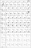
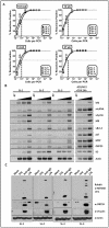
Human gammaherpesviruses are associated with the development of lymphoproliferative diseases and B cell lymphomas, particularly in immunosuppressed hosts. Understanding the molecular mechanisms by which human gammaherpesviruses cause disease is hampered by the lack of convenient small animal models to study them. However, infection of laboratory strains of mice with the rodent virus murine gammaherpesvirus 68 (MHV68) has been useful in gaining insights into how gammaherpesviruses contribute to the genesis and progression of lymphoproliferative lesions. In this report we make the novel observation that MHV68 infection of murine day 15 fetal liver cells results in their immortalization and differentiation into B plasmablasts that can be propagated indefinitely in vitro, and can establish metastasizing lymphomas in mice lacking normal immune competence. The phenotype of the MHV68 immortalized B cell lines is similar to that observed in lymphomas caused by KSHV and resembles the favored phenotype observed during MHV68 infection in vivo. All established cell lines maintained the MHV68 genome, with limited viral gene expression and little or no detectable virus production - although virus reactivation could be induced upon crosslinking surface Ig. Notably, transcription of the genes encoding the MHV68 viral cyclin D homolog (v-cyclin) and the homolog of the KSHV latency-associated nuclear antigen (LANA), both of which are conserved among characterized γ2-herpesviruses, could consistently be detected in the established B cell lines. Furthermore, we show that the v-cyclin and LANA homologs are required for MHV68 immortalization of murine B cells. In contrast the M2 gene, which is unique to MHV68 and plays a role in latency and virus reactivation in vivo, was dispensable for B cell immortalization. This new model of gammaherpesvirus-driven B cell immortalization and differentiation in a small animal model establishes an experimental system for detailed investigation of the role of gammaherpesvirus gene products and host responses in the genesis and progression of gammaherpesvirus-associated lymphomas, and presents a convenient system to evaluate therapeutic modalities.
Conflict of interest statement
The authors have declared that no competing interests exist.
Figures


Similar articles
-
Murine Gammaherpesvirus 68 Expressing Kaposi Sarcoma-Associated Herpesvirus Latency-Associated Nuclear Antigen (LANA) Reveals both Functional Conservation and Divergence in LANA Homologs.J Virol. 2017 Sep 12;91(19):e00992-17. doi: 10.1128/JVI.00992-17. Print 2017 Oct 1. J Virol. 2017. PMID: 28747501 Free PMC article.
-
Murine gammaherpesvirus 68 reactivation from B cells requires IRF4 but not XBP-1.J Virol. 2014 Oct;88(19):11600-10. doi: 10.1128/JVI.01876-14. Epub 2014 Jul 30. J Virol. 2014. PMID: 25078688 Free PMC article.
-
Identification of Viral and Host Proteins That Interact with Murine Gammaherpesvirus 68 Latency-Associated Nuclear Antigen during Lytic Replication: a Role for Hsc70 in Viral Replication.J Virol. 2015 Nov 18;90(3):1397-413. doi: 10.1128/JVI.02022-15. Print 2016 Feb 1. J Virol. 2015. PMID: 26581985 Free PMC article.
-
Murine Gammaherpesvirus 68: A Small Animal Model for Gammaherpesvirus-Associated Diseases.Adv Exp Med Biol. 2017;1018:225-236. doi: 10.1007/978-981-10-5765-6_14. Adv Exp Med Biol. 2017. PMID: 29052141 Review.
-
Gammaherpesviruses of New World primates.In: Arvin A, Campadelli-Fiume G, Mocarski E, Moore PS, Roizman B, Whitley R, Yamanishi K, editors. Human Herpesviruses: Biology, Therapy, and Immunoprophylaxis. Cambridge: Cambridge University Press; 2007. Chapter 60. In: Arvin A, Campadelli-Fiume G, Mocarski E, Moore PS, Roizman B, Whitley R, Yamanishi K, editors. Human Herpesviruses: Biology, Therapy, and Immunoprophylaxis. Cambridge: Cambridge University Press; 2007. Chapter 60. PMID: 21348092 Free Books & Documents. Review.
Cited by
-
Murine Models of Chronic Viral Infections and Associated Cancers.Mol Biol. 2022;56(5):649-667. doi: 10.1134/S0026893322050028. Epub 2022 Oct 5. Mol Biol. 2022. PMID: 36217336 Free PMC article.
-
Interleukin 16 contributes to gammaherpesvirus pathogenesis by inhibiting viral reactivation.PLoS Pathog. 2020 Jul 31;16(7):e1008701. doi: 10.1371/journal.ppat.1008701. eCollection 2020 Jul. PLoS Pathog. 2020. PMID: 32735617 Free PMC article.
-
CD4 and CD8 T cells directly recognize murine gammaherpesvirus 68-immortalized cells and prevent tumor outgrowth.J Virol. 2013 May;87(10):6051-4. doi: 10.1128/JVI.00375-13. Epub 2013 Mar 20. J Virol. 2013. PMID: 23514885 Free PMC article.
-
Animal models of tumorigenic herpesviruses--an update.Curr Opin Virol. 2015 Oct;14:145-50. doi: 10.1016/j.coviro.2015.09.006. Curr Opin Virol. 2015. PMID: 26476352 Free PMC article. Review.
-
CD154:CD11b blockade enhances CD8+ T cell differentiation during infection but not transplantation.JCI Insight. 2025 Jun 9;10(11):e184843. doi: 10.1172/jci.insight.184843. eCollection 2025 Jun 9. JCI Insight. 2025. PMID: 40485581 Free PMC article.
References
-
- Young LS, Rickinson AB. Epstein-Barr virus: 40 years on. Nat Rev Cancer. 2004;4:757–768. - PubMed
-
- Thorley-Lawson DA, Allday MJ. The curious case of the tumour virus: 50 years of Burkitt's lymphoma. Nat Rev Microbiol. 2008;6:913–924. - PubMed
-
- Chang Y, Cesarman E, Pessin MS, Lee F, Culpepper J, et al. Identification of herpesvirus-like DNA sequences in AIDS-associated Kaposi's sarcoma. Science. 1994;266:1865–1869. - PubMed
-
- Cesarman E, Chang Y, Moore PS, Said JW, Knowles DM. Kaposi's sarcoma-associated herpesvirus-like DNA sequences in AIDS-related body-cavity-based lymphomas. N Engl J Med. 1995;332:1186–1191. - PubMed
Publication types
MeSH terms
Substances
Grants and funding
LinkOut - more resources
Full Text Sources

