. 2011;6(9):e22356.
doi: 10.1371/journal.pone.0022356. Epub 2011 Sep 12.

cIAP1/2 are direct E3 ligases conjugating diverse types of ubiquitin chains to receptor interacting proteins kinases 1 to 4 (RIP1-4)

Affiliations

cIAP1/2 are direct E3 ligases conjugating diverse types of ubiquitin chains to receptor interacting proteins kinases 1 to 4 (RIP1-4)

Mathieu J M Bertrand et al. PLoS One. 2011.

Abstract

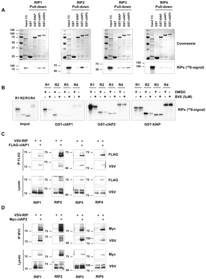
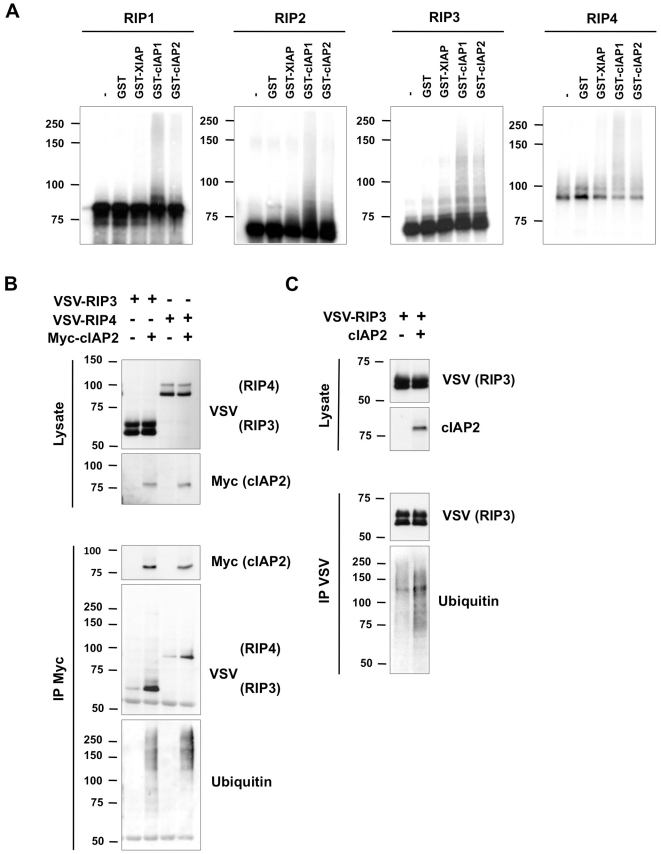
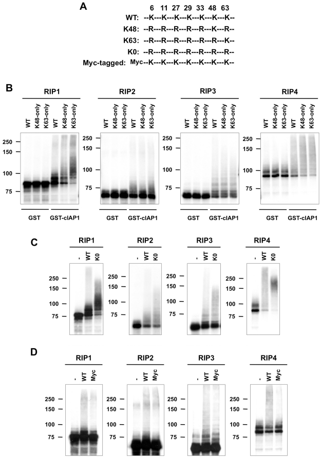
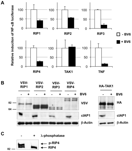
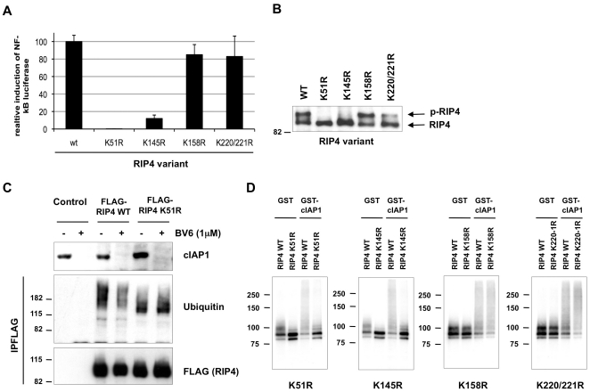
The RIP kinases have emerged as essential mediators of cellular stress that integrate both extracellular stimuli emanating from various cell-surface receptors and signals coming from intracellular pattern recognition receptors. The molecular mechanisms regulating the ability of the RIP proteins to transduce the stress signals remain poorly understood, but seem to rely only partially on their kinase activities. Recent studies on RIP1 and RIP2 have highlighted the importance of ubiquitination as a key process regulating their capacity to activate downstream signaling pathways. In this study, we found that XIAP, cIAP1 and cIAP2 not only directly bind to RIP1 and RIP2 but also to RIP3 and RIP4. We show that cIAP1 and cIAP2 are direct E3 ubiquitin ligases for all four RIP proteins and that cIAP1 is capable of conjugating the RIPs with diverse types of ubiquitin chains, including linear chains. Consistently, we show that repressing cIAP1/2 levels affects the activation of NF-κB that is dependent on RIP1, -2, -3 and -4. Finally, we identified Lys51 and Lys145 of RIP4 as two critical residues for cIAP1-mediated ubiquitination and NF-κB activation.


Conflict of interest statement

Competing Interests: The authors have declared that no competing interests exist.

Figures

Figure 1
Figure 1. XIAP, cIAP1 and cIAP2 bind RIP1-4 proteins.
(A) In vitro-transcribed and -translated RIP1, 2, 3 and 4 labeled with 35S-methionine (Input) was incubated overnight with bacterially produced GST, GST-XIAP, GST-cIAP1, or GST-cIAP2 bound to Sepharose beads. The beads were washed extensively and run on an acrylamide gel. Binding of RIPs was revealed by autoradiography (pull-down). (B) In vitro-transcribed and -translated RIP1, 2, 3 and 4 labeled with 35S-methionine (Input) was incubated overnight with bacterially produced GST-XIAP, GST-cIAP1, or GST-cIAP2 bound to Sepharose beads in presence of BV6 (5 µM) or DMSO as a control. The beads were washed extensively and run on an acrylamide gel. Binding of RIPs was revealed by autoradiography. (C) HEK293T cells were transfected with VSV-tagged RIP plasmids in the absence or presence of a FLAG-tagged cIAP1 plasmid. cIAP1 was immunoprecipitated in NP-40 buffer using anti-FLAG antibody and coimmunoprecipitated RIPs were revealed by immunoblotting with anti-VSV antibody. Protein expression in the lysates is shown. (D) HEK293T cells were transfected with VSV-tagged RIP plasmids in the absence or presence of a Myc-tagged cIAP2 plasmid. cIAP2 was immunoprecipitated in NP-40 buffer using anti-Myc antibody and coimmunoprecipitated RIPs were revealed by immunoblotting with anti-VSV antibody. Protein expression in the lysates is shown.
Figure 2
Figure 2. cIAP1 and cIAP2 are direct E3 ubiquitin ligases for RIP1–4 proteins.
(A) In vitro ubiquitination assays were performed on in vitro-transcribed and -translated RIP1–4 proteins labeled with 35S-methionine. GST, GST-XIAP, GST-cIAP1, or GST-cIAP2 was used as E3 component, UbcH5a as the E2 component, and using wild-type ubiquitin. RIP ubiquitination was revealed by autoradiography and appears as a smear in the figure. (B) HEK293T cells were transfected with VSV-tagged RIP3 and RIP4 plasmids in the absence or presence of a Myc-tagged cIAP2 plasmid. cIAP2 was immunoprecipitated in NP-40 buffer using anti-Myc antibody and coimmunoprecipitated ubiquitinated RIP3 and RIP4 were revealed by immunoblotting with anti-VSV and anti-ubiquitin antibodies. Protein expression in the lysates is shown. (C) HEK293T cells were transfected with VSV-tagged RIP3 plasmid in the absence or presence of a cIAP2 plasmid. RIP3 was immunoprecipitated in RIPA buffer using anti-VSV antibody and ubiquitinated RIP3 was revealed by immunoblotting with anti-VSV and anti-ubiquitin antibodies. Protein expression in the lysates is shown.
Figure 3
Figure 3. cIAP1 conjugates RIP1–4 proteins with Lys48-, Lys63-, and linear ubiquitin chains.
(A) Schematic representation of the different ubiquitin mutants used in the experiments. (B–D) In vitro ubiquitination assays were performed on in vitro-transcribed and -translated RIP1–4 proteins labeled with 35S-methionine. GST or GST-cIAP1 was used as E3 component and UbcH5a as the E2 component. The reaction was carried out in presence of (B) wild-type (WT) ubiquitin, K48-only ubiquitin and K63-only ubiquitin; (C) no ubiquitin, wild-type (WT) ubiquitin and lysine free ubiquitin (KO); (D) no ubiquitin, wild-type (WT) ubiquitin and Myc-tagged ubiquitin. Ubiquitination of the RIPs was revealed by autoradiography.
Figure 4
Figure 4. Requirement of cIAP1/2 for RIP1–4-mediated NF-κB activation.
(A) NF-κB luciferase assays were performed on lysates from HEK293T cells transfected with a plasmid encoding the indicated RIP kinase or TAK1. Cells were left untreated or were treated with 1 µM of BV6. Stimulation with hTNF was used as a positive control. (B) Western blot showing expression of the tagged-RIP kinases, cIAP1 and TAK1 in the lysates. (C) Phosphorylation of RIP4 was revealed by λ-phosphatase treatment of immunopurified Flag-RIP4 overexpressed in HEK293T cells. Samples were analyzed by western blot using anti-Flag antibody.
Figure 5
Figure 5. Lysine residues K51 and K145 of RIP4 are critical for cIAP1-mediated ubiquitination and NF-κB activation.
(A) Flag-tagged wild type RIP4, K51R, K145R, K158R and K220/221R mutants were ectopically expressed in HEK293T cells together with a NF-κB luciferase reporter. 24h after transfection, lysates were prepared and tested for luciferase activity. Relative luciferase activity is depicted after correction for transfection efficiency, ± S.D. (B) Lysates described in (B) were used for western blotting analysis with an anti-Flag antibody in order to detect expression of the different Flag-tagged RIP4 variants. (C) HEK293T cells were transfected with FLAG-tagged wild-type RIP4, the K51R mutant or with empty vector. The medium was replaced 8h post-transfection with medium containing, or not, BV6 (1 µM). The RIP4 proteins were immunoprecipitated 18h later in RIPA buffer using anti-FLAG antibody and ubiquitinated RIP4 proteins were revealed by immunoblotting with anti-FLAG and anti-ubiquitin antibodies. (D) In vitro ubiquitination assays were performed on in vitro-transcribed and -translated wild-type RIP4, K51R, K145R, K158R and K220/221R mutants labeled with 35S-methionine. GST or GST-cIAP1 was used as E3 component and UbcH5a as E2 component. The reaction was carried out in presence of wild-type ubiquitin. RIP4 ubiquitination was revealed by autoradiography.

Similar articles

Cited by

References

    1. Meylan E, Tschopp J. The RIP kinases: crucial integrators of cellular stress. Trends Biochem Sci. 2005;30:151–159. - PubMed
    1. Cusson-Hermance N, Khurana S, Lee TH, Fitzgerald KA, Kelliher MA. Rip1 mediates the Trif-dependent toll-like receptor 3- and 4-induced NF-{kappa}B activation but does not contribute to interferon regulatory factor 3 activation. J Biol Chem. 2005;280:36560–36566. - PubMed
    1. Meylan E, Burns K, Hofmann K, Blancheteau V, Martinon F, et al. RIP1 is an essential mediator of Toll-like receptor 3-induced NF-kappa B activation. Nat Immunol. 2004;5:503–507. - PubMed
    1. Janssens S, Tinel A, Lippens S, Tschopp J. PIDD mediates NF-kappaB activation in response to DNA damage. Cell. 2005;123:1079–1092. - PubMed
    1. Park JH, Kim YG, McDonald C, Kanneganti TD, Hasegawa M, et al. RICK/RIP2 mediates innate immune responses induced through Nod1 and Nod2 but not TLRs. J Immunol. 2007;178:2380–2386. - PubMed

Publication types

MeSH terms

Substances