. 2011;6(9):e23082.
doi: 10.1371/journal.pone.0023082. Epub 2011 Sep 13.

Egr-1 induces a profibrotic injury/repair gene program associated with systemic sclerosis

Affiliations

Egr-1 induces a profibrotic injury/repair gene program associated with systemic sclerosis

Swati Bhattacharyya et al. PLoS One. 2011.

Abstract

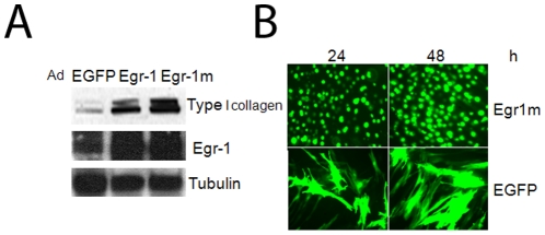
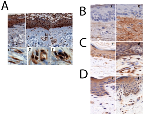
Transforming growth factor-ß (TGF-ß) signaling is implicated in the pathogenesis of fibrosis in scleroderma or systemic sclerosis (SSc), but the precise mechanisms are poorly understood. The immediate-early gene Egr-1 is an inducible transcription factor with key roles in mediating fibrotic TGF-ß responses. To elucidate Egr-1 function in SSc-associated fibrosis, we examined change in gene expression induced by Egr-1 in human fibroblasts at the genome-wide level. Using microarray expression analysis, we derived a fibroblast "Egr-1-responsive gene signature" comprising over 600 genes involved in cell proliferation, TGF-ß signaling, wound healing, extracellular matrix synthesis and vascular development. The experimentally derived "Egr-1-responsive gene signature" was then evaluated in an expression microarray dataset comprising skin biopsies from 27 patients with localized and systemic forms of scleroderma and six healthy controls. We found that the "Egr-1 responsive gene signature" was substantially enriched in the "diffuse-proliferation" subset comprising exclusively of patients with diffuse cutaneous SSc (dcSSc) of skin biopsies. A number of Egr-1-regulated genes was also associated with the "inflammatory" intrinsic subset. Only a minority of Egr-1-regulated genes was concordantly regulated by TGF-ß. These results indicate that Egr-1 induces a distinct profibrotic/wound healing gene expression program in fibroblasts that is associated with skin biopsies from SSc patients with diffuse cutaneous disease. These observations suggest that targeting Egr-1 expression or activity might be a novel therapeutic strategy to control fibrosis in specific SSc subsets.


Conflict of interest statement

Competing Interests: The authors have declared that no competing interests exist.

Figures

Figure 1
Figure 1. Adenovirus-mediated Egr-1 expression in human skin fibroblasts.
Confluent dermal fibroblasts were infected with Ad-EGFP, Egr-1 or Egr-1m (100 MOI) for 48 h. A. Cultures were harvested and whole cell lysates were subjected to Western analysis. Representative immunoblots. B. Fibroblasts were examined by fluorescence microscopy. Representative image. (Original magnification ×400).
Figure 2
Figure 2. Egr-1-induced transcriptional responses in skin fibroblasts.
Dermal fibroblasts were infected with Ad-EGFP or Ad-Egr-1m (100 MOI). At the end of 24 or 48 h incubation, total RNA was isolated and subjected to genome-wide transcriptional analysis using Illumina Microarray chips (A,B) or real-time qPCR (C). A. Heatmap of differentially expressed genes (FDR<0.01 and >2- fold-change) (48 h). The color represents the fold-change of Egr-1 in comparison with the average of control sample (red = increased, green = decreased). Each row represents a probe and each column represents one sample. Genes with similar changes in expression pattern compared to the control are clustered together for 24 and 48 h. B. Comparing a subset of biological processes significantly enriched (p<0.001) with Egr-1-regulated genes at 24 and 48 h. The number in the plot indicates the differentially expressed genes belonging to individual GO categories (row) at corresponding time point (column). The total number of genes at each time point (column) is shown below the Table. The background color represents the statistical significance of a particular biological process overrepresented in the differential gene list as estimated by Hyper-geometric test. C. Real-time qPCR. Results, normalized with GAPDH mRNA, are the means ± S.D. of triplicate determinations from a representative experiment.
Figure 3
Figure 3. Genes regulated by both TGF-ß and AdEgr-1.
Skin fibroblasts were infected with Ad-EGFP or incubated with TGF-ß in parallel for 48 h. Total RNA was subjected to microarray analysis using Illumina chips. A. Venn diagrams of the genes regulated by TGF-ß and Egr-1. Left, up-regulated genes; right, down-regulated genes. B. Ingenuity Pathway Analysis showing canonical signaling pathways enriched with 98 fibroblast genes regulated by both Egr-1 and TGF-ß. The Y axis shows the pathway enrichment.
Figure 4
Figure 4. Expression of the Egr-1-responsive gene signature in SSc skin biopsies.
A. Egr-1-responsive genes are aligned with the gene expression data from dcSSc and healthy control skin biopsies. The samples were clustered using the 647 genes that comprise Egr-1-responsive gene signature. Dendrogram shows clear differences in skin biopsies. The left branch of the dendogram (highlighted in red and blue), comprises solely dcSSc biopsies clustering with the diffuse-proliferation intrinsic subsets (diffuse 1 and diffuse 2). The right branch includes remaining dcSSc samples, as well as lcSSc, localized scleroderma and all healthy controls. Quantitation of Egr-1 signaling in each biopsy by Pearson correlation is shown below the heatmap. B. Genes showing high expression in dcSSc skin biopsies and in Egr-1-expressing fibroblasts. C. Genes associated with the inflammatory and normal-like intrinsic subsets, or low expression in proliferation subset and Egr-1-expressing fibroblasts. D. Genes showing low expression in the proliferation subset but high expression in Egr-1-expressing fibroblasts and inflammatory subset.
Figure 5
Figure 5. Expression of the Egr-1 and its target genes in SSc skin biopsies.
Immunohistochemistry. Skin biopsies from SSc patients and healthy controls were immunostained with antibodies. Representative images. A. Egr-1. a healthy control, b–f SSc. (a–c original magnification, ×100; d–f original magnification, ×400). B–D. COMP, E2F7 and GDF6. a,c,e healthy controls; b,d,f SSc.(Original magnification ×400).

Similar articles

Cited by

References

    1. Jimenez SA, Derk CT. Following the molecular pathways toward an understanding of the pathogenesis of systemic sclerosis. Ann Intern Med. 2004;140:37–45. - PubMed
    1. Rosenbloom J, Castro SV, Jimenez SA. Narrative review: fibrotic diseases Cellular and molecular mechanisms and novel therapies. Ann Intern Med. 2010;152:159–166. - PubMed
    1. Varga J, Whitfield ML. Transforming growth factor-beta in systemic sclerosis (scleroderma). Front Biosci. 2009;1:226–235. - PubMed
    1. Varga J, Pasche B. Transforming growth factor beta as a therapeutic target in systemic sclerosis. Nat Rev Rheumatol. 2009;5:200–206. - PMC - PubMed
    1. Thiel G, Cibelli G. Regulation of life and death by the zinc finger transcription factor Egr-1. J Cell Physiol. 2002;193:287–292. - PubMed

Publication types

MeSH terms

Substances