. 2011;6(9):e23341.
doi: 10.1371/journal.pone.0023341. Epub 2011 Sep 9.

Lithium suppresses astrogliogenesis by neural stem and progenitor cells by inhibiting STAT3 pathway independently of glycogen synthase kinase 3 beta

Affiliations

Lithium suppresses astrogliogenesis by neural stem and progenitor cells by inhibiting STAT3 pathway independently of glycogen synthase kinase 3 beta

Zhenzhong Zhu et al. PLoS One. 2011.

Abstract

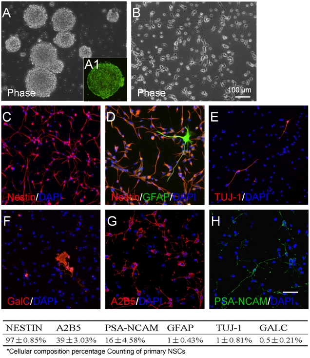
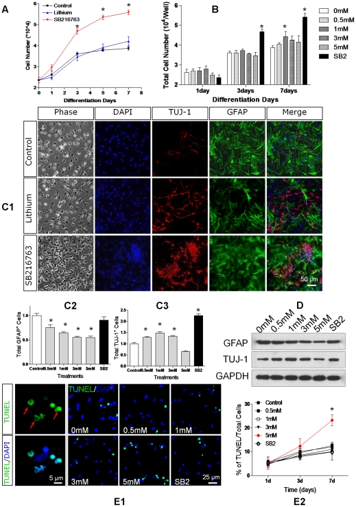
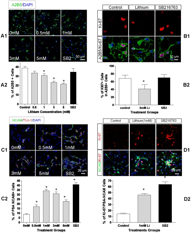
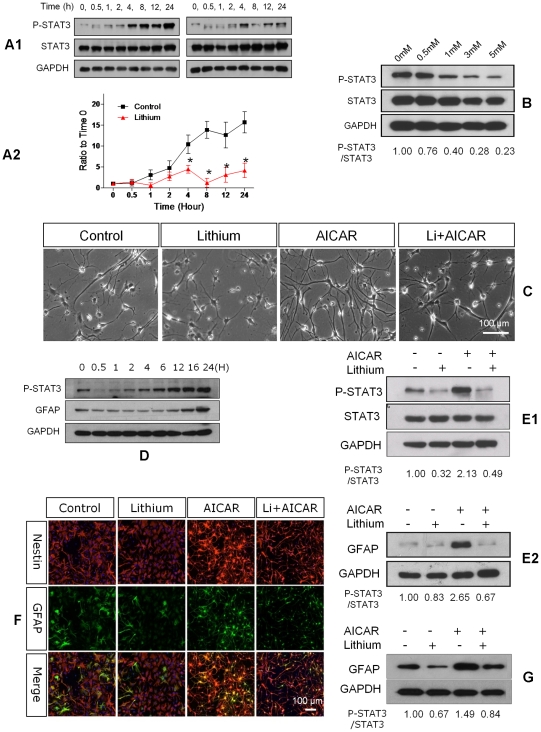
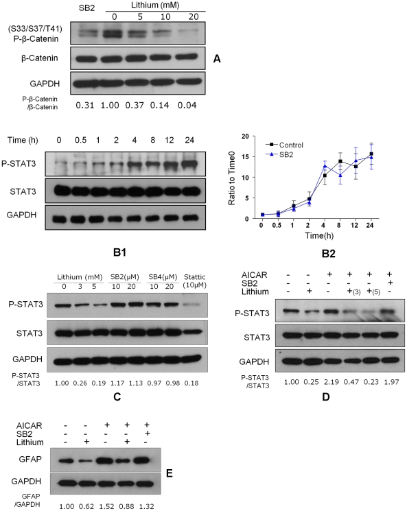
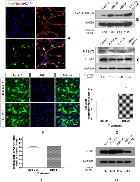
Transplanted neural stem and progenitor cells (NSCs) produce mostly astrocytes in injured spinal cords. Lithium stimulates neurogenesis by inhibiting GSK3b (glycogen synthetase kinase 3-beta) and increasing WNT/beta catenin. Lithium suppresses astrogliogenesis but the mechanisms were unclear. We cultured NSCs from subventricular zone of neonatal rats and showed that lithium reduced NSC production of astrocytes as well as proliferation of glia restricted progenitor (GRP) cells. Lithium strongly inhibited STAT3 (signal transducer and activator of transcription 3) activation, a messenger system known to promote astrogliogenesis and cancer. Lithium abolished STAT3 activation and astrogliogenesis induced by a STAT3 agonist AICAR (5-aminoimidazole-4-carboxamide 1-beta-D-ribofuranoside), suggesting that lithium suppresses astrogliogenesis by inhibiting STAT3. GSK3β inhibition either by a specific GSK3β inhibitor SB216763 or overexpression of GID5-6 (GSK3β Interaction Domain aa380 to 404) did not suppress astrogliogenesis and GRP proliferation. GSK3β inhibition also did not suppress STAT3 activation. Together, these results indicate that lithium inhibits astrogliogenesis through non-GSK3β-mediated inhibition of STAT. Lithium may increase efficacy of NSC transplants by increasing neurogenesis and reducing astrogliogenesis. Our results also may explain the strong safety record of lithium treatment of manic depression. Millions of people take high-dose (>1 gram/day) lithium carbonate for a lifetime. GSK3b inhibition increases WNT/beta catenin, associated with colon and other cancers. STAT3 inhibition may reduce risk for cancer.


Conflict of interest statement

Competing Interests: The authors have declared that no competing interests exist.

Figures

Figure 1
Figure 1. Heterogeneity of primary NSCs.
Primary rat NSCs were cultured in growth medium containing 10 ng/ml bFGF and 10 ng/ml EGF for 7 days. A. Neurospheres formed and expressed nestin (A1, green). B. A phase image of adherent NSCs. C–H. Immunostaining of nestin (C, red), GFAP (D, green), Tuj1 (E, red), and GalC (F, red), A2B5 (G, red) and PSA-NCAM (H, green). Nuclei were stained with Hoechst 33342 (DAPI, blue). The table lists percentages of each cell subpopulation relative to total cell count. Data are expressed as mean ± sem averaged from four independent experiments. The scale bar indicates 100 µm.
Figure 2
Figure 2. Inhibition of GSK3β regulates NSC differentiation.
A. Rat NSCs were cultured in NB27 medium supplemented with LiCl (1 mM), SB216763 (10 µM), or no treatment (control). Cell numbers were estimated with the CyQUANT assay. B. The total cell numbers of untreated (0 mM), LiCl-treated (0.5, 1.0, 3.0, 5.0 mM) and SB216763-treated (10 µM) cultures at 1, 3 and 7 days after passage. C. NSCs were grown for 7 days in NB27 with LiCl (0.5, 1.0, 3.0, 5.0 mM) or SB216763 (10 µM) and then stained for DAPI (blue), Tuj1 (red), and GFAP (green). The photomicrographs (C1) show representative fields from each treatment group (3 mM Lithium, scale bar = 50 µm). The graphs show actual number counts of GFAP+ cells (C2) and Tuj1+ cells (C3), normalized to untreated control counts. D. The expression of GFAP and Tuj1 on NSCs treated with LiCl (0, 0.5, 1.0, 3.0, 5.0 mM) or SB216763 (SB2, 10 µM) after growing 7 days. E1. NSCs were treated with LiCl (0, 0.5, 1.0, 3.0, 5.0 mM) or SB216763 (SB2, 10 µM) for 7 days and apoptotic cells were detected by TUNEL assay (E1, green). Red arrows indicate the typical morphology of apoptotic cells (left two panels, scale bar = 5 µm), the right 6 panels show the staining of TUNEL+ cells in different treatment groups. Nuclei were stained with Hoechst 33342 (blue, scale bar = 25 µm). E2. Percentages of TUNEL+ cells treated with LiCl and SB216763 at indicated time. Data are expressed as mean ± sem from three independent experiments (n = 3, * denotes P<0.05 vs. control, one way ANOVA with Dunnett's post-test).
Figure 3
Figure 3. Inhibition of GSK3β differentially regulates progenitor cell proliferation.
A1. Immunostaining for A2B5 (green) and Hoechst 33342 (blue). NSCs were grown for 2 days in NB27 media containing LiCl (0, 0.5, 1.0, 3.0, 5.0 mM), or SB216763 (SB2, 10 µM). A2. Quantification of A2B5+ cells as a percentage of total cell count for each LiCl dose and SB216763. B1. The proliferating A2B5+ cells identified by immunostaining for Ki-67 (red) and A2B5 (green). Cells were treated with LiCl or SB216763 for 24 h. B2. Percentages of Ki-67+ cells amongst A2B5+ cells cultured for 24 h in control, 3 mM LiCl, or SB216763-treated NSCs. C1. PSA-NCAM+ (green) co-localized with Tuj1 (red) at this stage, pictures show the percentage of PSA-NCAM+ cells in NSCs treated with LiCl (0, 0.5, 1, 3, 5 mM) or SB216763 (10 µM) for 5 days. C2. The percentage of PSA-NCAM+ cells relative to total cell count. D1. The proliferating PSA-NCAM+ cells identified by immunostaining with Ki-67 (red) and PSA-NCAM (green) after 4 days. D2. The percentage of double Ki-67+/PSA-NCAM+/cells among total PSA-NCAM+ cells. Data are expressed as mean ± sem from three independent experiments, * denotes P<0.05 vs. control (one way ANOVA with Dunnett's post-test).
Figure 4
Figure 4. Lithium suppresses STAT3 activation.
A1. LiCl inhibits serum-induced STAT3 activity in a time-dependent manner. Primary rat NSCs were cultured in NB27 medium with 0.5% of FBS in the absence (left) or presence of 3 mM lithium (right) for the indicated time. The STAT3 activity was assessed by detection of phospho-Tyr705-STAT3 (p-STAT3). Similar results were obtained from three independent experiments. B. LiCl inhibits serum-induced STAT3 activity in a dose-dependent manner. NSCs were cultured with various concentrations of LiCl (0.5, 1, 3, 5 mM) in the presence of serum for 24 h. C. Morphological changes of NSCs treated with LiCl, AICAR and LiCl+AICAR, respectively. NSCs received no treatment (Control), lithium (3 mM), AICAR (1 mM), or 45 minutes of lithium pretreatment and addition of AICAR (Li+AICAR). Phase contrast images indicate typical astroglia morphology. D. AICAR induced STAT3 activation and GFAP expression in a time dependent manner. NSCs were treated with 1 mM AICAR for the indicated time and P-STAT3, GFAP and GAPDH were assessed by Western Blot analysis. E. STAT3 (E1) activation and GFAP (E2) expression on NSCs treated with AICAR, lithium or both for 24 hours. F. Expression of Nestin (red) and GFAP (green) on NSCs treated with AICAR, lithium or both for 3 days. G. GFAP expression on NSCs treated with AICAR, lithium or both for 3 days.
Figure 5
Figure 5. Specific GSK3β blockade has no effect on STAT3 activation and astrogliogenesis.
A. Lithium and GSK3β blocker SB216763 inhibit beta catenin phosphorylation (p-beta-Catenin). NSCs were treated with SB216763 (SB2, 10 µM) and LiCl (0, 5, 10, 20 mM) for 30 min. The GSK3β activity was assessed by detection of p-beta-Catenin. B1. SB216763 had no effect on serum-induced STAT3 activation. NSCs were cultured in NB27 medium with 0.5% FBS in the presence of 10 µM SB216763 for the indicated time. B2. Serum increased p-STAT3 over time and SB216763 did not change this curve. Data are expressed as mean ± sem, averaged from three independent experiments and normalized to control values (n = 3, * P<0.05 vs. control, # P<0.05 vs. SB2 treatment group, one way ANOVA with Dunnett's post-test). C. STAT3 activation on NSCs treated with lithium and specific GSK3β inhibitors SB216763 and SB415286. NSCs were treated with LiCl, SB216763, SB415286 and STAT3 inhibitor Stattic at indicated concentrations for 24 h. D. STAT3 activation on NSCs incubated with 1 mM AICAR for 24 h with or without a 45-minute pretreatment of LiCl (3/5 mM) or SB216763 (SB2, 10 µM). E. GFAP expression on NSCs stimulated with AICAR for 3 days in the presence or absence of lithium.
Figure 6
Figure 6. GSK3β inhibition by GID 5-6 does not mimic lithium effect.
NSCs were transfected by electrophoresis with liposomes containing DNA to make Myc-labelled GID5-6, which binds GSK3β and prevents its docking to the cytoplasmic protein axin and phosphorylating beta-catenin. GID5-6/LP is an ineffective analog of GID5-6. A. Transfection efficiency was assessed by immunostaining the cells for Myc (green) after 24 h. Most of the cells were nestin+ (red). B. The effect of GID 5-6 transfection on GSK3β activity. GSK3β activity was assessed by immunoblotting for GSK3β phosphorylated at Ser9 (Ser-P-GSK3β). C. The effect of GID 5-6 transfection on STAT3 activation on NSCs incubated with 0.5% FBS for 24 hours. D. GID5-6 transfection increased cell numbers by 1.2 fold versus GID5-6 LP transfection group as measured by CyQUANT Assay. E. Neither GID5-6 nor GID5-6 LP affected the number of GFAP expressing cells. F. GID5-6 transfection had no effect on number of GFAP-expressing cells. Data were expressed as mean ± sem obtained from three independent experiments (n = 3, * p<0.05 vs. control, t -test). G. GID5-6 transfection did not affect GFAP level on NSCs but lithium (3 mM) markedly reduced GFAP level.

Similar articles

Cited by

References

    1. Cao QL, Zhang YP, Howard RM, Walters WM, Tsoulfas P, et al. Pluripotent stem cells engrafted into the normal or lesioned adult rat spinal cord are restricted to a glial lineage. Exp Neurol. 2001;167:48–58. - PubMed
    1. Nakamura M, Okada S, Toyama Y, Okano H. Role of IL-6 in spinal cord injury in a mouse model. Clin Rev Allergy Immunol. 2005;28:197–204. - PubMed
    1. Mukaino M, Nakamura M, Okada S, Toyama Y, Liu M, et al. [Role of IL-6 in regulation of inflammation and stem cell differentiation in CNS trauma]. Nihon Rinsho Meneki Gakkai Kaishi. 2008;31:93–98. - PubMed
    1. Lee MY, Kim CJ, Shin SL, Moon SH, Chun MH. Increased ciliary neurotrophic factor expression in reactive astrocytes following spinal cord injury in the rat. Neurosci Lett. 1998;255:79–82. - PubMed
    1. Tripathi RB, McTigue DM. Chronically increased ciliary neurotrophic factor and fibroblast growth factor-2 expression after spinal contusion in rats. J Comp Neurol. 2008;510:129–144. - PMC - PubMed

Publication types

MeSH terms