. 2011;6(9):e23859.
doi: 10.1371/journal.pone.0023859. Epub 2011 Sep 8.

Key role of polyphosphoinositides in dynamics of fusogenic nuclear membrane vesicles

Affiliations

Key role of polyphosphoinositides in dynamics of fusogenic nuclear membrane vesicles

Vanessa Zhendre et al. PLoS One. 2011.

Abstract

The role of phosphoinositides has been thoroughly described in many signalling and membrane trafficking events but their function as modulators of membrane structure and dynamics in membrane fusion has not been investigated. We have reconstructed models that mimic the composition of nuclear envelope precursor membranes with naturally elevated amounts of phosphoinositides. These fusogenic membranes (membrane vesicle 1(MV1) and nuclear envelope remnants (NER) are critical for the assembly of the nuclear envelope. Phospholipids, cholesterol, and polyphosphoinositides, with polyunsaturated fatty acid chains that were identified in the natural nuclear membranes by lipid mass spectrometry, have been used to reconstruct complex model membranes mimicking nuclear envelope precursor membranes. Structural and dynamic events occurring in the membrane core and at the membrane surface were monitored by solid-state deuterium and phosphorus NMR. "MV1-like" (PC∶PI∶PIP∶PIP(2), 30∶20∶18∶12, mol%) membranes that exhibited high levels of PtdIns, PtdInsP and PtdInsP(2) had an unusually fluid membrane core (up to 20% increase, compared to membranes with low amounts of phosphoinositides to mimic the endoplasmic reticulum). "NER-like" (PC∶CH∶PI∶PIP∶PIP(2), 28∶42∶16∶7∶7, mol%) membranes containing high amounts of both cholesterol and phosphoinositides exhibited liquid-ordered phase properties, but with markedly lower rigidity (10-15% decrease). Phosphoinositides are the first lipids reported to counterbalance the ordering effect of cholesterol. At the membrane surface, phosphoinositides control the orientation dynamics of other lipids in the model membranes, while remaining unchanged themselves. This is an important finding as it provides unprecedented mechanistic insight into the role of phosphoinositides in membrane dynamics. Biological implications of our findings and a model describing the roles of fusogenic membrane vesicles are proposed.


Conflict of interest statement

Competing Interests: The authors have declared that no competing interests exist.

Figures

Figure 1
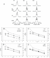
Figure 1. Core fluidity of ptdcho/ptdins model membranes by deuterium wide-line nmr spectroscopy.
A-Representative deuterium wide-line NMR spectra of POPC-2H31 in the absence (bottom) or presence of different PtdIns. The molar ratios are representative of the lipid composition found in MV1: POPC/PtdIns (30/20), POPC/PtdIns/PtdInsP2 (30/20/12), and MV1-like: POPC/PtdIns/PtdInsP/PtdInsP2 (30/20/18/12). Temperatures are indicated on spectra. Sample hydration (water mass/water + lipid mass) is 95%. Depending on POPC amounts (1–3 mg), each spectrum is the result of 10 k to 80 k cumulative scans. A Lorentzian filtering (LB) of 200–300 Hz was applied prior to Fourier transformation. FIG. 1B–D Ordering of chain segments close to glycerol backbone, MV1-like membranes. Thermal variation of the plateau (k = 2 to 8–10) quadrupolar splittings of POPC, POPC/PtdIns (30/20 and 10/40) panel B; POPC/PtdIns/PtdInsP (30/20/18), panel C); POPC/PtdIns/PtdInsP2 (30/20/12), panel D; and MV1-like model membranes POPC/PtdIns/PtdInsP/PtdInsP2 (30/20/18/12), panel E. For comparison, data for pure POPC and POPC/PtdIns (30/20) membranes was added to the graph for the three last compositions. Accuracy of the measure is ±1 kHz. On the double Y-axis the corresponding Carbon-Deuterium order parameter (SCD) is shown. Because the average orientation of all plateau C–D bonds is at 90° with respect to the long lipid axis, 2 |SCD| is plotted to express residual ordering information relative to the bilayer normal.
Figure 2
Figure 2. Core fluidity of PtdCho/Chol/PtdIns model membranes deuterium wide-line NMR spectra.
A-Representative deuterium wide-line NMR spectra of POPC-2H with cholesterol in the absence or presence of different PtdIns. The molar ratios are representative of the lipid composition found in NER: POPC/Chol (58/42), POPC/Chol/PtdIns (28/42/30), and NERs-like membranes POPC/Chol/PtdIns/PtdInsP/PtdInsP2 (28/42/23/16/7/7). Temperatures are indicated on the spectra. Sample hydration (water mass/water + lipid mass) is 95%. Depending on deuterated POPC amounts (1–3 mg), each spectrum is the result of 10 k to 100 k cumulative scans. A Lorentzian filtering (LB) of 200–300 Hz was applied prior to Fourier transformation. FIG. 2B–D . Ordering of chain segments close to glycerol backbone, NER-like membranes. Thermal variation of the plateau (k = 2 to 8–10) quadrupolar splittings of POPC/Chol/PtdIns (28/42/30) and POPC/Chol/PtdIns/PtdInsP (28/42/23/7), panel B; POPC/Chol/PtdIns (28/42/30) and POPC/Chol/PtdIns/PtdInsP2 (28/42/23/7), panel C: POPC/Chol/PtdIns (28/42/30) and NERs-like model membranes POPC/Chol/PtdIns/PtdInsP/PtdInsP2 (28/42/23/16/7/7), panel D. For comparison, data for pure POPC and POPC/Chol (58/42) membranes was added to the graphs. Accuracy of the measure is ±1 kHz. On the double Y-axis the corresponding Carbon-Deuterium order parameter is shown. Because the average orientation of all plateau C–D bonds is at 90° with respect to the long lipid axis, twice |SCD| is plotted to express residual ordering information relative to the bilayer normal.
Figure 3
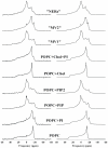
Figure 3. Orientational dynamics at the membrane surface determined by phosphorus-31 NMR spectra.
Left Column: representative experimental wide-line phosphorus-31 NMR spectra of different model membranes containing POPC, PtdIns and cholesterol. The molar ratios are representative of the lipid composition found in MV1, MV2 and NERs, from bottoms to top: pure POPC, POPC/PtdIns (30/20), POPC/PtdInsP (30/18), POPC/PtdInsP2 (30/12), POPC/Chol (58/42), POPC/Chol/PtdIns (28/42/30), “MV1”: POPC/PtdIns/PtdInsP/PtdInsP2 (30/20/18/12), “MV2”: POPC/POPE/PtdIns/POPS (30/25/20/5) and NERs: POPC/Chol/PtdIns/PtdInsP/PtdInsP2 (28/42/23/16/7/7). Temperature is 10°C. Sample hydration (lipid mass/lipid + water mass) is 95%. Each spectrum is the result of 5 k cumulative scans. A Lorentzian filtering (LB) of 50–100 Hz was applied prior to Fourier transformation. Chemical shifts are expressed relative to 85% H3PO4 (0 ppm). Right column: simulated spectra according to procedures described in text. Initial guesses, as measured on de-Paked spectra, of chemical shielding anisotropies, Δσ, line widths, isotropic chemical shifts and relative weights of each subspectrum were supplied to the simulation procedure and iterative changes were performed until the best fit of experimental spectra was obtained. Δσ and isotropic chemical shifts are reported in Table 3.
Figure 4
Figure 4. A- Distinct ordering of chain segments close to the glycerol backbone in MV1, MV2 and NERs like membranes.
Thermal variation of the plateau (k = 2 to 8–10) quadrupolar splittings of MV1 MV2 and NERs model membranes. Data for POPC and POPC/Chol is also shown for comparison. Accuracy of the measure is ±1 kHz. On the double Y-axis the corresponding Carbon-Deuterium order parameter is shown. Because the average orientation of all plateau C–D bonds is at 90° with respect to the long lipid axis, twice |SCD| is plotted to express residual ordering information relative to the bilayer normal. FIG. 4B. Scheme describing how the physical properties of NER, MV1 and MV2 membranes could affect nuclear envelope assembly. Left: NERs (relatively-rigid) are located at the poles of the sperm nucleus and play the role of anchorage to chromatin. “MV1- like” membranes are very fluid and hence may have the role to “prime” the process of fusion. PLCγ in a second step hydrolyses PtdInsP2 into DAG, which initiates fusion of vesicles. Right: experimental deuterium NMR spectra of MV1, MV2 and NERs: the wider the trace the more rigid the system.

Similar articles

Cited by

References

    1. Mima J, Hickey CM, Xu H, Jun Y, Wickner W. Reconstituted membrane fusion requires regulatory lipids, SNAREs and synergistic SNARE chaperones. EMBO J. 2008;27:2031–2042. - PMC - PubMed
    1. Poccia D, Larijani B. Phosphatidylinositol metabolism and membrane fusion. Biochem J. 2009;418:233–246. - PubMed
    1. Wang L, Seeley ES, Wickner W, Merz AJ. Vacuole fusion at a ring of vertex docking sites leaves membrane fragments within the organelle. Cell. 2002;108:357–369. - PubMed
    1. Wickner W, Schekman R. Membrane fusion. Nat Struct Mol Biol. 2008;15:658–664. - PMC - PubMed
    1. James DJ, Khodthong C, Kowalchyk JA, Martin TF. Phosphatidylinositol 4,5-bisphosphate regulates SNARE-dependent membrane fusion. J Cell Biol. 2008;182:355–366. - PMC - PubMed

Publication types