. 2011;6(9):e23870.
doi: 10.1371/journal.pone.0023870. Epub 2011 Sep 8.

Mycobacterium tuberculosis lineage influences innate immune response and virulence and is associated with distinct cell envelope lipid profiles

Affiliations

Mycobacterium tuberculosis lineage influences innate immune response and virulence and is associated with distinct cell envelope lipid profiles

Nitya Krishnan et al. PLoS One. 2011.

Abstract

The six major genetic lineages of Mycobacterium tuberculosis are strongly associated with specific geographical regions, but their relevance to bacterial virulence and the clinical consequences of infection are unclear. Previously, we found that in Vietnam, East Asian/Beijing and Indo-Oceanic strains were significantly more likely to cause disseminated tuberculosis with meningitis than those from the Euro-American lineage. To investigate this observation we characterised 7 East Asian/Beijing, 5 Indo-Oceanic and 6 Euro-American Vietnamese strains in bone-marrow-derived macrophages, dendritic cells and mice. East Asian/Beijing and Indo-Oceanic strains induced significantly more TNF-α and IL-1β from macrophages than the Euro-American strains, and East Asian/Beijing strains were detectable earlier in the blood of infected mice and grew faster in the lungs. We hypothesised that these differences were induced by lineage-specific variation in cell envelope lipids. Whole lipid extracts from East Asian/Beijing and Indo-Oceanic strains induced higher concentrations of TNF-α from macrophages than Euro-American lipids. The lipid extracts were fractionated and compared by thin layer chromatography to reveal a distinct pattern of lineage-associated profiles. A phthiotriol dimycocerosate was exclusively produced by East Asian/Beijing strains, but not the phenolic glycolipid previously associated with the hyper-virulent phenotype of some isolates of this lineage. All Indo-Oceanic strains produced a unique unidentified lipid, shown to be a phenolphthiocerol dimycocerosate dependent upon an intact pks15/1 for its production. This was described by Goren as the 'attenuation indictor lipid' more than 40 years ago, due to its association with less virulent strains from southern India. Mutation of pks15/1 in a representative Indo-Oceanic strain prevented phenolphthiocerol dimycocerosate synthesis, but did not alter macrophage cytokine induction. Our findings suggest that the early interactions between M. tuberculosis and host are determined by the lineage of the infecting strain; but we were unable to show these differences are driven by lineage-specific cell-surface expressed lipids.


Conflict of interest statement

Competing Interests: The authors have declared that no competing interests exist.

Figures

Figure 1
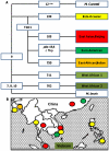
Figure 1. The phylogeny of M. tuberculosis in South East Asia.
Large sequence polymorphisms define six major lineages of M. tuberculosis (A) which are strongly associated with specific geographical regions (B). In Vietnam, three lineages cause the majority of disease: the East Asian/Beijing, the Indo-Oceanic, and the Euro-American (adapted from reference 1). Numercial values in the figure represent regions of deletions (RD) that define each of the lineages.
Figure 2
Figure 2. M. tuberculosis lineage influences cytokine expression from infected macrophages.
Bone marrow derived macrophages were infected with 6 Euro-American, 7 East Asian/Beijing and 5 Indo-Oceanic strains at a M.O.I of 5, for 72 hours. Cytokine concentrations in the cell supernatants were determined using ELISA. TNF-α concentration at 24 hours (A), IL-1β at 48 hours (B), IL-10 levels at 48 hours (C) and IL-12p40 levels at 72 hours (D) are displayed. Beijing and Indo-Oceanic strains induced more pro-inflammatory TNF-α (Beijing, p = 0.001; Indo-Oceanic, p = 0.003) and IL-1β (Beijing, p = 0.049; Indo-Oceanic, p<0.001) than Euro-American strains. Data represents mean ± standard deviation (SD) of three independent experiments.
Figure 3
Figure 3. The cytokine profiles from macrophages infected with individual strains over 72 hours.
Bone-marrow derived macrophages were infected with 6 Euro-American, 7 East Asian/Beijing and 5 Indo-Oceanic strains at a M.O.I of 5, for 72 hours. Cytokine concentrations for TNF-α and IL-1β (A) and IL-10 and IL-12p40 (B) were determined in the cell supernatants using ELISA. Lines represent the mean cytokine expression at 24, 48, and 72 hours of infection from three independent experiments, each performed in triplicate. The individual strains and their spoligotypes are labelled within the figure (see with table 1).
Figure 4
Figure 4. M. tuberculosis lineage does not significantly influence cytokine expression from infected dendritic cells.
Bone-marrow-derived dendritic cells were infected with 3 Euro-American, 3 East Asian/Beijing and 3 Indo-Oceanic strains at a M.O.I of 5, for 72 hours. Cytokine concentrations in the cell supernatant were determined using ELISA. TNF-α concentration at 24 hours (A), IL-1β at 48 hours (B), IL-10 at 48 hours (C), IL-12p40 (D) and IL-12p70 levels (E) at 72 hours are displayed. There were no significant (p<0.05) differences in cytokine expression between any of the lineages, although there was a trend for the Indo-Oceanic strains to induce more IL-10 than the Euro-American strains. Data represents mean ± SD of three independent experiments.
Figure 5
Figure 5. The cytokine profiles from dendritic cells infected with individual strains over 72 hours.
Bone-marrow derived dendritic cells were infected with 3 Euro-American (all T1 spoligotype), 3 East Asian/Beijing (all Beijing spoligotype) and 3 Indo-Oceanic (all EA14_VNM spoligotype) strains at a M.O.I of 5, for 72 hours. Cytokine concentrations for TNF-α and IL-1β (A) and IL-10 and IL-12p40 (B) were determined in the cell supernatants using ELISA. Lines represent the mean cytokine expression of three independent experiments at 24, 48, and 72 hours of infection from 3 independent experiments, each performed in triplicate. The individual strains are labelled within the figure (see with table 1).
Figure 6
Figure 6. East Asian/Beijing lineage is associated with increased bacterial load in the lungs and spleen at early time points and bacteraemia.
BALB/c mice were infected with 104 CFU of H37Rv and three representative isolates of the Euro-American, East Asian/ Beijing and Indo-Oceanic lineages of M. tuberculosis. At the indicated time-points bacterial load was measured in the lungs, spleen, and blood. Mean CFU or the percentages of mice with bacteria cultured from blood (5 mice per group per time-point) are given for individual strains (A) (with the strain numbers indicated within each graph). The data are combined and presented by lineage in (B). Asterix (*) indicate significant (p<0.05) differences between lineages: * Indo-Oceanic and East Asian/Beijing > Euro-American (p = 0.01 and p = 0.08, respectively), ** East Asian/Beijing > Euro-American (p = 0.004), *** East Asian/Beijing > Euro-American (p = 0.002). Data represents mean ± 1 SD of three independent experiments, or percentage of 5 mice per group per time-point.
Figure 7
Figure 7. Pulmonary cytokine expression varies according to the infecting lineage of M. tuberculosis.
Indicated cytokines were measured in the BAL using ELISA. Data are representative of three independent experiments, each with 5 mice per group. Bars represent mean +/- 1 SD. * signifies p-value of <0.05.
Figure 8
Figure 8. Histology of lungs infected with different lineages of M. tuberculosis.
Sections of the lungs were stained with H&E stain. Representative lung sections for each M. tuberculosis infected group at day 14 and day 28 post-infection are displayed (A). Lung sections were scored (blind to the strain lineage) for lymphocytic infiltrates (B) and granulomas (C) and mean scores for each group presented. A score of 0 =  normal lung; a score of 2 =  moderate lymphocytic infiltrate/granuloma formation seen; a score of 3 =  extensive lymphocytic infiltrate/granuloma formation seen. The displayed histology images are from one experiment only.
Figure 9
Figure 9. Total lipid extracts from East Asian/Beijing and Indo-Oceanic strains induce higher concentrations of TNF-α from macrophages than Euro-American extracts.
Macrophages were incubated with 0.1 µg (A) and 1 µg (B) of total lipid extracts from M. tuberculosis strains from the different lineages. Culture supernatants were analysed by ELISA at 24 hours. Data represents means ±1SD of 6 Euro-American, 7 East Asian/Beijing and 5 Indo-Oceanic strains performed in triplicate.
Figure 10
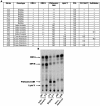
Figure 10. The cell envelope lipid profile varies according to M. tuberculosis lineage.
The lipid profile of each strain was examined by thin layer chromatography (TLC) of the crude lipid extract using various solvents (A). TLC analysis of lipids from representative Indo-Oceanic, East-Asian/Beijing or Euro-American strains is displayed. Lipids were radiolabelled with 14C- propionate. The TLC was run in petroleum ether/diethylether (90∶10, v/v) (B).
Figure 11
Figure 11. Lipid Y is a phenolphthiocerol dimycocerosate.
One representative Indo-Oceanic strain (no. 367) and H37Rv Rv2962c::km: pPET1 (complemented pks15/1, disrupted Rv2962c) were incubated with either 14C- propionate (lane ‘P’) or 14C-p-hydroxybenzoic acids (lane ‘B’) to label methyl-branched fatty acids (such as those found in PGL and DIM) or the phenol ring of PGL respectively. Thin layer chromatography revealed super-imposable spots (A). Lipid Y and lipids exhibiting R f values similar to DIM B were then separated on a SepPak silica column and the enriched fractions analyzed by MALDI-TOF MS and 1H-NMR. The mass spectrum of lipid Y showed a series of pseudomolecular ion (M + Na+) peaks centered at m/z 1412 (B), identical to the ones obtained for phenolphthiocerol dimycocerosates purified from H37Rv ΔRv2962c::km: pPET1. A SNP at position nt 880 of gene Rv2962c introduces a stop codon and a truncated open reading frame (C) in Indo-Oceanic strains.
Figure 12
Figure 12. Phenolphthiocerol dimycocerosate does not influence TNF-α and IL-1β production from macrophages.
TLC analysis of lipids from Indo-Oceanic strain 367, its isogenic pks15/1::km mutant (PMM162) and pks15/1 complemented strain (PMM162:pPET1). Lipids were radiolabelled with 14C- propionate. The TLC was run in petroleum ether/diethylether (90:10, v/v) (A). Bone marrow derived macrophages were infected at an M.O.I of 5 for 72 hours with a representative Indo-Oceanic strain (no. 367) , mutant (PMM162) strain incapable of making lipid Y (pks15/1 knock-out mutant) and the complemented strain . Cytokine concentrations in the cell supernatants were determined using ELISA. TNF-α (B) and IL-1β concentrations (C) are shown across the 3 time-points studied. Data represents mean ± standard deviation (SD) of three independent experiments.

Similar articles

Cited by

References

    1. Gagneux S, Small PM. Global phylogeography of Mycobacterium tuberculosis and implications for tuberculosis product development. Lancet Infect Dis. 2007;7:328–337. - PubMed
    1. Nicol MP, Wilkinson RJ. The clinical consequences of strain diversity in Mycobacterium tuberculosis. Trans R Soc Trop Med Hyg. 2008;102:955–965. - PubMed
    1. Mitchison DA, Wallace JG, Bhatia AL, Selkon JB, Subbaiah TV, et al. A comparison of the virulence in guinea-pigs of South Indian and British tubercle bacilli. Tubercle. 1960;41:1–22. - PubMed
    1. Dunn PL, North RJ. Virulence ranking of some Mycobacterium tuberculosis and Mycobacterium bovis strains according to their ability to multiply in the lungs, induce lung pathology, and cause mortality in mice. Infect Immun. 1995;63:3428–3437. - PMC - PubMed
    1. Lopez B, Aguilar D, Orozco H, Burger M, Espitia C, et al. A marked difference in pathogenesis and immune response induced by different Mycobacterium tuberculosis genotypes. Clin Exp Immunol. 2003;133:30–37. - PMC - PubMed

Publication types

MeSH terms