. 2011;6(9):e23971.
doi: 10.1371/journal.pone.0023971. Epub 2011 Sep 8.

Functional dissection of the TBK1 molecular network

Affiliations

Functional dissection of the TBK1 molecular network

Adriana Goncalves et al. PLoS One. 2011.

Abstract

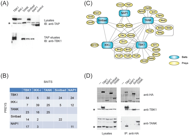
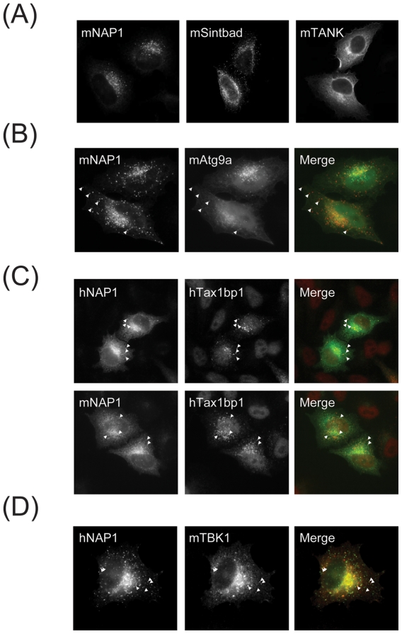
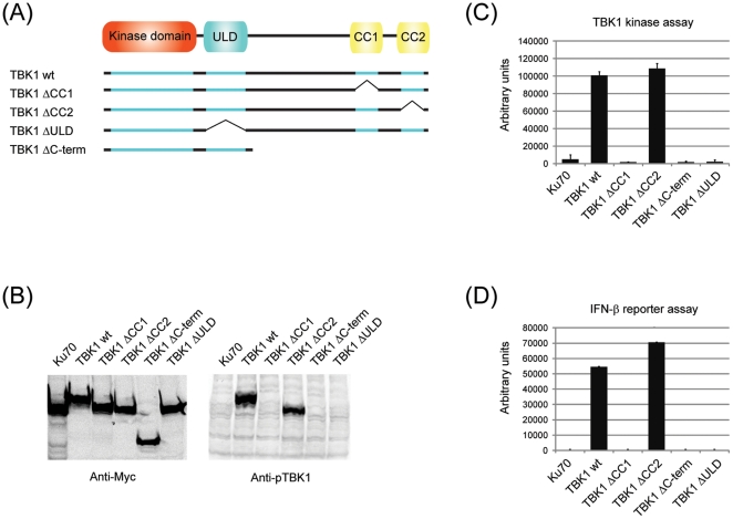
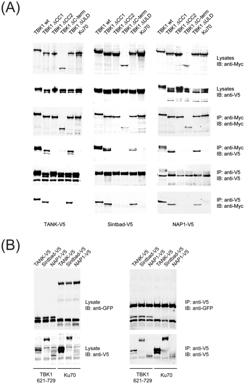
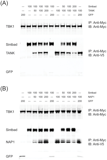
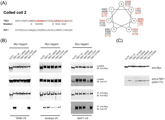
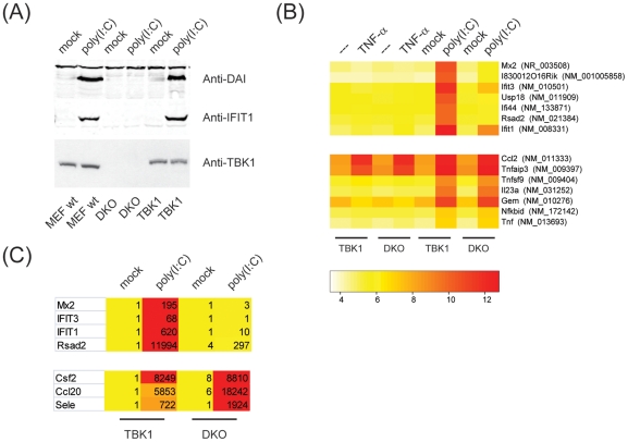
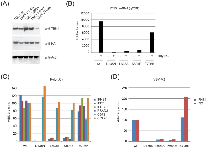
TANK-binding kinase 1 (TBK1) and inducible IκB-kinase (IKK-i) are central regulators of type-I interferon induction. They are associated with three adaptor proteins called TANK, Sintbad (or TBKBP1) and NAP1 (or TBKBP2, AZI2) whose functional relationship to TBK1 and IKK-i is poorly understood. We performed a systematic affinity purification-mass spectrometry approach to derive a comprehensive TBK1/IKK-i molecular network. The most salient feature of the network is the mutual exclusive interaction of the adaptors with the kinases, suggesting distinct alternative complexes. Immunofluorescence data indicated that the individual adaptors reside in different subcellular locations. TANK, Sintbad and NAP1 competed for binding of TBK1. The binding site for all three adaptors was mapped to the C-terminal coiled-coil 2 region of TBK1. Point mutants that affect binding of individual adaptors were used to reconstitute TBK1/IKK-i-deficient cells and dissect the functional relevance of the individual kinase-adaptor edges within the network. Using a microarray-derived gene expression signature of TBK1 in response virus infection or poly(I∶C) stimulation, we found that TBK1 activation was strictly dependent on the integrity of the TBK1/TANK interaction.


Conflict of interest statement

Competing Interests: The authors have declared that no competing interests exist.

Figures

Figure 1
Figure 1. Protein networks associated with TBK1, IKK-i, TANK, Sintbad and NAP1.
(A) RAW264.7 cells stably expressing GS-TAP-tagged TBK1, IKK-I, TANK, Sintbad or NAP1 were purified by tandem affinity purification. Lysates and eluates were analyzed by immunoblotting as indicated. (*) indicates a non-specific band detected by the TAP-specific antiserum. (B) Representation of the number of unique peptides identified in each mass spectrometry run (average of 2 technical repeats). Bait proteins are listed in columns, while prey proteins identified by MS are listed in rows. (C) Schematic representation of the protein-protein interaction network assembled around TBK1, IKK-i and the adaptor proteins TANK, Sintbad and NAP1. Baits are represented in blue, preys are represented in yellow. (D) NIH3T3 cells stably expressing Strep-HA-tagged TBK1, NAP1, Sintbad or TANK were lysed and subjected to immunoprecipitation using anti-HA agarose. Lysates and eluates were analyzed by immunoblotting for anti-HA.11 (Covance), anti-TBK1 (Cell Signaling) or anti-TANK (custom made). (*) indicates the band representing endogenous TBK1 and TANK, respectively.
Figure 2
Figure 2. NAP1, SINTBAD and TANK display distinct localization patterns.
HeLa cells were transiently transfected with the indicated constructs and stained as described under Materials and Methods. (A) V5-tagged NAP1, SINTBAD and TANK. (B) Co-expression of V5 tagged NAP1 (red) and Atg9a-GFP (green). (C) Co-expression of human or murine V5-tagged NAP1 (green) with myc-tagged Tax1bp1 (red). (D) Co-expression of V5-tagged NAP1 (red) with HA-tagged TBK1 (green).
Figure 3
Figure 3. Structural requirements for TBK1 activity.
(A) (A) Individual domains of murine TBK1 were either truncated or deleted as indicated in the schematic (kinase domain 1–297, ubiquitin-like domain 304–382, coiled coil 1 619–657, coiled coil 2 682–713). Myc-tagged TBK1 wt or deletion mutants (schematically represented in (A)) were expressed as indicated by transient transfection of 293T cells. Lysates were analyzed by immunoblotting for phospho-TBK1 (B) or submitted to immunoprecipitation using anti-Myc agarose (C). Immunoprecipitates were incubated with a biotinylated IRF3-derived peptide (DLHISNSHPLSLC) in the presence of [γ32-P]ATP and incorporated radioactivity was quantified as described in the methods section. (D) Myc-tagged TBK1 wt or deletion mutants were expressed as indicated by transient transfection of 293T cells in the presence of pIFN-Luc and pRL-TK (Promega). Lysates were analyzed using the DualGlo Luciferase Assay System (Promega). Results were normalized to Ku70-induced IFN-β induction.
Figure 4
Figure 4. TANK, Sintbad and NAP1 all bind to the coiled-coil 2 region in TBK1.
Myc-tagged TBK1 wt or deletion mutants were coexpressed with V5-tagged TBK1 adaptors as indicated by transient transfection of HEK293 cells. Lysates were subjected to immunoprecipitation using anti-Myc agarose (Sigma) or anti-V5 agarose (Sigma). Lysates and eluates were analyzed by immunoblotting for anti-Myc-IRDye800 (Rockland) or anti-V5 (Invitrogen).
Figure 5
Figure 5. Sintbad competes with TANK or NAP1 for TBK1 binding.
(A,B) Individual proteins (Myc-tagged TBK1, V5-tagged GFP, TANK, Sintbad or NAP1) were expressed in HEK293 cells by transient transfection. Cell extracts were mixed as indicated (numbers represent the volume (µl) of each cell extract in each condition) and subjected to immunoprecipitation using anti-Myc agarose (Sigma). Eluates were analyzed by immunoblotting for anti-Myc-IRDye800 (Rockland) or anti-V5 (Invitrogen).
Figure 6
Figure 6. Point mutants in the coiled-coil 2 region of TBK1 lead to selective impairment in adaptor binding.
(A) Schematic representation of the coiled-coil 2 region in TBK1 and IKK-i. A helical wheel representation of the coiled coil 2 (M690-E712) was generated according to (B) TBK1 wt or corresponding point mutants in the coiled-coil 2 region were expressed in HEK293 cells, subjected to immunoprecipitation and analyzed as in Fig. 3. (C) TBK1 wt or corresponding point mutants were expressed in HEK293 cells and analyzed for (auto-)phosphorylation at Ser172 by immunoblotting.
Figure 7
Figure 7. A molecular signature for TBK1.
(A) Different batches of MEFs (MEF wt, TBK1/IKK-i-deficient MEFs (DKO for double-KO) reconstituted with TBK1 wt or left untreated) were stimulated with transfected poly(I∶C) for 16 h. Lysates were analyzed by immunoblotting for anti-DAI (Millipore), anti-IFIT1 or anti-TBK1 (Cell Signaling). (B) DKO MEFs reconstituted with TBK1 or left untreated were stimulated with 10 ng/ml TNF-α or 10 µg/ml poly(I∶C) for 4 h. Total RNA was extracted and global changes in transcription were monitored by microarray analysis. Genes were clustered according to recurring patterns. (C) Total RNA obtained for cells stimulated with poly(I∶C) was reverse transcribed and corresponding cDNA levels were analyzed by qPCR. Numbers represent fold changes over TBK1/mock.
Figure 8
Figure 8. Coiled-coil 2 mutants of TBK1 fail to reconstitute TBK1-/IKK-i-deficient cells.
TBK1/IKK-i-deficient MEFs were reconstituted with Strep-HA fusions of TBK1 wt, D135N, L693A, K694E and E706K. (A) Lysates from reconstituted cells were analyzed for immunoblotting using anti-TBK1 (Cell Signaling), anti-HA.11 (Covance) and anti-Actin (Cytoskeleton). (B) Cells as described in (A) were transfected with 10 µg/ml poly(I∶C) for 4 h. Total RNA was extracted, reverse transcribed and analyzed for the IFN-β mRNA by qPCR. Data are represented as fold changes over unstimulated TBK1 wt cells. (C) cDNA from (B) was analyzed by qPCR for the following mRNAs: IFN-β, IFIT1, IFIT3, RSAD3, CSF2, CCL20. Data were normalized such that the average values obtained for TBK1 wt and TBK1 E706K were arbitrarily defined as 100. (D) Cells as described in (A) were infected with Vesicular Stomatitis Virus M2 for 8 h. Total RNA was extracted and processed as described in (B).

Similar articles

Cited by

References

    1. Pichlmair A, Reis ESC. Innate Recognition of Viruses. Immunity. 2007;27:370–383. - PubMed
    1. Panne D, Maniatis T, Harrison SC. An atomic model of the interferon-beta enhanceosome. Cell. 2007;129:1111–1123. - PMC - PubMed
    1. Hemmi H, Takeuchi O, Sato S, Yamamoto M, Kaisho T, et al. The roles of two IkappaB kinase-related kinases in lipopolysaccharide and double stranded RNA signaling and viral infection. J Exp Med. 2004;199:1641–1650. - PMC - PubMed
    1. Perry AK, Chow EK, Goodnough JB, Yeh WC, Cheng G. Differential requirement for TANK-binding kinase-1 in type I interferon responses to toll-like receptor activation and viral infection. J Exp Med. 2004;199:1651–1658. - PMC - PubMed
    1. Miyahira AK, Shahangian A, Hwang S, Sun R, Cheng G. TANK-binding kinase-1 plays an important role during in vitro and in vivo type I IFN responses to DNA virus infections. J Immunol. 2009;182:2248–2257. - PMC - PubMed

Publication types