. 2011;6(9):e23993.
doi: 10.1371/journal.pone.0023993. Epub 2011 Sep 13.

Triptolide (TPL) inhibits global transcription by inducing proteasome-dependent degradation of RNA polymerase II (Pol II)

Affiliations

Triptolide (TPL) inhibits global transcription by inducing proteasome-dependent degradation of RNA polymerase II (Pol II)

Ying Wang et al. PLoS One. 2011.

Abstract

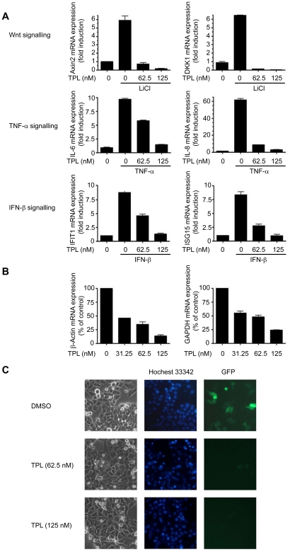
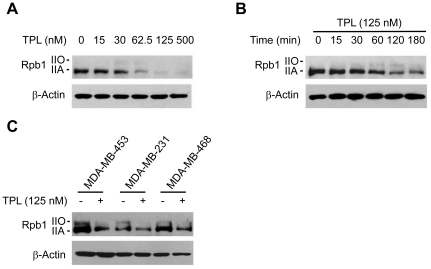
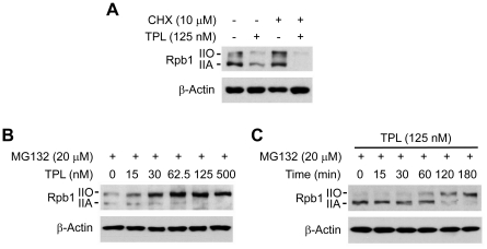
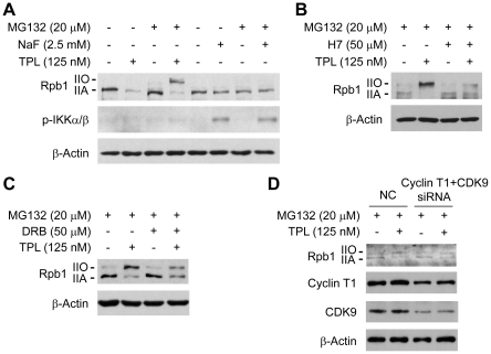
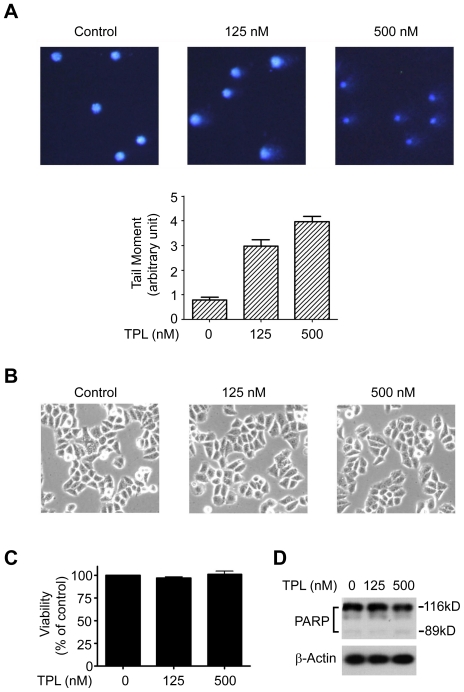
Triptolide (TPL), a key biologically active component of the Chinese medicinal herb Tripterygium wilfordii Hook. f., has potent anti-inflammation and anti-cancer activities. Its anti-proliferative and pro-apoptotic effects have been reported to be related to the inhibition of Nuclear Factor κB (NF-κB) and Nuclear Factor of Activated T-cells (NFAT) mediated transcription and suppression of HSP70 expression. The direct targets and precise mechanisms that are responsible for the gene expression inhibition, however, remain unknown. Here, we report that TPL inhibits global gene transcription by inducing proteasome-dependent degradation of the largest subunit of RNA polymerase II (Rpb1) in cancer cells. In the presence of proteosome inhibitor MG132, TPL treatment causes hyperphosphorylation of Rpb1 by activation of upstream protein kinases such as Positive Transcription Elongation Factor b (P-TEFb) in a time and dose dependent manner. Also, we observe that short time incubation of TPL with cancer cells induces DNA damage. In conclusion, we propose a new mechanism of how TPL works in killing cancer. TPL inhibits global transcription in cancer cells by induction of phosphorylation and subsequent proteasome-dependent degradation of Rpb1 resulting in global gene transcription, which may explain the high potency of TPL in killing cancer.


Conflict of interest statement

Competing Interests: The authors have declared that no competing interests exist.

Figures

Figure 1
Figure 1. TPL inhibited global transcription.
(A) Effects of TPL on the transcription of inducible genes. Hela cells were pretreated with TPL at indicated concentrations for 2 h before stimulation by LiCl (20 mM), TNF-α (100ng/ml) or IFN-β (100ng/ml) for another 3 h. The mRNA expression of the indicated genes was analyzed by Real-time PCR. All values were normalized to the mRNA level of β-Actin because the mRNA level of β-Actin was not affected at this time point. (B) Effects of TPL on the transcription of housekeeping genes. Hela cells were treated with TPL for 12 h. The mRNA expression of the indicated genes was analyzed by Real-time PCR. Equal amounts of RNA were used to normalize the loadings. (C) Effects of TPL on expression of transiently transfected genes. Hela cells were transiently transfected with a pEGFP-N1 plasmid. 6 h after transfection, the medium was replaced with the fresh medium containing TPL at indicated concentrations and the Hela cells were incubated for another 15h. GFP expression was visualized by fluorescent microscopy. The picture is a representative of four visual fields. Original magnification: ×200.
Figure 2
Figure 2. Effects of TPL on Rpb1 protein levels.
(A) Hela cells were treated with TPL at indicated concentrations for 3 h. Whole cell lysates were processed for SDS-PAGE and Western blot analysis using antibodies as indicated. (B) Hela cells were treated with 125 nM TPL for various lengths of time (0–180 min) as indicated. Whole cell lysates were subjected to SDS-PAGE and Western blot analysis. (C) MDA-MB-453, MDA-MB-231 or MDA-MB-468 cells were treated with 125 nM TPL for 3 h. Whole cell lysates were subjected to SDS-PAGE and Western blot analysis. β-Actin served as a loading control.
Figure 3
Figure 3. TPL induced phosphorylation and proteasome-dependent degradation of Rpb1.
(A) Hela cells were pretreated with CHX (10 µM) for 0.5 h before addition of TPL (125 nM) and treated for another 3 h. Whole cell lysates were processed for Western blot analysis using antibodies as indicated. (B) Hela cells were pretreated with MG132 (20 µM) for 1 h and then treated with TPL at indicated concentrations for another 3 h. Whole cell lysates were subjected to SDS-PAGE and Western blot analysis. (C) Hela cells were treated with 125 nM TPL for various lengths of time (0–180 min) in the presence of MG132 (20 µM). Whole cell lysates were subjected to SDS-PAGE and Western blot analysis. β-Actin served as a loading control.
Figure 4
Figure 4. TPL induced phosphorylation of Rpb1 by activation of protein kinases.
(A) Comparison of the effect of TPL with NaF on Rpb1. Hela cells were treated with TPL or NaF for 3 h in the presence/absence of MG132. Whole cell lysates were processed for SDS-PAGE and Western blot analysis using antibodies as indicated. (B) Hela cells were pretreated with MG132 for 1 h, and then treated with TPL in the presence/absence of 50 µM H7 for 3 h. Whole cell lysates were subjected to SDS-PAGE and Western blot analysis. (C) Hela cells were pretreated with MG132 for 1 h, and then treated with TPL in the presence/absence of 50 µM DRB for 3 h. Whole cell lysates were subjected to SDS-PAGE and Western blot analysis. (D) Analysis of effect of CDK9 and Cyclin T1 siRNA on TPL-induced Rpb1 phosphorylation. Hela cells were transfected with NC (negative control) or CDK9 and Cyclin T1 siRNA. 48 h after transfection, the Hela cells were pretreated with 20 µM MG132 for 1 h. Then the cells were treated with DMSO or 125 nM TPL for another 3 h before collected to SDS-PAGE and Western blot analysis. β-Actin served as a loading control.
Figure 5
Figure 5. TPL induced DNA damage.
(A) Comet assay. Hela cells were treated with TPL at indicated concentrations for 3 h, and then subjected to comet assay. Quantification analysis of the results was presented below the graph. The pictures are representatives of four fields. Original magnification: ×200. (B) Morphology of Hela cells treated with TPL at indicated concentrations for 3 h. The pictures are representatives of four fields. Original magnification: ×200. (C) Viability of Hela cells treated with TPL at indicated concentrations for 3 h. (D) Apoptosis analysis of Hela cells treated with TPL at indicated concentrations for 3 h. Whole cell lysates were processed for Western blot analysis using anti-PARP antibody. β-Actin served as a loading control.

Similar articles

Cited by

References

    1. Corson TW, Crews CM. Molecular understanding and modern application of traditional medicines: triumphs and trials. Cell. 2007;130:769–774. - PMC - PubMed
    1. Efferth T, Li PC, Konkimalla VS, Kaina B. From traditional Chinese medicine to rational cancer therapy. Trends Mol Med. 2007;13:353–361. - PubMed
    1. Bao X, Cui J, Wu Y, Han X, Gao C, et al. The roles of endogenous reactive oxygen species and nitric oxide in triptolide-induced apoptotic cell death in macrophages. J Mol Med. 2007;85:85–98. - PubMed
    1. Fidler JM, Li K, Chung C, Wei K, Ross JA, et al. PG490-88, a derivative of triptolide, causes tumor regression and sensitizes tumors to chemotherapy. Mol Cancer Ther. 2003;2:855–862. - PubMed
    1. Kiviharju TM, Lecane PS, Sellers RG, Peehl DM. Antiproliferative and proapoptotic activities of triptolide (PG490), a natural product entering clinical trials, on primary cultures of human prostatic epithelial cells. Clin Cancer Res. 2002;8:2666–2674. - PubMed

Publication types

MeSH terms