. 2011;6(9):e24152.
doi: 10.1371/journal.pone.0024152. Epub 2011 Sep 8.

Evolution of the TGF-β signaling pathway and its potential role in the ctenophore, Mnemiopsis leidyi

Affiliations

Evolution of the TGF-β signaling pathway and its potential role in the ctenophore, Mnemiopsis leidyi

Kevin Pang et al. PLoS One. 2011.

Abstract

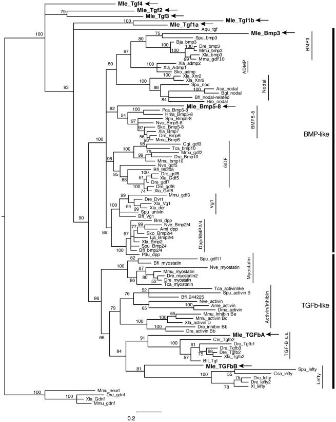
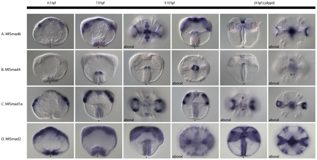
The TGF-β signaling pathway is a metazoan-specific intercellular signaling pathway known to be important in many developmental and cellular processes in a wide variety of animals. We investigated the complexity and possible functions of this pathway in a member of one of the earliest branching metazoan phyla, the ctenophore Mnemiopsis leidyi. A search of the recently sequenced Mnemiopsis genome revealed an inventory of genes encoding ligands and the rest of the components of the TGF-β superfamily signaling pathway. The Mnemiopsis genome contains nine TGF-β ligands, two TGF-β-like family members, two BMP-like family members, and five gene products that were unable to be classified with certainty. We also identified four TGF-β receptors: three Type I and a single Type II receptor. There are five genes encoding Smad proteins (Smad2, Smad4, Smad6, and two Smad1s). While we have identified many of the other components of this pathway, including Tolloid, SMURF, and Nomo, notably absent are SARA and all of the known antagonists belonging to the Chordin, Follistatin, Noggin, and CAN families. This pathway likely evolved early in metazoan evolution as nearly all components of this pathway have yet to be identified in any non-metazoan. The complement of TGF-β signaling pathway components of ctenophores is more similar to that of the sponge, Amphimedon, than to cnidarians, Trichoplax, or bilaterians. The mRNA expression patterns of key genes revealed by in situ hybridization suggests that TGF-β signaling is not involved in ctenophore early axis specification. Four ligands are expressed during gastrulation in ectodermal micromeres along all three body axes, suggesting a role in transducing earlier maternal signals. Later expression patterns and experiments with the TGF-β inhibitor SB432542 suggest roles in pharyngeal morphogenesis and comb row organization.


Conflict of interest statement

Competing Interests: The authors have declared that no competing interests exist.

Figures

Figure 1
Figure 1. Basic overview of TGF-β signaling pathway.
Binding of a ligand to a Type II receptor initiates signaling. The sequestering of a Type I receptor results in the activation of a Receptor-Smad (Smad1/5, Smad2/3). Together with the Co-Smad (Smad4), this complex enters the nucleus and activates the transcription of target genes. The pathway can be inhibited by extracelluar antagonists, or intracellularly via Inhibitor-Smad (Smad6/7) or the ubiquitin ligase SMURF.
Figure 2
Figure 2. Bayesian analysis of TGF-β ligands.
Analyses were performed using only the TGF-β peptide domain, with Mnemiopsis members bolded and marked by arrows. Representative taxa from deuterostomes, protostomes, and non-bilaterians were used (for full list of taxa, see Table S1). Four independent runs of five million generations were run using the “mixed” model, with the strict consensus tree shown. Nodes are labeled with posterior probabilities.
Figure 3
Figure 3. TGF-β protein structures and motifs.
(A) Predicted amino acid sequences of the TGF-β peptide domain and flanking region. Adjacent to the peptide domain is the cleavage site, showing the conserved RXXR motif. Asterisks below the sequence mark the seven conserved cysteine residues. The arrow indicates the conserved cysteine found in TGF-β-like class of ligands. (B) Conserved protein domains of Mnemiopsis TGF-β ligands. The red boxes indicate signal sequences, while the other shaded boxes represent TGF-β propeptide and TGF-β peptide domains, as predicted by SMART.
Figure 4
Figure 4. Bayesian analysis of TGF-β receptors.
Mnemiopsis members are bolded and marked by arrows. Representative taxa from deuterostomes, protostomes, and non-bilaterians were used (for full list of taxa, see Table S1). Four independent runs of 5 million generations were run using the “mixed” model, with the strict consensus tree shown. Nodes are labeled with posterior probabilities.
Figure 5
Figure 5. Bayesian analysis of Smad proteins.
Mnemiopsis members are bolded and marked by arrows. Representative taxa from deuterostomes, protostomes, and non-bilaterians were used (for full list of taxa, see Table S1). Four independent runs of 5 million generations were run using the “mixed” model, with the strict consensus tree shown. Nodes are labeled with posterior probabilities.
Figure 6
Figure 6. Early TGF-β mRNA expression.
Four of the TGF-β genes are detected early in development, prior to and during gastrulation. The schematic at the top depicts the stages of embryos during cleavage and gastrulation, at 1–2 and 3 hours post fertilization (hpf), respectively. Embryos are lateral views, otherwise oral/aboral as stated. The asterisk marks the position of the blastopore. (A) MlBmp5–8 expression in the aboral ectoderm, with more expression detected in the sagittal plane (black arrows). (B) MlTgf1a expression is detected in late cleavage stages around the nuclei of aboral micromeres. By gastrulation, the aboral expression remains, however there expression is primarily along the tentacular plane (white arrows). (C) MlBmp3 is detected in four groups of ectodermal cells from early to mid-gastrulation. (D) MlTGFbA is detected in four groups of ectodermal cells just adjacent to the blastopore at gastrulation.
Figure 7
Figure 7. Late TGF-β mRNA expression.
MlBmp5–8, MlTgf1a, MlTgf2, MlTGFbB and MlTolloid are detected during later stages of development. The diagram at the top depicts the stages of development in the columns below, identifying some of the major features and structures. Views are lateral, unless otherwise specified as oral or aboral. The asterisks marks the position of the blastopore or mouth. (A) MlBmp5–8 expression in the aboral ectoderm and later in the invaginating pharynx. The aboral expression later becomes part of the apical organ and the anal canals. There is also an additional domain of expression in the tentacle bulbs. (B) MlTgf1a is expressed in parts of the tentacle bulbs, pharynx, and apical organ. (C) MlTgf2 is expressed faintly in part of the tentacle bulbs, similar to that of MlTgf1a, however by cydippid stages, expression is barely detectable. (D) MlTGFbB is expressed after gastrulation in a fairly complex pattern. There are expression domains at the oral and aboral ends of the pharynx. There is also expression in parts of the tenacle bulbs and in the apical organ. (E) MlTolloid is expressed around the blastopore and later in the pharynx, as well as in mesodermal derivatives, in transtentacular muscle and parts of the tentacle bulbs.
Figure 8
Figure 8. TGF-β receptor expression patterns.
Expression of TGF-β receptors through development, from gastrulation (3 hpf) to cydippid (24 hpf). Views are lateral unless otherwise specified, and asterisks mark the position of the blastopore or mouth. (A) MlTgfRII, the lone Type II receptor, is expressed ubiquitously from egg through cypdippid stages. (B) MlTgfRIa is expressed in the aboral ectoderm as well as in the pharynx. The aboral ectoderm expression is confined to the developing comb rows and apical organ. (C) MlTgfRIb is detected in the pharynx, as well as in mesodermal derivatives. Cydippid expression is confined to parts of the tentacle bulb, as well as the endodermal part of the gut. (D) MlTgfRIc is expressed in the ectoderm, more towards the oral pole. Late expression is confined to parts of the tentacle bulbs.
Figure 9
Figure 9. Smad expression patterns.
mRNA expression of Mnemiopsis Smad genes during development. All views are lateral, unless otherwise specified. The asterisk marks the position of the blastopore or mouth. (A) MlSmad6, the I-Smad, is expressed in mesodermal derivatives of the tentacle bulb, as well as in the apical organ. (B) MlSmad4, the Co-Smad, is expressed in a discrete domain of the pharynx at the ectoderm-endoderm boundary. There is also late expression in the apical organ in four spots. (C) MlSmad1a, an R-Smad, is expressed in parts of the tentacle bulb and apical organ. (D) MlSmad2, another R-Smad, is expressed ubiquitously from egg to cydippid.
Figure 10
Figure 10. SB431542 treatment during Mnemiopsis development.
Effects of TGF-β inhibitor, SB431542, at 12 hours post fertilization. (A,B, D–F) are treated embryos, while (C, G–I) are controls. (A–C) Confocal projections of embryos stained with anti-tyrosinated tubulin (red) showing the cilia, Alexa-488 phalloidin (green) showing cell borders, and Hoechst 33342 (blue) showing the nuclei. All are aboral views, with the apical organ (ao) in the center. The white arrowheads point to individual comb plates, while the arrows in (C) show the eight comb rowsC. (D–I) are live embryos imaged under DIC. (D) Lateral view of SB431542-treated embryo, showing that pharynx has not invaginated (compare to (G)), and the tentacle bulbs (tb) have formed but are smaller in size. The apical organ appears normal. (E) Aboral view of the same embryo, mid-focal plane, again showing the smaller tentacle bulbs. The ectoderm (ecto) and endoderm (endo) both appear normal. (F) Aboral and surface view, showing the disorganized comb plates (arrowhead), compared to the eight comb rows (arrows) in the control (I).
Figure 11
Figure 11. Summary of presence and absence of TGF-β components.
The rows contain the different TGF-β components. The columns represent the four early-branching lineages of the Metazoa, plus the Bilateria. Each row represents the presence (black dot) or absence (grey dot) of a particular component in the corresponding lineage. The box shows the absences shared by Porifera and Ctenophora. Data is compiled from genomic data of Amphimedon queenslandica (Porifera), Mnemiopsis leidyi (Ctenophora), Trichoplax adhaerens (Placozoa), Nematostella vectensis and Hydra magnipapillata (Cnidaria), and Drosophila melanogaster and Homo sapiens (Bilateria).

Similar articles

Cited by

References

    1. Derynck R, Jarrett JA, Chen EY, Eaton DH, Bell JR, et al. Human transforming growth factor-beta complementary DNA sequence and expression in normal and transformed cells. Nature. 1985;316:701–705. - PubMed
    1. Kingsley DM. The TGF-β superfamily: new members, new receptors, and new genetic tests of function in different organisms. Genes Dev. 1995;8:133–146. - PubMed
    1. Massague J. TGF-β signal transduction. Annu Rev Biochem. 1998;67:753–791. - PubMed
    1. Massague J, Blain SW, Lo RS. TGFB signaling in growth control, cancer, and heritable disorders. Cell. 2000;103:295–309. - PubMed
    1. Herpin A, Lelong C, Favrel P. Transforming growth factor-β-related proteins: an ancestral and widespread superfamily of cytokines in metazoans. Dev Comp Immunol. 2004;28:461–485. - PubMed

Publication types

MeSH terms

Substances