. 2011;6(9):e24168.
doi: 10.1371/journal.pone.0024168. Epub 2011 Sep 8.

In vivo RNAi screen reveals neddylation genes as novel regulators of Hedgehog signaling

Affiliations

In vivo RNAi screen reveals neddylation genes as novel regulators of Hedgehog signaling

Juan Du et al. PLoS One. 2011.

Abstract

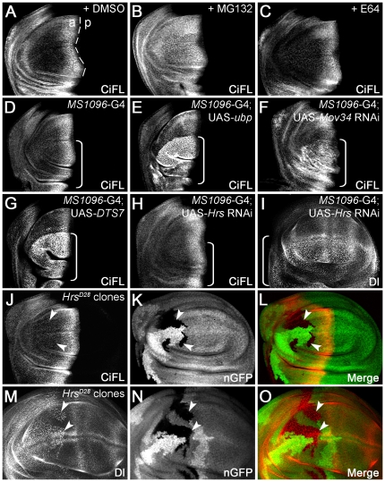
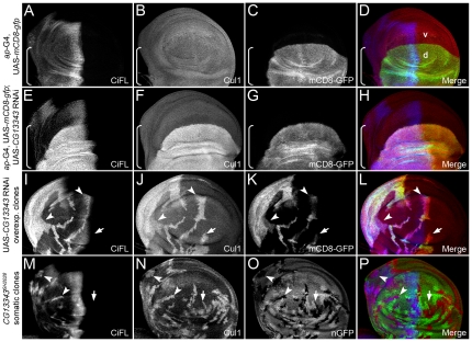
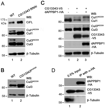
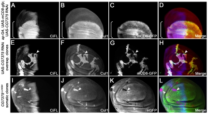
Hedgehog (Hh) signaling is highly conserved in all metazoan animals and plays critical roles in many developmental processes. Dysregulation of the Hh signaling cascade has been implicated in many diseases, including cancer. Although key components of the Hh pathway have been identified, significant gaps remain in our understanding of the regulation of individual Hh signaling molecules. Here, we report the identification of novel regulators of the Hh pathway, obtained from an in vivo RNA interference (RNAi) screen in Drosophila. By selectively targeting critical genes functioning in post-translational modification systems utilizing ubiquitin (Ub) and Ub-like proteins, we identify two novel genes (dUba3 and dUbc12) that negatively regulate Hh signaling activity. We provide in vivo and in vitro evidence illustrating that dUba3 and dUbc12 are essential components of the neddylation pathway; they function in an enzyme cascade to conjugate the ubiquitin-like NEDD8 modifier to Cullin proteins. Neddylation activates the Cullin-containing ubiquitin ligase complex, which in turn promotes the degradation of Cubitus interruptus (Ci), the downstream transcription factor of the Hh pathway. Our study reveals a conserved molecular mechanism of the neddylation pathway in Drosophila and sheds light on the complex post-translational regulations in Hh signaling.


Conflict of interest statement

Competing Interests: The authors have declared that no competing interests exist.

Figures

Figure 1
Figure 1. The stability of endogenous Ci is regulated by the UPS in vitro.
(A) Lysates extracted from cl-8 cells that were treated with 50 µg/ml cycloheximide (CHX) for the indicated hours (hrs) were immunoblotted (WB) with the 2A1 antibody, which specifically recognizes full-length Ci (CiFL) . β-Tubulin detection served as the loading control in all figures. All immunoblotting data presented in the figures are representative of independent experiments that were performed at least three times. (B) Endogenous CiFL degraded rapidly with a half-life of approximately two hours (indicated by dashed lines) as determined by Image J densitometry. (C) CHX-induced CiFL degradation was rescued upon pre-incubation with UPS inhibitors (MG132, ALLN and Lactacystin), but not with DMSO or lysosomal inhibitors (E64, Leupeptin and NH4Cl). (D) In the absence of CHX treatment, incubation with UPS inhibitors alone resulted in significant accumulation of CiFL, while lysosomal inhibitors had no effect, thus suggesting a physiological relevance of the UPS-mediated Ci degradation in Hh signaling.
Figure 2
Figure 2. The stability of endogenous Ci is regulated by the UPS in vivo.
(AC) UPS inhibition protected CiFL from degradation in the wing disc. CiFL, detected with the 2A1 antibody, accumulated abutting the anterior/posterior (a/p) boundary (marked by the dashed line) of a wildtype wing disc treated with DMSO (A). Incubation with the UPS inhibitor MG132 led to accumulation of CiFL in more anterior cells in the wing disc (B), while the lysosomal inhibitor E64 had no obvious effect (C). (D and E) Blockage of ubiquitination in the wing disc by overexpressing UAS-ubp resulted in accumulation of CiFL in more anterior cells (E). MS1096-Gal4 (G4), which was used in Figures 2, 3, 4 and 6 to drive transgene expression at a much higher level in the dorsal compartment of the wing disc (indicated by a box bracket), did not alter the stability of CiFL (D). (FO) Genetic manipulation to disrupt UPS- or lysosome-mediated protein degradation in wing discs. Knockdown of the 19S proteasome subunit Mov34 by RNAi (F) or disrupting the function of the 20S proteasome core particle b2 subunit by expression of a dominant negative temperature-sensitive DTS7 transgene (G) in the dorsal compartment of wing discs (indicated by box brackets) resulted in significant accumulation of CiFL. In contrast, the expression pattern of CiFL was not altered when the lysosomal function was disrupted by Hrs RNAi (H, box bracket) or in HrsD28 loss-of-function somatic clones (JL, arrowheads). As a control, accumulation of Delta protein (Dl), which normally undergoes endocytosis to the lysosome, was observed when Hrs function was disrupted (I, MO). HrsD28 loss-of-function clones were negatively marked by nuclear GFP (nGFP; K and N). Note that the MS1096-Gal4 driver alone did not alter the expression patterns of CiFL or Dl in wing discs (see Figure S1).
Figure 3
Figure 3. CG13343 negatively regulates CiFL stability and Hh signaling.
(AC) Stabilization of CiFL (A), induction of Hh signaling reporter dpp-lacZ (B) and Col protein (C) abutting the a/p boundary (indicated by the dashed line in A) in wing discs correlate with low-, intermediate- and high-threshold Hh signaling activity, respectively. Box brackets mark the dorsal compartment of wing discs where MS1096-Gal4 exhibits a much higher activity (also see Figure S1C). (DO) CG13343 negatively regulates Hh signaling. RNAi knockdown of CG13343 in the dorsal compartment of the wing disc (box bracket) led to accumulation of CiFL protein (D) and expansion of dpp-lacZ activity (E). Similarly, CiFL stabilization (G) and ectopic Col expression (J) were observed in CG13343 RNAi-overexpressing cells (positively marked by mCD8-GFP in H and K) in anterior clones (GL, arrowheads), but not in posterior clones (arrow). Ectopic Col activation was also evident in CG13343SH2028 somatic clones (negatively marked by nGFP in N) located in the anterior compartment of the wing disc (M, arrowheads). Note that Ci and Col are not expressed in posterior cells.
Figure 4
Figure 4. The stability of Cul1 is regulated by CG13343.
(AD) Uniform expression of Cul1 protein (B) in a wing disc expressing the ap-Gal4 driver. UAS-mCD8-gfp expression (C) reflects the ap-Gal4 activity in the dorsal (d) compartment (marked by the box bracket). (EP) Regulation of Cul1 protein stability by CG13343. Knockdown of CG13343 expression by RNAi in the dorsal compartment of the wind disc resulted in significant accumulation of CiFL (E) and Cul1 (F). Analysis of CG13343 RNAi-overexpressing clones (positively marked by mCD8-GFP in K) in the anterior compartment (arrowheads) confirmed that the stability of CiFL (I) and Cul1 (J) was cell-autonomously regulated by CG13343. Similarly, stabilized CiFL (M, arrowheads) and Cul1 (N, arrowheads and arrow) were observed in CG13343SH2028 loss-of-function somatic clones (negatively marked by nGFP in O) in the wing disc.
Figure 5
Figure 5. CG13343 protein functions as the E1 enzyme for Cullin neddylation.
(A) Immunoblot analysis (WB) of lysates extracted from wildtype (WT) wing discs (lane 1) or wing discs overexpressing CG13343 RNAi driven by the MS1096-Gal4 driver (lane 2). CG13343 RNAi led to accumulation of both CiFL and Cul1 (lane 2). Furthermore, stabilized Cul1 was predominantly un-neddylated (lane 2). Neddylation of Cul3, another Cullin family protein, was also reduced. However, there was no accumulation of un-neddylated Cul3 (lane 2). Note that this Cul3 antibody does not work for immunohistochemistry in wing discs. (B) Immunoblot analysis of lysates extracted from wildtype (lane 1) or homozygous loss-of-function CG13343SH2028 first-instar larvae (lane 2). Neddylation of Cul3 protein was abolished and un-neddylated Cul3 was stabilized. (C) The E1 activity of CG13343 for Cullin neddylation. In an in vitro neddylation assay, purified human Ubc12 was used as E2 and cl-8 cell lysate provided the source for Cullin proteins. In the absence of added CG13343-V5, minimal neddylation activity was observed (lane 1). Overexpressed CG13343-V5 in cl-8 cells was sufficient to function as an E1 enzyme to neddylate Cul1, but not Cul3 (lane 2). The neddylation activity of CG13343-V5 was greatly enhanced when CG13343-V5 was co-expressed with dAPPBP1-HA in cl-8 cells (lane 3), resulting in the neddylation of both Cul1 and Cul3. Note that equal amounts of plasmid DNA were transfected in cl-8 cells, i.e. half amount of CG13343-V5 plasmid was transfected in lane 3 compared to that in lane 2. (D) CG13343 protein forms an E1 complex with dAPPBP1. cl-8 cells were transiently transfected with dAPPBP1-HA and CG13343-V5, and 0.5% of the cell lysate was loaded as input (lane 1). An anti-HA antibody was used for immunoprecipitation (IP) (lane 2).
Figure 6
Figure 6. CG7375 negatively regulates CiFL stability and Hh signaling.
RNAi knockdown of CG7375 in the dorsal compartment of the wing disc (box bracket) led to accumulation of CiFL (A) and expansion of dpp-lacZ (B). Analysis of CG7375 RNAi-overexpressing clones (positively marked by mCD8-GFP in E and H) confirmed that CiFL stability (D, arrowhead) and Col expression (G, arrowhead) were cell-autonomously regulated by CG7375 in the anterior compartment of the wing disc. Similarly, ectopic Col expression (J, arrowheads) was observed in loss-of-function CG7375LL04684 somatic clones (negatively marked by nGFP in K) in the wing disc.
Figure 7
Figure 7. CG7375 controls Cul1 protein stability.
(AD) Knockdown of CG7375 expression by RNAi in the dorsal compartment of the wing disc (marked by mCD8-GFP expression in C) resulted in significant accumulation of CiFL (A) and Cul1 (B). (EL) Cell-autonomous stabilization of CiFL (E and I) and Cul1 (F and J) was observed when CG7375 function was disrupted in CG7375 RNAi-overexpressing clones (arrowhead in EH; positively marked by mCD8-GFP in G) or in CG7375LL04684 loss-of-function somatic clones in wing discs (arrowheads in IL; negatively marked by nGFP in K).
Figure 8
Figure 8. CG7375 protein functions as the E2 enzyme for Cullin neddylation.
(A) Immunoblot analysis (WB) of lysates extracted from wildtype (WT) wing discs (lane 1) or wing discs overexpressing CG7375 RNAi driven by the MS1096-Gal4 driver (lane 2). CG7375 RNAi led to accumulation of both CiFL and un-neddylated Cul1 (lane 2). Similarly, neddylation of Cul3 was reduced, but the amount of un-neddylated Cul3 was not obviously changed (lane 2). (B) Immunoblot analysis of lysates extracted from wildtype (lane 1) or homozygous CG7375LL04684 first-instar larvae (lane 2). Neddylation of Cul3 protein was abolished and un-neddylated Cul3 was stabilized. (C and D) The E2 activity of CG7375 for Cullin neddylation. In an in vitro neddylation assay, purified human Uba3/APPBP1 complex was used as E1 and lysates extracted from wing discs expressing CG7375 RNAi provided the source for Cullin proteins. CG7375 RNAi wing disc lysates did not display neddylation activity (as 90% endogenous CG7375 was knocked down by CG7375 RNAi; Figure S5C), unless purified GST-CG7375 protein was added (lane 2 in C; lane 3 in D): both Cu1 and Cul3 were neddylated. Purified GST protein was used as a negative control (lane 1 in C and D). The neddylation activity of GST-CG7375 was dependent on the presence of purified E1 complex (lane 3 in C) and ATP (lane 4 in C). The N-terminus of human ortholog of CG7375 (Ubc12) is required to selectively recruit NEDD8's E1 to promote thioester formation between E2 and NEDD8 (Figure S7). Deletion of this conserved N terminal motif in GST-CG7375DN abolished its neddylation E2 activity (lane 2 in D). (E) In vitro reconstitution of Drosophila neddylation cascade. Cul1 and Cul3 were neddylated when both E1 complex (CG13343-V5 and dAPPBP1-HA produced in cl-8 cells) and E2 enzyme (GST-CG7375) were added to cl-8 lysates, which provided the source of Cullins (lane 4). Adding E1 (lane 2) or E2 (lane 3) alone did not result in neddylation of Cul1 or Cul3.

References

    1. Hooper JE, Scott MP. Communicating with Hedgehogs. Nat Rev Mol Cell Biol. 2005;6:306–317. - PubMed
    1. Wilson CW, Chuang PT. Mechanism and evolution of cytosolic Hedgehog signal transduction. Development. 2010;137:2079–2094. - PMC - PubMed
    1. Barakat MT, Humke EW, Scott MP. Learning from Jekyll to control Hyde: Hedgehog signaling in development and cancer. Trends Mol Med. 2010;16:337–348. - PMC - PubMed
    1. Varjosalo M, Taipale J. Hedgehog: functions and mechanisms. Genes Dev. 2008;22:2454–2472. - PubMed
    1. Noureddine MA, Donaldson TD, Thacker SA, Duronio RJ. Drosophila Roc1a encodes a RING-H2 protein with a unique function in processing the Hh signal transducer Ci by the SCF E3 ubiquitin ligase. Dev Cell. 2002;2:757–770. - PubMed

Publication types

MeSH terms