. 2011;6(9):e24332.
doi: 10.1371/journal.pone.0024332. Epub 2011 Sep 12.

Phosphorylation provides a negative mode of regulation for the yeast Rab GTPase Sec4p

Affiliations

Phosphorylation provides a negative mode of regulation for the yeast Rab GTPase Sec4p

Christopher D Heger et al. PLoS One. 2011.

Abstract

The Rab family of Ras-related GTPases are part of a complex signaling circuitry in eukaryotic cells, yet we understand little about the mechanisms that underlie Rab protein participation in such signal transduction networks, or how these networks are integrated at the physiological level. Reversible protein phosphorylation is widely used by cells as a signaling mechanism. Several phospho-Rabs have been identified, however the functional consequences of the modification appear to be diverse and need to be evaluated on an individual basis. In this study we demonstrate a role for phosphorylation as a negative regulatory event for the action of the yeast Rab GTPase Sec4p in regulating polarized growth. Our data suggest that the phosphorylation of the Rab Sec4p prevents interactions with its effector, the exocyst component Sec15p, and that the inhibition may be relieved by a PP2A phosphatase complex containing the regulatory subunit Cdc55p.


Conflict of interest statement

Competing Interests: The authors have declared that no competing interests exist.

Figures

Figure 1
Figure 1. Mutation of Sec4p phosphorylation sites.
a. Schematic of Sec4p. The positions of the nucleotide-dependent conformational switches (SWI, SWII) and the catalytic Q79 residue are shown relative to the positions of the phosphorylated serines (S8, S11, S201, S204). b. Functionality of GFP-tagged Sec4p phosphorylation mutants. Sec4p phosphorylation sites were mutated as described in the text to alanine (Sec4pALA; 8AAAA11 201AINA204), aspartic acid (Sec4pASP; 8DADD11 201DIND204), glutamic acid (Sec4pGLU; 8EAEE11 201EINE204) or glutamine (Sec4pGLN; 8QAQQ11 201QINQ204) residues and transformed into the tester strain along with wild type Sec4p (Sec4pWT; 8SASS11 201SINS204) and vector (pRS315) as positive and negative controls, respectively. Transformants were spotted onto media with (+) or without (−) 5-Fluoroorotic acid (5-FOA) and incubated at 30°C for 3 days. c. Expression of Sec4p mutants without NH2-terminal tag. Cells containing episomal plasmids with the indicated SEC4 constructs as the sole cellular source of SEC4 were tested for growth ability in the sensitized background of sec15-1. d. Sec4p Q79L phosphomimetics are non-functional in sec15-1 cells. Constructs of Sec4pALAQ79L (1), Sec4p Q79L (2), GFP-Sec4p (3), vector (4), Sec4pGLUQ79L (5) and Sec4pASPQ79L (6) were transformed into sec15-1 SEC4Δ cells. Resulting transformants were struck onto media with (+) or without (−) 5-FOA and incubated at 25°C for 3 days. e. Expression of GFP-Sec4p phosphomutants. Cells containing GFP-tagged Sec4p phosphomutants were grown to mid-log phase prior to harvesting. Clarified supernatants of yeast extracts were generated by glass bead lysis, and subjected to SDS-PAGE and Western blot analysis with α-GFP. α-Sec26p was used separately as a lysate loading control. Lane 1 Sec4pGLU, Lane 2 Sec4pASP, Lane 3 Sec4pWT, Lane 4 Sec4pALA. f. Localization of GFP-tagged Sec4p phosphomutants. (i) GFP-Sec4pWT (RCY3122), (ii) GFP-Sec4pASP (RCY3124) (iii) GFP-Sec4pALA (RCY3126) cells were examined by fluorescence microscopy. The Sec4pWT and Sec4pALA constructs were expressed as the sole copy of SEC4. Bar = 5 µm.
Figure 2
Figure 2. Mutational analysis of phosphorylated serine residues.
a. Functional analysis of cells containing sec4 alleles that differ in the substitution of residues at the sites of serine phosphorylation as indicated. Each of the Sec4p mutant constructs was transformed into SEC4Δ along with a vector control (pRS315) and plated on SD media for 3d prior to streaking on either SD or SCD+5-FOA at the indicated temperatures to assess functionality. b–e. Thin section electron microscopy of sec4 phosphomimetic alleles. Cells containing phosphomimetic sec4 alleles sec4 8DASD11 201DIND204 (b) and sec4 8DADS11 201DIND204 (d,e), and an isogenic wild type control (c), were examined by thin-section electron microscopy. Representative examples of each strain are shown. Bar = 1 µm. White arrows indicate examples of accumulated vesicles at the restrictive temperature in the sec4 phosphomimetic alleles.
Figure 3
Figure 3. Invovement of PP2A in pathways of exocytosis.
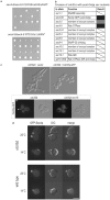
a. CDC55 and RTS1 genetic interactions with sec4-8. sec4-8/sec4-8 CDC55/cdc55ΔKANR and sec4-8/sec4-8 RTS1/rts1ΔKANR diploid cells were sporulated and tetrads dissected on YPD media. Five representative tetrad dissections are shown in each case. b. Crosses of cdc55Δ with post-Golgi sec mutants. White boxes with diagonal stripe indicate no interaction, black boxes indicate synthetic lethality at 25°C, and gray boxes indicate that the restrictive temperature of the double mutant was at least 3°C lower than that of the single mutant. c. Localization of Cdc55p to the sites of exocytosis is abolished in sec4-8 mutant cells. The Cdc55p construct is functional when tagged at the COOH-terminus as it can restore normal growth morphology to the cdc55Δ cells; (i) cdc55Δ cells + vector only (ii) cdc55Δ cells + plasmid expressing Cdc55p-GFP under control of the endogenous promoter and terminator (pRC3897A CDC55-GFP pRS315). Isogenic (iii) wild type and (iv) sec4-8 haploid cells containing COOH-terminally tagged Cdc55p-GFP as the only source of Cdc55p were examined for Cdc55p to the sites of exocytosis (white arrow). Cdc55p-GFP is normally distributed throughout the cytosol and also at sites of exocytosis. The enrichment of Cdc55p-GFP at the sites of exocytosis is lost in sec4-8 cells after a shift to the restrictive temperature when compared to the isogenic wild type cells. d. Localization of Sec4p to the sites of exocytosis is abolished in cdc55Δ null cells. GFP-Sec4p localization was examined in cdc55Δ cells at the permissive (25°C) and restrictive (14°C) conditions in comparison to a wild type control.
Figure 4
Figure 4. Structural and bioinformatic predictions for the sites of Sec4p phosphorylation.
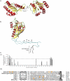
a. Diagram showing the relative positions of the boundary residues of the core GTPase domain relative to the nucleotide binding pocket. Structural determinations for the core GTPase domain of Sec4p reveal that the boundary residues of the core GTPase domain are physically located closely in three-dimensional space and are on the opposite face of the protein to the nucleotide binding cleft and switch regions. The terminal residues are shown in blue (the NH2 residue at position 19 and the COOH-terminal residue of the core domain at position 187) with the nucleotide in orange. Cartoon representation generated using MacPyMOL (DeLano Scientific) with PDB accession number 1G17. b. Hypothetical model showing the peptide extensions from the core GTPase domain of Sec4p. The ribbon diagram shows the relative size of the modeled NH2 and COOH tails (colored turquoise) for Sec4p including the geranylated di-cysteine motif at the extreme COOH-terminus. The positions of S8, S11, S201, and S204 are indicated with an asterisk. The tails are modeled using MacPyMOL (DeLano Scientific), extending from the core GTPase domain with approximately the same trajectory as Ypt1p for which a structure is available that includes these regions of the protein , . c. (i) Graphical output of the predictive algorithm NetPhos Yeast , (http://www.cbs.dtu.dk/services/NetPhosYeast/) for the primary sequence of Sec4p showing a clustering of sites with high probability (p>0.6, dotted grey line), at the protein termini, and includes the residues verified experimentally and analyzed in this study. (ii) Alignment of peptide extensions at the NH2- and COOH-termini reveals phosphorylated serines and other potentially phosphorylated residues in these termini are also apparent in the exocytic Rab proteins Rab13, Rab8 and Rab3A. Also shown are the equivalent regions of the yeast Rab proteins Ypt1p and Vps21p that contain phosphorylated serine residues. Shaded in orange are serine residues for which phosphorylation has been experimentally determined, underlined are residues that score with p>0.6 as potential phosphorylation sites with NetPhos 2.0 (http://www.cbs.dtu.dk/services/NetPhos/). Note the length of the peptide extensions of these Rab GTPases compared to other Ras superfamily members such as Cdc42Hs with a total protein length of 191 residues, or K-Ras (protein length 188 residues).

Similar articles

Cited by

References

    1. Chi A, Huttenhower C, Geer LY, Coon JJ, Syka JE, et al. Analysis of phosphorylation sites on proteins from Saccharomyces cerevisiae by electron transfer dissociation (ETD) mass spectrometry. Proc Natl Acad Sci U S A. 2007;104:2193–2198. - PMC - PubMed
    1. Ficarro SB, McCleland ML, Stukenberg PT, Burke DJ, Ross MM, et al. Phosphoproteome analysis by mass spectrometry and its application to Saccharomyces cerevisiae. Nat Biotechnol. 2002;20:301–305. - PubMed
    1. Gruhler A, Olsen JV, Mohammed S, Mortensen P, Faergeman NJ, et al. Quantitative phosphoproteomics applied to the yeast pheromone signaling pathway. Mol Cell Proteomics. 2005;4:310–327. - PubMed
    1. Li X, Gerber SA, Rudner AD, Beausoleil SA, Haas W, et al. Large-scale phosphorylation analysis of alpha-factor-arrested Saccharomyces cerevisiae. J Proteome Res. 2007;6:1190–1197. - PubMed
    1. Bodenmiller B, Wanka S, Kraft C, Urban J, Campbell D, et al. Phosphoproteomic analysis reveals interconnected system-wide responses to perturbations of kinases and phosphatases in yeast. Sci Signal. 3:rs4. - PMC - PubMed

Publication types

MeSH terms