. 2011;6(9):e24376.
doi: 10.1371/journal.pone.0024376. Epub 2011 Sep 8.

Specific interaction of Gαi3 with the Oa1 G-protein coupled receptor controls the size and density of melanosomes in retinal pigment epithelium

Affiliations

Specific interaction of Gαi3 with the Oa1 G-protein coupled receptor controls the size and density of melanosomes in retinal pigment epithelium

Alejandra Young et al. PLoS One. 2011.

Abstract

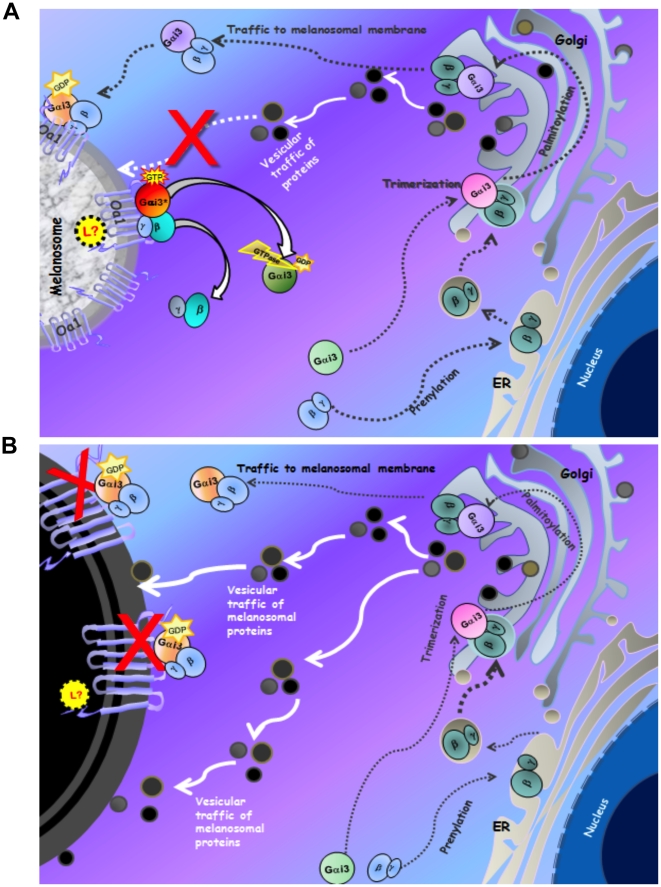
Background: Ocular albinism type 1, an X-linked disease characterized by the presence of enlarged melanosomes in the retinal pigment epithelium (RPE) and abnormal crossing of axons at the optic chiasm, is caused by mutations in the OA1 gene. The protein product of this gene is a G-protein-coupled receptor (GPCR) localized in RPE melanosomes. The Oa1-/- mouse model of ocular albinism reproduces the human disease. Oa1 has been shown to immunoprecipitate with the Gαi subunit of heterotrimeric G proteins from human skin melanocytes. However, the Gαi subfamily has three highly homologous members, Gαi1, Gαi2 and Gαi3 and it is possible that one or more of them partners with Oa1. We had previously shown by in-vivo studies that Gαi3-/- and Oa1-/- mice have similar RPE phenotype and decussation patterns. In this paper we analyze the specificity of the Oa1-Gαi interaction.

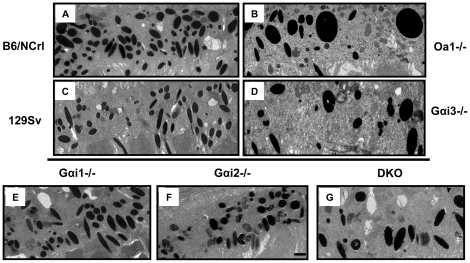
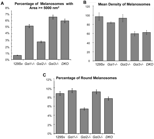
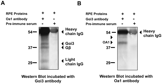
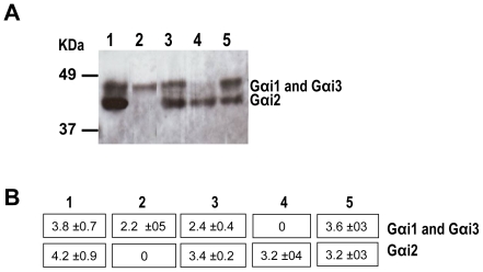
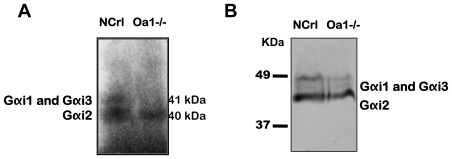
Methodology: By using the genetic mouse models Gαi1-/-, Gαi2-/-, Gαi3-/- and the double knockout Gαi1-/-, Gαi3-/- that lack functional Gαi1, Gαi2, Gαi3, or both Gαi1 and Gαi3 proteins, respectively, we show that Gαi3 is critical for the maintenance of a normal melanosomal phenotype and that its absence is associated with changes in melanosomal size and density. GST-pull-down and immunoprecipitation assays conclusively demonstrate that Gαi3 is the only Gαi that binds to Oa1. Western blots show that Gαi3 expression is barely detectable in the Oa1-/- RPE, strongly supporting a previously unsuspected role for Gαi3 in melanosomal biogenesis.

Conclusion: Our results identify the Oa1 transducer Gαi3 as the first downstream component in the Oa1 signaling pathway.


Conflict of interest statement

Competing Interests: The authors have declared that no competing interests exist.

Figures

Figure 1
Figure 1. Appearance of RPE melanosomes from Gαi1-/-, Gαi2-/- and DKO Gαi1-/-, Gαi3-/- mice.
RPEs from Oa1-/- (B) and their control B6/NCrl (A) mice and Gαi3-/- (D) and their control 129 Sv (C) mice have been published before and are shown for comparison. The ultrathin RPE sections show that Gαi1-/- (E) and Gαi2 -/- (F) mice do not have macromelanosomes as those present in Oa1-/- (B), Gαi3-/- (D) and DKO Gαi1-/-, Gαi3-/- (G) mice. Electron micrographs of ultrathin sections, 16,000× magnification; scale bars for all micrographs, 01 µm, is shown only in (F).
Figure 2
Figure 2. RPE melanosomal size, density and morphology of all Gαi-/- mice compared to control 129 Sv mice.
(A) Percentage of melanosomes larger than 5000 nm2 in each of the mouse lines analyzed. The Gαi3-/- mice have the highest percentage of melanosomes larger than 5000 nm2 in the RPE. (B) Mean density of melanosomes: The Gαi3-/- group had the lowest mean number of melanosomes per RPE area (59±5.2), and the wild-type and Gαi2-/- groups had the largest mean density of melanosomes (96.8±8.2 and 93.4±8.1, respectively). (C) Percentage of round melanosomes: The Gαi1-/- and Gαi3-/- groups have the larger number of round melanosomes (9.37±0.77 and 9.12±1.85 respectively), followed by the 129 Sv wild-type (8.75%±0.25), DKO (7.65%±0.31) and Gαi2-/- (5.39%±0.37).
Figure 3
Figure 3. Representative ERG responses of Gαi1-/-, Gαi2-/-, Gαi3-/- and DKO mice.
Standardized ERG was performed in experimental and control mice in two different sessions. A) ERGs from wild-type, Gαi1-/-, Gαi3-/- and DKO mice. B) ERGs from wild-type and Gαi2-/- mice.
Figure 4
Figure 4. Analysis of interactions between Oa1 and heterotrimeric Gα proteins by in- vitro pull-down assay.
A) Schematic representation of the vector used to make the GST-fusion polypeptides GST::Oa1-i3 and GST::Oa1-CT. Each amplified Oa1 sequence (i3 and CT) was cloned into the PGEXT4-2 vector containing GST between the selected restriction enzymes (BamH I and Sal I). B) Coomassie-blue stained gel showing the SDS-PAGE-separated fusion proteins used in the pull-down experiment. GST by itself has an apparent molecular weight of 26 kDa, the GST::Oa1-i3 fusion protein of 29 kDa and the GST::Oa1-CT fusion protein of 37 kDa in agreement with their estimated molecular masses. C) Autoradiogram of in-vitro transcribed and translated 35S-labeled Gα proteins that will be tested for Oa1 binding activity below (D–E), after separation by SDS-PAGE on a 10% Tris-Glycine polyacrylamide gel (D–E). The apparent molecular weight of each Gα protein is in agreement with its predicted molecular mass, Gαi1: 41 kDa, Gαi2: 40 kDa, Gαi3: 41 kDa, Gαq: 42 kDa, Gαs: 46 kDa and luciferase, run as a control: 61 kDa. D) Commassie blue staining of the gel used for SDS-PAGE of recombinant GST::Oa1-CT and GST::Oa1-i3 fusion proteins immobilized on glutathione-agarose beads and incubated with the indicated 35S-labeled Gα proteins. Lanes 1 and 14 had the GST protein incubated with one of the 35S-labeled Gα proteins in this experiment, 35S-Gαi3. Lanes 2–7: GST::Oa1-CT incubated with the indicated 35S-labeled Gα. Lanes 8–13: GST::Oa1-i3 incubated with the indicated 35S-Gα. Since the amount of 35S-Gα protein is minimal, only the fusion proteins are observed in this gel. Lanes 7 and 13: GST-fusion proteins immobilized on glutathione-agarose beads (Neg. C: negative control). All lanes with the same fusion protein have comparable amounts of protein. E) Autoradiograph of the same gel showing that of all 35S-labeled Gα proteins, Gαi3 is the only one that binds specifically to Oa1(lanes 4 and 10). Molecular weight markers are expressed in kDa.
Figure 5
Figure 5. Gαi3 is the specific Oa1-interacting protein.
A) Western blot of proteins from human RPE immunoprecipitated with the anti-OA1 antibody as described in Materials and Methods. SDS–PAGE was followed by incubation with anti-Gαi3 antibodies. Left lane: RPE extract incubated with OA1 antibody; right lane: RPE extract incubated with pre-immune serum. B) Western blot analysis of the proteins from human RPE immunoprecipitated with anti-Gαi3 antibody. SDS–PAGE was followed by incubation with anti-OA1 antibody. Left lane: RPE extract incubated with Gαi3 antibody; right lane: RPE extract incubated with pre-immune serum. Two isoforms of OA1 have apparent molecular weights of 45 and 48 kDa (arrowheads).
Figure 6
Figure 6. Western blot of RPE proteins from all Gαi-/- mice using an anti- Gαcommon antibody.
A) RPE membrane proteins from 3-month-old mice were separated by SDS-PAGE, blotted and reacted with Gαcommon antibodies. Protein bands were visualized using the ECL detection reagent. The RPE proteins are from: Gαi1-/- (lane 1), Gαi2-/- (lane 2), Gαi3-/- (lane 3), DKO (lane 4) and 129 Sv (lane 5) mice. The membrane was then stripped and reacted with α-tubulin antibodies to confirm that the amount of protein loaded on each lane was the same. B) Analysis of the immunoblot by densitometry using the Quantity One 1-D Software. Numbers represent the optical density units per mm2 (OD U/mm2) of each Gαi protein band.
Figure 7
Figure 7. ADP-ribosylation of Gαi proteins and Western blot of Oa1-/- and control B6/NCrl RPE proteins.
A) RPE membrane proteins from 3-month-old mice were subjected to 32P-ADP ribosylation with PTX, separated by SDS-PAGE and visualized by autoradiography. Lane 1: control B6/NCrl RPE; lane 2: Oa1-/- RPE. B) Western blot using the same RPE membrane proteins separated by SDS-PAGE and reacted with the Gαcommon antibody. Lane 1: B6/NCrl RPE and lane 2: Oa1-/- RPE.
Figure 8
Figure 8. Proposed hypothetical model of the Gαi3 regulation of RPE melanosomal size.
A) An unknown luminal ligand (L?) turns on wild-type Oa1, which activates Gαi3. The active Gαi3 in turn inhibits vesicular traffic of proteins from the TGN to the melanosome, controlling in this way its size. B) Mutated OA1 is unable to activate Gαi3 and, thus, the continuous supply of melanin-related proteins to the melanosome results in the formation of very big organelles, the macromelanosomes.

Similar articles

Cited by

References

    1. King RC, Stansfield WD, Mulligan PK. A Dictionary of Genetics, Seventh Edition. Oxford University Press; 2006.
    1. Oetting WS, Summers CG, King RA. Albinism and the associated ocular defects. Metab Pediatr Syst Ophthalmol. 1994;17:5–9. - PubMed
    1. Garner A, Jay BS. Macromelanosomes in X-linked ocular albinism. Histopathology. 1980;4:243–54. - PubMed
    1. O'Donnell FE, Jr, Hambrick GW, Jr, Green WR, Iliff WJ, Stone DL. X-linked ocular albinism: An oculocutaneous macromelanosomal disorder. Arch Ophthalmol. 1976;94:1883–92. - PubMed
    1. Schiaffino MV, Tacchetti C. The ocular albinism type 1 (OA1) protein and the evidence for an intracellular signal transduction system involved in melanosome biogenesis. Pigment Cell Res. 2005;18:227–33. - PubMed

Publication types

MeSH terms