. 2011;6(9):e24392.
doi: 10.1371/journal.pone.0024392. Epub 2011 Sep 8.

Cyclic AMP control measured in two compartments in HEK293 cells: phosphodiesterase K(M) is more important than phosphodiesterase localization

Affiliations

Cyclic AMP control measured in two compartments in HEK293 cells: phosphodiesterase K(M) is more important than phosphodiesterase localization

Karina Matthiesen et al. PLoS One. 2011.

Abstract

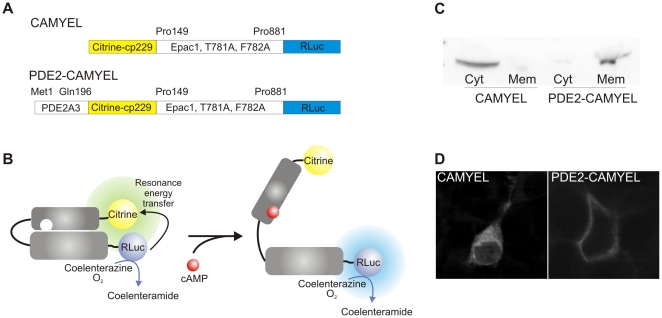
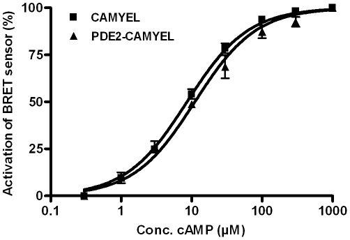
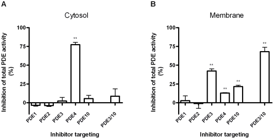
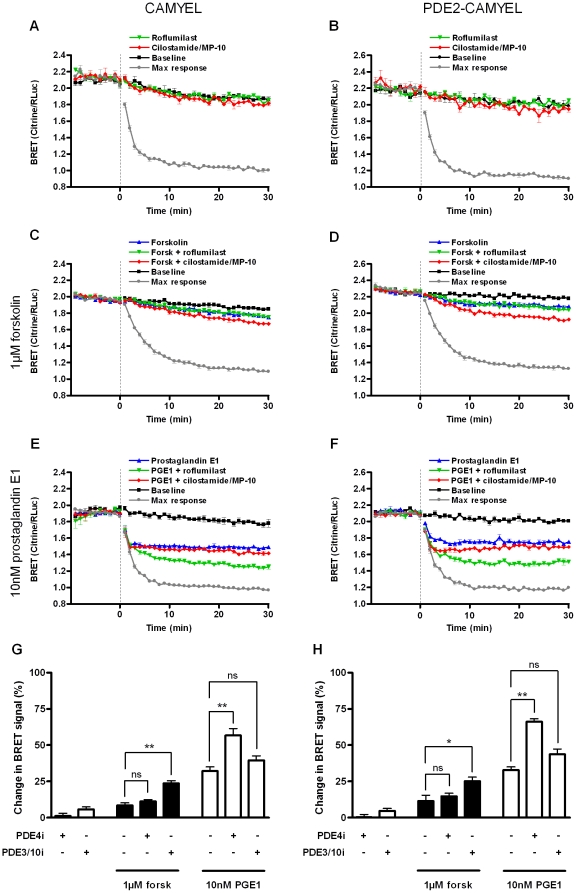
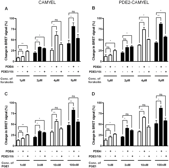
The intracellular second messenger cyclic AMP (cAMP) is degraded by phosphodiesterases (PDE). The knowledge of individual families and subtypes of PDEs is considerable, but how the different PDEs collaborate in the cell to control a cAMP signal is still not fully understood. In order to investigate compartmentalized cAMP signaling, we have generated a membrane-targeted variant of the cAMP Bioluminiscence Resonance Energy Transfer (BRET) sensor CAMYEL and have compared intracellular cAMP measurements with it to measurements with the cytosolic BRET sensor CAMYEL in HEK293 cells. With these sensors we observed a slightly higher cAMP response to adenylyl cyclase activation at the plasma membrane compared to the cytosol, which is in accordance with earlier results from Fluorescence Resonance Energy Transfer (FRET) sensors. We have analyzed PDE activity in fractionated lysates from HEK293 cells using selective PDE inhibitors and have identified PDE3 and PDE10A as the major membrane-bound PDEs and PDE4 as the major cytosolic PDE. Inhibition of membrane-bound or cytosolic PDEs can potentiate the cAMP response to adenylyl cyclase activation, but we see no significant difference between the potentiation of the cAMP response at the plasma membrane and in cytosol when membrane-bound and cytosolic PDEs are inhibited. When different levels of stimulation were tested, we found that PDEs 3 and 10 are mainly responsible for cAMP degradation at low intracellular cAMP concentrations, whereas PDE4 is more important for control of cAMP at higher concentrations.


Conflict of interest statement

Competing Interests: JN is an employee of H. Lundbeck A/S and KM was an employee of H. Lundbeck A/S when the study was performed. The authors confirm that this does not alter their adherence to all the PloS ONE policies on sharing data and material. No Lundbeck products can be considered competing interests.

Figures

Figure 1
Figure 1. Schematic overview of BRET sensors.
(A) CAMYEL is comprised of cytosolic, catalytic inactive Epac1 sandwiched between Citrine-cp229 and the Renilla luciferase (RLuc). PDE2-CAMYEL is targeted to the membrane by fusion of the N-terminal part of PDE2A3 to CAMYEL. (B) Binding of cAMP to CAMYEL/PDE2-CAMYEL induces a conformational change in the Epac1 part resulting in a decrease of energy transfer from the luciferase to Citrine. (C and D) Western blot and confocal images showing the distribution of CAMYEL in the cytosol and the targeting of PDE2-CAMYEL to the membrane. Cyt, cytosolic fraction; Mem, membrane fraction.
Figure 2
Figure 2. Characterization of the BRET sensors.
cAMP dose-response graphs showing the relative activation of CAMYEL and PDE2-CAMYEL from HEK293 lysates. EC50 values were 8.4±0.7 µM and 10.6±0.9 µM, respectively (n = 3–4).
Figure 3
Figure 3. cAMP PDE activity in HEK293 cytosol and membrane fractions.
Lysate from either the cytosolic fraction (A) or the membrane fraction (B) was tested in the PDE activity with 1 µM cAMP. Selective PDE inhibitors were added alone or in combination as indicated. The results represent the mean ± SEM of three experiments. Compounds and concentrations used: PDE1i, 250 nM (see M&M section); PDE2i, 20 nM BAY 60-7550; PDE3i, 400 nM cilostamide; PDE4i, 50 nM roflumilast; PDE10i, 300 nM MP-10. The ratio of total cAMP PDE activity in the cytosolic fraction and membrane fraction compared to whole cells was 40.4±5.3% and 59.6±5.3%, respectively.
Figure 4
Figure 4. Effect of selective PDE inhibitors on cAMP response in HEK293 cells.
Changes in BRET signal were measured in HEK293 cells transfected with either CAMYEL or PDE2-CAMYEL. (A and B) BRET was measured in response to addition of PDE inhibitors. 50 nM roflumilast or 400 nM cilostamide and 300 nM MP-10 was added at time 0. (C–F) BRET was measured in response to stimulation with 1 µM forskolin (C, D) or 10 nM PGE1 (E, F). Forskolin or PGE1 was added at time 0 alone or in combination with 50 nM roflumilast or 400 nM cilostamide and 300 nM MP-10. For baselines, buffer was added, and for maximum response, 10 µM forskolin plus all three PDE inhibitors were added. Each point represents the mean ± SEM of triplicate determinations in a representative experiment. (G and H) Summary of BRET measurements. Relative BRET responses are averaged for the time period 21–30 min after addition of drugs. The results represent the mean ± SEM of three experiments. ns (not significant), P>0.05; *, 0.01
Figure 5
Figure 5. Different PDEs dominate at different cAMP concentrations.
Average relative BRET response 21–30 min after addition of forskolin (A, B) or PGE1 (C, D) in combination with PDE inhibitors to HEK293 cells transfected with CAMYEL or PDE2-CAMYEL. Different concentrations of forskolin or PGE1 was added alone or in combination with 50 nM roflumilast or 400 nM cilostamide and 300 nM MP-10 as indicated. From curves similar to those shown in Figure 4, the change in BRET signal was calculated relative to the maximum response (10 µM forskolin plus all three PDE inhibitors). The results represent the mean ± SEM of three experiments. ns (not significant), P>0.05; *, 0.01

References

    1. Beavo JA, Brunton LL. Cyclic nucleotide research – still expanding after half a century. Nat Rev Mol Cell Biol. 2002;3:710–718. - PubMed
    1. Chin KV, Yang WL, Ravatn R, Kita T, Reitman E, et al. Reinventing the wheel of cyclic AMP: novel mechanisms of cAMP signaling. Ann N Y Acad Sci. 2002;968:49–64. - PubMed
    1. Cooper DM. Compartmentalization of adenylate cyclase and cAMP signalling. Biochem Soc Trans. 2005;33:1319–1322. - PubMed
    1. McKnight GS. Cyclic AMP second messenger systems. Curr Opin Cell Biol. 1991;3:213–217. - PubMed
    1. Beavo JA. Cyclic nucleotide phosphodiesterases: functional implications of multiple isoforms. Physiol Rev. 1995;75:725–748. - PubMed

Publication types

MeSH terms