. 2011;6(9):e24434.
doi: 10.1371/journal.pone.0024434. Epub 2011 Sep 8.

Determinants of GBP recruitment to Toxoplasma gondii vacuoles and the parasitic factors that control it

Affiliations

Determinants of GBP recruitment to Toxoplasma gondii vacuoles and the parasitic factors that control it

Sebastian Virreira Winter et al. PLoS One. 2011.

Abstract

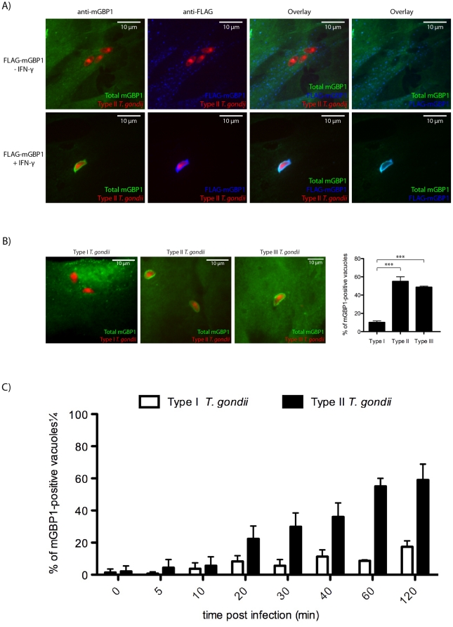
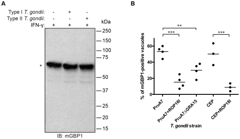
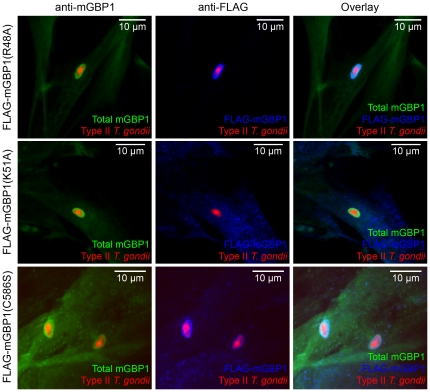
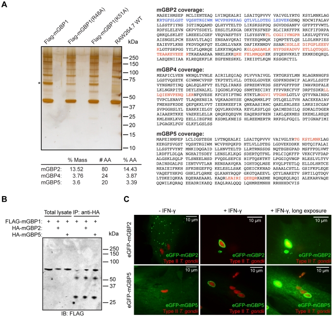
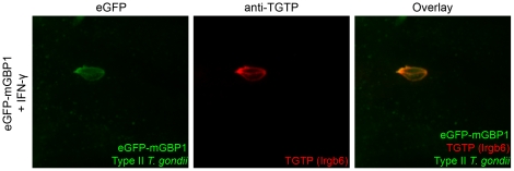
IFN-γ is a major cytokine that mediates resistance against the intracellular parasite Toxoplasma gondii. The p65 guanylate-binding proteins (GBPs) are strongly induced by IFN-γ. We studied the behavior of murine GBP1 (mGBP1) upon infection with T. gondii in vitro and confirmed that IFN-γ-dependent re-localization of mGBP1 to the parasitophorous vacuole (PV) correlates with the virulence type of the parasite. We identified three parasitic factors, ROP16, ROP18, and GRA15 that determine strain-specific accumulation of mGBP1 on the PV. These highly polymorphic proteins are held responsible for a large part of the strain-specific differences in virulence. Therefore, our data suggest that virulence of T. gondii in animals may rely in part on recognition by GBPs. However, phagosomes or vacuoles containing Trypanosoma cruzi did not recruit mGBP1. Co-immunoprecipitation revealed mGBP2, mGBP4, and mGBP5 as binding partners of mGBP1. Indeed, mGBP2 and mGBP5 co-localize with mGBP1 in T. gondii-infected cells. T. gondii thus elicits a cell-autonomous immune response in mice with GBPs involved. Three parasitic virulence factors and unknown IFN-γ-dependent host factors regulate this complex process. Depending on the virulence of the strains involved, numerous GBPs are brought to the PV as part of a large, multimeric structure to combat T. gondii.


Conflict of interest statement

Competing Interests: Commercial funding has been received from Bayer HealthCare. This does not alter the authors' adherence to all the PLoS ONE policies on sharing data and materials. The authors have no patents or products in development or marketed products to declare.

Figures

Figure 1
Figure 1. mGBP1 preferentially accumulates on type II (Pru) and III (CEP) but not on type I (RH) vacuoles.
(A) MEFs that overexpress FLAG-mGBP1 were stimulated with 200 U/ml IFN-γ overnight and infected with mCherry-expressing type II T. gondii at an MOI between 5 and 10 for 1 h. (B) Representative pictures of IFN-γ-induced wild-type MEFs infected with either type I, II or III 1 h post-infection show that mGBP1 is preferentially recruited to nonvirulent types II and III vacuoles. The right panel shows the frequencies of mGBP1-positive vacuoles. However, when mGBP1 was recruited to the virulent type I (11% of the vacuoles), the intensity of recruitment was comparable to the recruitment to types II and III. The differences in extent of recruitment of mGBP1 to PVs between type I and types II and III are significant with *** p<0.001. Error bars represent standard deviations of three independent experiments. (C) The frequency of mGBP1-positive vacuoles of type I and type II T. gondii was determined at various time points. MEFs that overexpress FLAG-mGBP1 were induced with 200 U/ml IFN-γ overnight and infected with type I or type II T. gondii at an MOI between 5 and 10 before they were fixed after the indicated time. For each strain, at least 100 vacuoles of invaded T. gondii were checked for recruitment of mGBP1. Error bars represent standard deviations of three independent experiments. A rabbit polyclonal anti-mGBP1 antiserum and a mouse monoclonal anti-FLAG antibody (Sigma-Aldrich) were used for stainings. Pictures were taken with a spinning disk confocal microscope.
Figure 2
Figure 2. T. gondii cannot degrade mGBP1 but parasitic virulence factors interfere with recruitment of mGBP1.
(A) Degradation of mGBP1 after invasion of T. gondii is not detectable by IB. RAW264.7 cells were induced with 200 U/ml IFN-γ overnight and infected with mCherry-expressing T. gondii (Pru or RH). Infected cells were separated from uninfected cells by FACS 2 h post-infection. For each lysate, 50 µg of protein, which was equivalent to 300,000 cells, were loaded per lane. The mGBP1 protein was detected by rabbit polyclonal anti-mGBP1 antiserum (*). (B) Type II strains with deleted GRA15, type II strains transgenic for the type I version of ROP16, and type III strains transgenic for the type I version of ROP18 show less mGBP1-positive vacuoles compared to the parental strain. MEFs that overexpress eGFP-mGBP1 were induced with 200 U/ml IFN-γ and infected with PruA7, a Pru type II T. gondii strain, PruA7 either lacking GRA15 or transgenic for the type I version of ROP16, CEP, a type II strain, or CEP transgenic for the type I version of ROP18. For each strain, at least 100 vacuoles of invaded T. gondii were checked for recruitment of mGBP1. Data represent four independent experiments. Decrease of mGBP1 recruitment is significant with ** p<0.01 and *** p<0.001.
Figure 3
Figure 3. Nucleotide-dependent multimerization is required while farnesylation is dispensable for recruitment of mGBP1 to the PV.
(A) Confocal pictures show MEFs that overexpress FLAG-tagged mutants of mGBP1 stimulated with 200 U/ml IFN-γ overnight and infected with mCherry-expressing type II T. gondii (Pru) at an MOI between 5 and 10 for 1 h. The R48A and C586S mGBP1 mutants were targeted to the PV like the wild-type counterpart. In contrast, the K51A mGBP1 mutant was unable to accumulate around the PV. Rabbit polyclonal anti-mGBP1 antiserum and a mouse monoclonal anti-FLAG antibody were used for stainings.
Figure 4
Figure 4. mGBP2 and mGBP5 interact with mGBP1 and accumulate on type II T. gondii (Pru) vacuoles.
(A) mGBP2, mGBP4, and mGBP5 were identified as interactors of mGBP1 in a immunoprecipitation with subsequent MS/MS analysis (A). The left panel shows a silver-stained SDS-PAGE gel following an anti-FLAG IP from wild-type RAW264.7 cells (control lane) or RAW264.7 that overexpress wild-type or various mGBP1 mutants. The right panel shows three GBPs that were identified in the mass spectrometry analysis as binding partners of mGBP1. Wild-type RAW264.7 cells or RAW264.7 cells that overexpress FLAG-mGBP1, FLAG-mGBP1(R48A) or FLAG-mGBP1(K51A) were stimulated with 200 U/ml IFN-γ overnight and infected with type II T. gondii at an MOI of 5–10 for 1 h. The mGBP1 protein was recovered from all lanes except from the control lane (*). Unique peptides recovered from mGBP2, mGBP4, and mGBP5 are shown in red and peptides common with mGBP1 are shown in blue. The total number of unique amino acids (# AA), the percentage of total mass (% Mass), and the percentage of total amino acids (% AA) recovered are also shown. For mGBP4 and mGBP5, the sequence coverage was 3–4%. 14% of the mGBP2-specific amino acids were found in the gel (21% when including amino acids common to mGBP1). (B) Interaction of HA-mGBP2 and HA-mGBP5 with FLAG-mGBP1 was confirmed by co-IP with subsequent immunoblot analysis. RAW264.7 cells that overexpress either FLAG-mGBP1 and HA-mGBP2, FLAG-mGBP1 and HA-mGBP5, or FLAG-mGBP1 alone were lysed in 0.5% NP-40 and proteins were immunoprecipitated with an anti-HA antibody. Co-IP of FLAG-mGBP1 was confirmed by immunoblot with an anti-FLAG antibody. A mouse monoclonal anti-FLAG antibody (Sigma-Aldrich) and a rat monoclonal anti-HA antibody (Roche Applied Science) were used. (C) mGBP2 and mGBP5 are recruited to type II T. gondii vacuoles in an IFN-γ dependent manner. Confocal pictures show MEFs that overexpress either eGFP-mGBP2 or eGFP-mGBP5 pre-induced with 200 U/ml IFN-γ and infected with type II T. gondii. Live imaging was performed with a spinning disk confocal microscope and revealed that both mGBP2 and mGBP5 were targeted to T. gondii vacuoles in an IFN-γ-dependent manner.
Figure 5
Figure 5. mGBP1 co-localizes with TGTP after infection with T. gondii.
Confocal pictures show that mGBP1 co-localizes with TGTP after infection with T. gondii in vitro (B). MEFs that overexpress eGFP-mGBP1 were stimulated with 200 U/ml IFN-γ overnight and infected with GFP-expressing Type II T. gondii at a MOI between 5 and 10 for 1 h. A goat polyclonal anti-TGTP antibody was used for staining.

Similar articles

Cited by

References

    1. Ehrt S, Schnappinger D, Bekiranov S, Drenkow J, Shi S, et al. Reprogramming of the macrophage transcriptome in response to interferon-gamma and Mycobacterium tuberculosis: signaling roles of nitric oxide synthase-2 and phagocyte oxidase. J Exp Med. 2001;194:1123–1140. - PMC - PubMed
    1. de Veer MJ, Holko M, Frevel M, Walker E, Der S, et al. Functional classification of interferon-stimulated genes identified using microarrays. J Leukoc Biol. 2001;69:912–920. - PubMed
    1. MacMicking JD. IFN-inducible GTPases and immunity to intracellular pathogens. Trends Immunol. 2004;25:601–609. doi: 10.1016/j.it.2004.08.010. - DOI - PubMed
    1. Staeheli P, Danielson P, Haller O, Sutcliffe JG. Transcriptional activation of the mouse Mx gene by type I interferon. Mol Cell Biol. 1986;6:4770–4774. - PMC - PubMed
    1. Taylor GA. IRG proteins: key mediators of interferon-regulated host resistance to intracellular pathogens. Cell Microbiol. 2007;9:1099–1107. doi: 10.1111/j.1462-5822.2007.00916.x. - DOI - PubMed

Publication types

MeSH terms