Randomized Controlled Trial
. 2011;6(9):e24522.
doi: 10.1371/journal.pone.0024522. Epub 2011 Sep 8.

Self-regulation of amygdala activation using real-time FMRI neurofeedback

Affiliations
Randomized Controlled Trial

Self-regulation of amygdala activation using real-time FMRI neurofeedback

Vadim Zotev et al. PLoS One. 2011.

Abstract

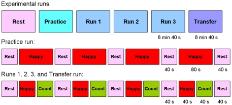
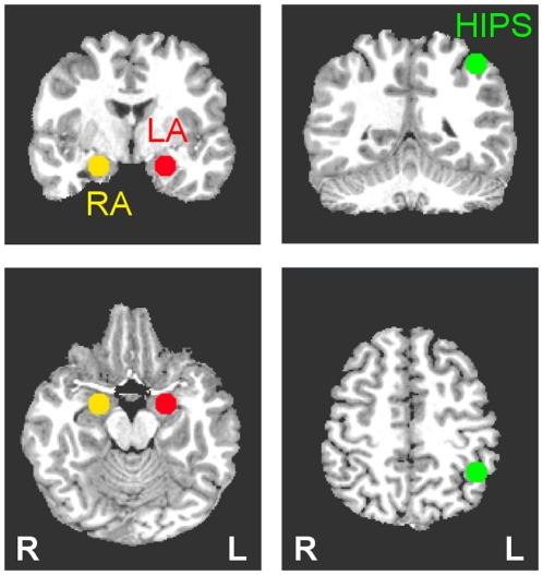
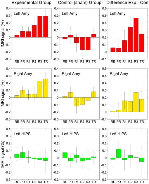
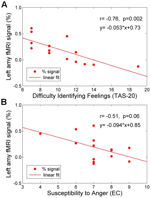
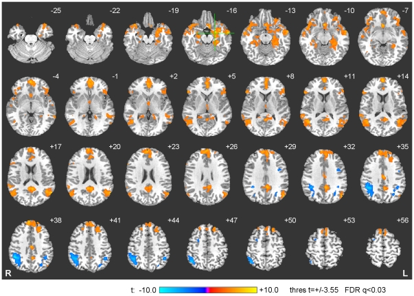
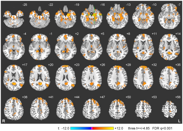
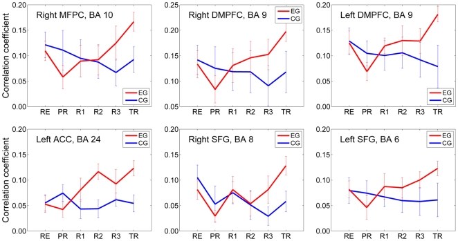
Real-time functional magnetic resonance imaging (rtfMRI) with neurofeedback allows investigation of human brain neuroplastic changes that arise as subjects learn to modulate neurophysiological function using real-time feedback regarding their own hemodynamic responses to stimuli. We investigated the feasibility of training healthy humans to self-regulate the hemodynamic activity of the amygdala, which plays major roles in emotional processing. Participants in the experimental group were provided with ongoing information about the blood oxygen level dependent (BOLD) activity in the left amygdala (LA) and were instructed to raise the BOLD rtfMRI signal by contemplating positive autobiographical memories. A control group was assigned the same task but was instead provided with sham feedback from the left horizontal segment of the intraparietal sulcus (HIPS) region. In the LA, we found a significant BOLD signal increase due to rtfMRI neurofeedback training in the experimental group versus the control group. This effect persisted during the Transfer run without neurofeedback. For the individual subjects in the experimental group the training effect on the LA BOLD activity correlated inversely with scores on the Difficulty Identifying Feelings subscale of the Toronto Alexithymia Scale. The whole brain data analysis revealed significant differences for Happy Memories versus Rest condition between the experimental and control groups. Functional connectivity analysis of the amygdala network revealed significant widespread correlations in a fronto-temporo-limbic network. Additionally, we identified six regions--right medial frontal polar cortex, bilateral dorsomedial prefrontal cortex, left anterior cingulate cortex, and bilateral superior frontal gyrus--where the functional connectivity with the LA increased significantly across the rtfMRI neurofeedback runs and the Transfer run. The findings demonstrate that healthy subjects can learn to regulate their amygdala activation using rtfMRI neurofeedback, suggesting possible applications of rtfMRI neurofeedback training in the treatment of patients with neuropsychiatric disorders.


Conflict of interest statement

Competing Interests: The authors have declared that no competing interests exist.

Figures

Figure 1
Figure 1. Real-time Display Screens for the Real-time fMRI Neurofeedback Procedure.
Visual cues (i.e. text, color bars, and icons) were presented for each of the experimental conditions. During the Happy Memories condition, the word “Happy”, two color bars, and a number indicating the neurofeedback fMRI signal level were displayed on the screen. The participants were instructed to evoke happy autobiographical memories to make themselves feel happy while trying to increase the level of the red bar to a given target level (indicated by the fixed height blue bar). During the Count condition, the subjects saw the cue with a counting instruction, e.g. “Count 100, 99, 98 … (−1)”, and were instructed to mentally count backwards from 100 by subtracting a given integer number (shown in parentheses). During the Rest condition, the participants saw the cue “Rest” and were asked to relax while looking at the screen. For the Happy Memories condition without neurofeedback, no bars were displayed, and the cue “As Happy as possible” was presented instead.
Figure 2
Figure 2. Protocol for the Real-time fMRI Neurofeedback Experiment.
The experimental procedure consisted of six runs each lasting 8 min 40 sec. During the Rest run, the participants were instructed to rest. During the Practice run, the subjects were given the opportunity to become comfortable with the rtfMRI neurofeedback procedure. During Runs 1, 2, and 3, the participants underwent rtfMRI neurofeedback training consisting of alternating blocks of Rest, Happy, and Count conditions, each lasting 40 seconds. During the Transfer Run, the subjects were instructed to perform the same task as during the neurofeedback training, but neurofeedback information (bars, number) was not be provided.
Figure 3
Figure 3. Regions of Interest (ROIs) for the Real-time fMRI Neurofeedback Procedure.
Three regions of interest (spheres of 7 mm radius) were used to assess changes in BOLD activity in the left amygdala (LA, red), right amygdala (RA, yellow), and left horizontal segment of the intraparietal sulcus (HIPS, green). The ROI placements are illustrated on T1-weighted coronal (upper row) and axial (lower row) human brain sections in the Talairach space. Following the radiological notation, the left side (L) of the brain is shown on the right, and the right side (R) of the brain – on the left.
Figure 4
Figure 4. Learned Enhancement of Control over BOLD fMRI Activation and Mood Induction.
A significant training effect was observed for the left amygdala for the subjects in the experimental group. The control of BOLD fMRI activation in the left amygdala ROI monotonically increased over training runs and persisted during the Transfer run. Each bar represents mean percent signal change in the BOLD signal (± s.e.m.) averaged across Happy Memories conditions during a given run (see text for details) for each ROI (left amygdala, red; right amygdala, yellow; left HIPS, green) and group (experimental, control). The difference between the corresponding average fMRI percent signal change values for the experimental and control (sham) groups is also shown.
Figure 5
Figure 5. Relationship between the Neurofeedback Training Effect on the Left Amygdala Activation and Individual Psychological Scores.
A) Correlation with the Difficulty Identifying Feelings (TAS-20). The training effect for the left amygdala was correlated with the participants' insight into their feelings. Thus the more highly the participants rated their capacity for identifying their own feelings (based on the Difficulty Identifying Feelings sub-scale of the Toronto Alexithymia Scale, TAS-20), the more they increased the BOLD signal in the left amygdala during training. B) Correlation with the Susceptibility to Anger (EC). The higher the participants rated their sensitivity to other peoples' anger (based on the Susceptibility to Anger sub-scale of the Emotional Contagion scale, EC), the less BOLD activation was observed in their left amygdala during training. The activation levels shown (in both A and B) are averages across the three neurofeedback training runs (Runs 1–3) for each subject in the Experimental group.
Figure 6
Figure 6. Cardiac and Respiratory Rate Variations during the Neurofeedback Experiment.
A) Average Cardiac Rate. The experimental group (EG, red) and control (sham) group (CG, blue) exhibited no statistically significant differences in mean cardiac rates for either Happy Memories or Rest conditions for any of the six experimental runs. B) Average Respiratory Rate. Similarly, no statistically significant differences were observed in mean respiratory rates of the two groups (EG and CG) for either condition for any of the runs.
Figure 7
Figure 7. Activation Network for Happy Memories and Count Conditions.
The group activation analysis for Happy>Count contrast revealed significant BOLD signal changes in a fronto-temporo-limbic network, while the Count>Happy contrast revealed activations in a parietal network (see text for details and Table 1 for coordinates). The activation maps are projected on a representative single-subject T1 template in the Talairach space with 3 mm separation between axial slices (the number adjacent to each slice indicates the z coordinate in mm from the bicommissural plane, with positive z indicating dorsal). The left hemisphere (L) is to the reader's right. The green crosshairs mark the center of the left amygdala ROI.
Figure 8
Figure 8. Functional Connectivity Analysis for the Amygdala Network.
The group functional connectivity analysis using a seed ROI in the left amygdala region revealed a fronto-temporo-limbic network (see text for details and Table 3 for coordinates). The connectivity maps are projected on a representative single-subject T1 template in the Talairach space. The coordinates and orientation of each slice are described in the legend for Figure 7. The green crosshairs mark the center of the seed ROI for the connectivity analysis.
Figure 9
Figure 9. Enhancement in Functional Connectivity with the Left Amygdala during the Neurofeedback Training.
For the subjects in the experimental group (EG, red), the functional connectivity with the left amygdala increased across the neurofeedback training runs (PR, R1, R2, R3) and the Transfer run (TR) for the right medial frontal polar cortex (MFPC, BA 10), bilateral dorsomedial prefrontal cortex (DMPFC, BA 9), left pregenual anterior cingulate cortex (ACC, BA 24), and bilateral superior frontal gyrus (SFG, BA 6,8). In contrast, no significant connectivity changes were observed for the same regions for the Control (sham) group (CG, blue).

Similar articles

Cited by

References

    1. Cox RW, Jesmanowicz A, Hyde JS. Real-time functional magnetic resonance imaging. Magn Reson Med. 1995;33(2):230–236. - PubMed
    1. deCharms RC. Applications of real-time fMRI. Nat Rev Neurosci. 2008;9(9):720–729. - PubMed
    1. Sterman MB. Basic concepts and clinical findings in the treatment of seizure disorders with EEG operant conditioning. Clin Electroencephalogr. 2000;31(1):45–55. - PubMed
    1. Nowlis DP, Kamiya J. The control of electroencephalographic alpha rhythms through auditory feedback and the associated mental activity. Psychophysiology. 1970;6(4):476–484. - PubMed
    1. Levesque J, Beauregard M, Mensour B. Effect of neurofeedback training on the neural substrates of selective attention in children with attention-deficit/hyperactivity disorder: a functional magnetic resonance imaging study. Neurosci Lett. 2006;394:216–224. - PubMed

Publication types