. 2011;6(9):e24531.
doi: 10.1371/journal.pone.0024531. Epub 2011 Sep 9.

A Gateway MultiSite recombination cloning toolkit

Affiliations

A Gateway MultiSite recombination cloning toolkit

Lena K Petersen et al. PLoS One. 2011.

Abstract

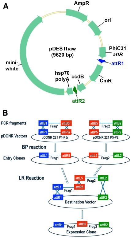
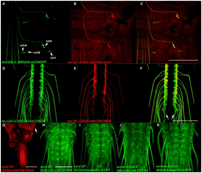
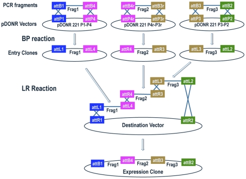
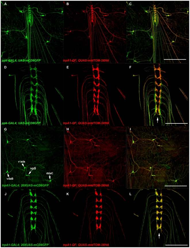
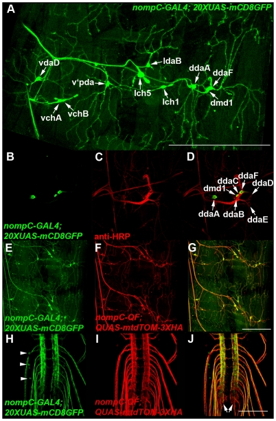
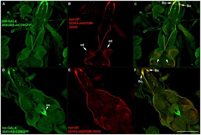
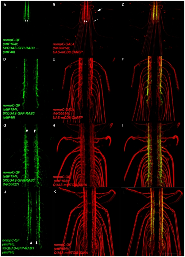
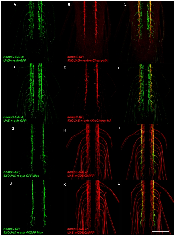
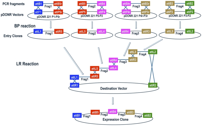
The generation of DNA constructs is often a rate-limiting step in conducting biological experiments. Recombination cloning of single DNA fragments using the Gateway system provided an advance over traditional restriction enzyme cloning due to increases in efficiency and reliability. Here we introduce a series of entry clones and a destination vector for use in two, three, and four fragment Gateway MultiSite recombination cloning whose advantages include increased flexibility and versatility. In contrast to Gateway single-fragment cloning approaches where variations are typically incorporated into model system-specific destination vectors, our Gateway MultiSite cloning strategy incorporates variations in easily generated entry clones that are model system-independent. In particular, we present entry clones containing insertions of GAL4, QF, UAS, QUAS, eGFP, and mCherry, among others, and demonstrate their in vivo functionality in Drosophila by using them to generate expression clones including GAL4 and QF drivers for various trp ion channel family members, UAS and QUAS excitatory and inhibitory light-gated ion channels, and QUAS red and green fluorescent synaptic vesicle markers. We thus establish a starter toolkit of modular Gateway MultiSite entry clones potentially adaptable to any model system. An inventory of entry clones and destination vectors for Gateway MultiSite cloning has also been established (www.gatewaymultisite.org).


Conflict of interest statement

Competing Interests: The authors have declared that no competing interests exist.

Figures

Figure 1
Figure 1. Drosophila destination vector and two-fragment schematic for Gateway MultiSite cloning.
A) The Drosophila destination vector pDESThaw. This destination vector contains an hsp70 polyadenylation sequence, a PhiC31 attB site for PhiC31 integrase-mediate site-specific transgenesis, and mini-white as a transformation marker. Appropriate combinations of two, three, or four entry clones recombine in a position and orientation-specific manner with ampicillin-resistant pDESThaw in the LR reaction to generate expression clones. The LR reactions for two, three, and four fragment Gateway MultiSite recombination all use the LR Clonase II Plus enzyme mix. Both the BP and LR reactions take advantage of the Gateway cassette that includes a chloramphenicol resistance marker and the ccdB gene. The Gateway cassette is located between the attP sites in the pDONR vectors (Figure S1) and between the attR1 and attR2 sites of the pDESThaw destination vector. The ccdB gene is toxic to any bacterial strain that does not contain a genetic suppressor including most common laboratory bacterial strains used for cloning such as DH10B and DH5α. ccdB containing clones were propagated using the now discontinued bacterial strain DB3.1 that has been replaced by Invitrogen with strain ccdB Survival 2 T1R. When BP and LR reactions are transformed into non-ccdB suppressor strains, only bacteria containing clones in which the Gateway cassette has been recombined out (and presumably the fragment(s) of interest recombined in) survive on kanamycin or ampicillin-selective plates. This results in a low frequency of colonies that do not contain the insert(s) of interest. B) Schematic diagram of two-fragment Gateway MultiSite recombination cloning. Fragment 1 and fragment 2 are amplified by PCR using oligonucleotides that incorporate flanking attB1 and attB5r sites in fragment 1 and flanking attB5 and attB2 sites in fragment 2. Fragment 1 is combined with pDONR 221 P1-P5r and fragment 2 is combined with pDONR 221 P5-P2 in separate BP reactions. The products of the BP reactions are pENTR attL1-Frag1-attR5 and pENTR attL5-Frag2-attL2. In the LR reaction both of these entry clones are combined with a destination vector to produce an expression clone containing fragment 1 and fragment 2 in a position and orientation-specific manner. Note that pENTR L1-R5 entry clones are also used in four-fragment Gateway MultiSite cloning. Schematic modified from the Invitrogen MultiSite Gateway Pro user manual.
Figure 2
Figure 2. Expression of drivers and reporters generated using two-fragment Gateway MultiSite cloning.
A-F) Representative confocal images of yw; iav-GAL4/QUAS-mtdTOM-3XHA; iav-QF/20XUAS-mCD8GFP third instar larval body wall segment (A-C) and ventral nerve cord (D-F) double-labeled with anti-GFP and anti-HA. iav-GAL4 expresses in the chordotonal organs vchA, vchB, lch5, and lch1. iav-QF expresses in lch5, variably in vchA and vchB, but not in lch1. The chordotonal organ neurons of both drivers project to the lateral regions of the ventral nerve cord neuropil (arrows, F). G) Representative confocal image of yw; synj-QF/+; QUAS-mtdTOM-3XHA/+ third instar larva labeled with anti-HA. synj-QF exhibits broad expression in the nervous system, but also shows strong expression in the imaginal discs (arrow, eye-antennal disc), and moderate expression in at least the salivary gland and gut (not shown). H-K) Representative confocal images of yw; synj-QF/5XQUAS-CHETA-YFP (H), yw; synj-QF/5XQUAS-eNpHR3.0-YFP (I), yw; scratch-GAL4/20XUAS-CHETA-YFP (J), and yw; scratch-GAL4/20XUAS-eNpHR3.0-YFP (K) third instar larval ventral nerve cords labeled with anti-GFP. Each combination exhibits broad expression in the ventral nerve cord with subcellular localization observed in the cell bodies and the axo-dendritic neuropil. Scale bars: C) 500 µm, F)100 µm, G) 150 µm, H) 75 µm.
Figure 3
Figure 3. Schematic diagram of three-fragment Gateway MultiSite recombination cloning.
Fragments 1, 2, and 3 are amplified by PCR using oligonucleotides that incorporate flanking attB1 and attB4 sites in fragment 1, flanking attB4r and attB3r sites in fragment 2, and flanking attB3 and attB2 sites in fragment 3. Fragment 1 is combined with pDONR 221 P1-P4, fragment 2 with pDONR 221 P4r-P3r, and fragment 3 with pDONR 221 P2-P2 in separate BP reactions. The products of the BP reactions are pENTR attL1-Frag1-attL4, pENTR attR4-Frag2-attR3, and pENTR attL3-Frag3-attL2. In the LR reaction these three entry clones are combined with a destination vector to produce an expression clone containing fragment 1, fragment 2, and fragment 3 in a position and orientation-specific manner. Note that the pENTR R4-R3 and pENTR L3-L2 entry clones are also used in four-fragment Gateway MultiSite recombination cloning. Schematic modified from the Invitrogen MultiSite Gateway Pro user manual.
Figure 4
Figure 4. The trpA1-GAL4 and trpA1-QF drivers express in type IV larval sensory neurons.
A-F) Representative confocal images of yw; ppk-GAL4, UAS-mCD8GFP/QUAS-mtdTOM-3XHA; trpA1-QF/+ third instar larva. trpA1-QF exhibits overlapping expression with the class IV sensory neuron-specific driver ppk-GAL4 (A-C). ppk-GAL4 and trpA1-QF neurons project to the central region of the VNC (D-F; arrow, F). G-L) Representative confocal images of a yw; trpA1-GAL4/QUAS-mtdTOM-3XHA; trpA1-QF/20XUAS-mCD8GFP larva. trpA1-GAL4 and trpA1-QF exhibit nearly indistinguishable patterns of expression in the three class IV neurons vdaB, v′ada, and ddaC and the external sensory organ neuron vp5 (G-I, arrow, G). trpA1-GAL4 and trpA1-QF both project to the central region of the VNC (J-L, arrow, L). Both animals were double-labeled with anti-GFP and anti-HA. Scale bars: C) and I) 350 µm, F) and L) 100 µm.
Figure 5
Figure 5. The nompC-GAL4 and nompC-QF drivers express in chordotonal organs and class III larval sensory neurons.
A) Representative composite confocal image of a yw; nompC-GAL4/+; 20XUAS-mCD8GFP/+ third instar larva abdominal body wall segment labeled with anti-GFP. nompC-GAL4 expresses in the chordotonal organs vchA, vchB, lch1 and lch5, the class III sensory neurons vdaD, v′pda, ldaB, ddaA, and ddaF, and the md neuron dmd1. B-D) Higher resolution confocal images of the dorsal cluster of sensory neurons from A) labeled with anti-GFP and anti-HRP. Expression of nompC-GAL4 can be distinguished in ddaF, dmd1, and ddaA. E-J) Representative confocal images of a yw; nompC-GAL4/QUAS-mtdTOM-3XHA; nompC-QF/20XUAS-CD8GFP third instar larval body wall segment (E-G) and VNC (H-I) double labeled with anti-GFP and anti-HA. The nompC-QF expression pattern is very similar to nompC-GAL4. nompC sensory neurons project to the lateral regions of the VNC (arrows, J). A small number of presumptive interneurons with cell bodies in the VNC exhibit weak expression with nompC-GAL4 (arrowheads, H). Scale bars: A) 500 µm, G) 350 µm, J) 100 µm.
Figure 6
Figure 6. The trpl-QF and trp-GAL4 drivers express in Bolwig's organ larval photoreceptors.
A-C) Representative confocal images of a yw; trpl-QF/20XUAS-mCD8GFP; Rh6-GAL4/ QUAS-mtdTOM-3XHA third instar larva. trpl-QF exhibits overlapping expression with Rh6-GAL4 in Bolwig's organ. Bolwig's organ photoreceptor cell bodies are indicated (arrows, C). These photoreceptor neurons project to the central region of the larval brain hemispheres (arrowheads, C). In addition, trpl-QF expresses in the eye imaginal disc (arrows, B). D-F) Representative confocal images of a yw; trp-GAL4/QUAS-mtdTOM-3XHA; trpl-QF/20XUAS-mCD8GFP third instar larva. trp-GAL4 exhibits overlapping expression with trpl-QF in Bolwig's organ photoreceptors (arrows, F). trp-GAL4 also exhibits strong expression in the corpus cardiacum region of the ring gland (arrowhead, D). Both images were double-labeled with anti-GFP and anti-HA. Bo-Bolwig's organ; cc-corpus cardiacum; ed-eye imaginal disc. Scale bar: 350 µm.
Figure 7
Figure 7. 5XQUAS-GFP-Rab3 is a green fluorescent reporter of synaptic vesicles for the Q system.
A-F) Representative confocal images of a yw; nompC-GAL4/5XQUAS-GFP-Rab3 (attP40); nompC-QF (attP154)/UAS-mCD8.ChRFP third instar larva double-labeled with anti-GFP and anti-CD8. mCD8.ChRFP localizes to the cell body (large arrow, B), axon (small arrow, B), and lateral neuropil (arrowheads, B) regions of nompC neurons. GFP-Rab3 localization is only observed in the lateral neuropil region of the ventral nerve cord (arrowheads, A) where the presynaptic terminals of nompC neurons are located. The image in A) was collected at higher than usual signal intensity to detect possible weak signals in the cell body or axonal regions but none were observed. D-F) Higher magnification images of the ventral nerve cord. G-I) Representative confocal images of a yw; QUAS-mtdTOM-3XHA/+; nompC-QF (attP154)/5XQUAS-GFP-Rab3 (VK00027) third instar larva ventral nerve cord. The VK00027 insertion site of 5XQUAS-GFP-Rab3 exhibits increased expression levels relative to attP40 in the more medial portion of the nompC neuron projection region (arrowheads, G). J-L) Representative confocal images of a yw; nompC-QF (attP40)/5XQUAS-GFP-Rab3 (attP40); QUAS-mtdTOM-3XHA/+ third instar ventral nerve cord. The attP40 insertion site of nompC-QF exhibits increased expression levels relative to attP154 in the more medial portion of the nompC neuron projection region (arrowheads, J). The images in G-L were double-labeled with anti-GFP and anti-HA. Scale bar: 100 µm.
Figure 8
Figure 8. Fusion variants of neuronal-synaptobrevin function as red and green fluorescent synaptic vesicle reporters for the Q system.
A-F) Representative confocal images of yw; nompC-GAL4/5XQUAS-n-syb-mCherry-HA; nompC-QF (attP154)/UAS-n-syb-GFP (A-C) and yw; nompC-GAL4/5XQUAS-n-syb-4XmCherry-HA; nompC-QF (attP154)/UAS-n-syb-GFP (D-F) third instar larval ventral nerve cords double-labeled with anti-GFP and anti-HA. Expression of n-syb-mCherry-HA and n-syb-4XmCherry-HA is restricted to the lateral regions of the neuropil where nompC neuron presynaptic terminals are located. G-L) Representative confocal images of yw; nompC-GAL4/5XQUAS-n-syb-GFP-Myc (G-I) and yw; nompC-QF (attP154)/UAS-mCD8.ChRFP third instar larval ventral nerve cords double-labeled with anti-GFP and anti-CD8. Expression of n-syb-GFP-Myc and n-syb-4XGFP-Myc is restricted to the lateral regions of the neuropil where nompC neuron presynaptic terminals are located. Scale bar: 100 µm.
Figure 9
Figure 9. GFP-Rab3 distributes broadly within the larval neuropil when driven by synj-QF.
A-C) Representative confocal images of synj-QF/5XQUAS-GFP-Rab3 (attP40); QUAS-mtdTOM-3XHA/+ third instar larval ventral nerve cords double-labeled with anti-GFP and anti-HA. GFP-Rab3 localization is highly preferential to the neuropil region of synj-QF neurons (arrows, A) and is broadly distributed within the neuropil similar to mtdTOM-3XHA. However, a small amount of GFP-Rab3 is detected in the cell bodies (arrowheads, A) that that is probably a result of the high level of expression of syjn-QF. In contrast to mtdTOM-3XHA that distributes robustly to axons and dendrites within larval nerves (arrow, B), GFP-Rab3 is nearly undetectable in these regions of larval sensory and motor neurons. Bottom panels are orthogonal views of the images above. White bars in A) indicate the location from which the orthogonal views were taken. Scale bar: 100 µm.
Figure 10
Figure 10. Schematic diagram of four-fragment Gateway MultiSite recombination cloning.
Fragment 1, fragment 2, fragment 3, and fragment 4 are amplified by PCR using oligonucleotides that incorporate flanking attB1 and attB5r sites in fragment 1, flanking attB5 and attB4 sites in fragment 2, flanking attB4r and attB3r sites in fragment 3, and flanking atB3 and attB2 sites in fragment 4. Fragment 1 is combined with pDONR 221 P1-P5r, fragment 2 with pDONR 221 P5-P4, fragment 3 with pDONR 221 P4r-P3r, and fragment 4 with pDONR 221 P3-P2 in separate BP reactions. The products of the BP reactions are pENTR attL1-Frag1-attR5, pENTR attL5-Frag2-attL4, pENTR attR4-Frag3-attR3, and pENTR attL3-Frag4-attL2. In the LR reaction these four entry clones are combined with a destination vector to produce an expression clone containing fragment 1, fragment 2, fragment 3, fragment 4 in a position and orientation-specific manner. Note that pENTR L1-R5 entry clones are used in two-fragment and pENTR R4-R3 and pENTR L3-L2 entry clones in three-fragment Gateway MultiSite recombination cloning. Schematic modified from the Invitrogen MultiSite Gateway Pro user manual.
Figure 11
Figure 11. The CD4-4XmCherry-HA reporter preferentially localizes to presynaptic terminals.
A-F) Representative confocal images of yw; nompC-GAL4/20XUAS-hCD4-4XmCherry-HA; 20XUAS-mCD8GFP/+ third instar larva double-labeled with anti-GFP and anti-HA. A-C) mCD8-GFP is easily visualized in cell bodies (larger arrowhead, A), axons (arrow, A) and presynaptic terminals (smaller arrowheads, A). In contrast, CD4-4X-mCherry-HA (B) is barely visible in the cell bodies and axons, but exhibits preferential localization to presynaptic terminals (labeling same as in A). D-F) Higher magnification images of the VNC. Scale bars: C) 350 µm, F) 100 µm.

References

    1. Hartley JL, Temple GF, Brasch MA. DNA cloning using in vitro site-specific recombination. Genome Res. 2000;10:1788–95. - PMC - PubMed
    1. Gong W, Shen YP, Ma LG, Pan Y, Du YL, et al. Genome-wide ORFeome cloning and analysis of Arabidopsis transcription factor genes. Plant Physiol. 2004;125:773–82. - PMC - PubMed
    1. Luan CH, Qiu S, Finley JB, Carson M, Gray RJ, et al. High-throughput expression of C. elegans proteins. Genome Res. 2004;14:2101–10. - PMC - PubMed
    1. Pfeiffer BD, Jenett A, Hammonds AS, Ngo TT, Misra S, et al. Tools for neuroanatomy and neurogenetics in Drosophila. Proc Natl Acad Sci. 2008;105:9715–20. - PMC - PubMed
    1. Cheo DL, Titus SA, Byrd DR, Hartley JL, Temple GF, et al. Concerted assembly and cloning of multiple DNA segments using in vitro site-specific recombination: functional analysis of multi-segment expression clones. Genome Res. 2004;14:2111–20. - PMC - PubMed

Publication types