. 2011;6(9):e24535.
doi: 10.1371/journal.pone.0024535. Epub 2011 Sep 12.

Hydrogen sulfide and neurogenic inflammation in polymicrobial sepsis: involvement of substance P and ERK-NF-κB signaling

Affiliations

Hydrogen sulfide and neurogenic inflammation in polymicrobial sepsis: involvement of substance P and ERK-NF-κB signaling

Seah-Fang Ang et al. PLoS One. 2011.

Abstract

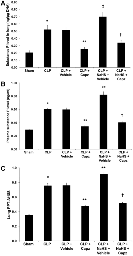
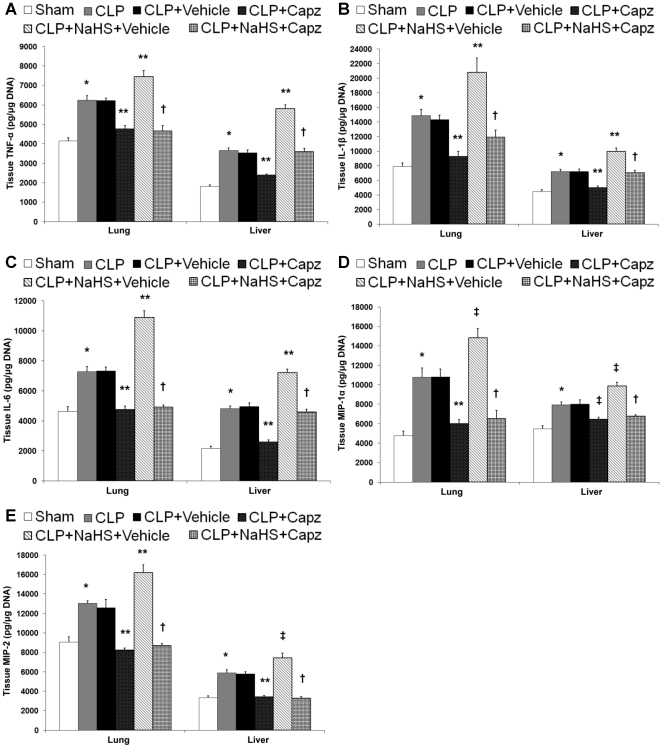
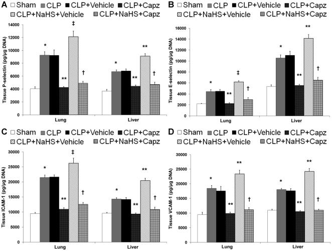
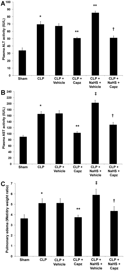
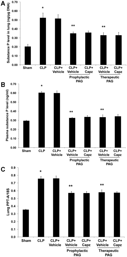
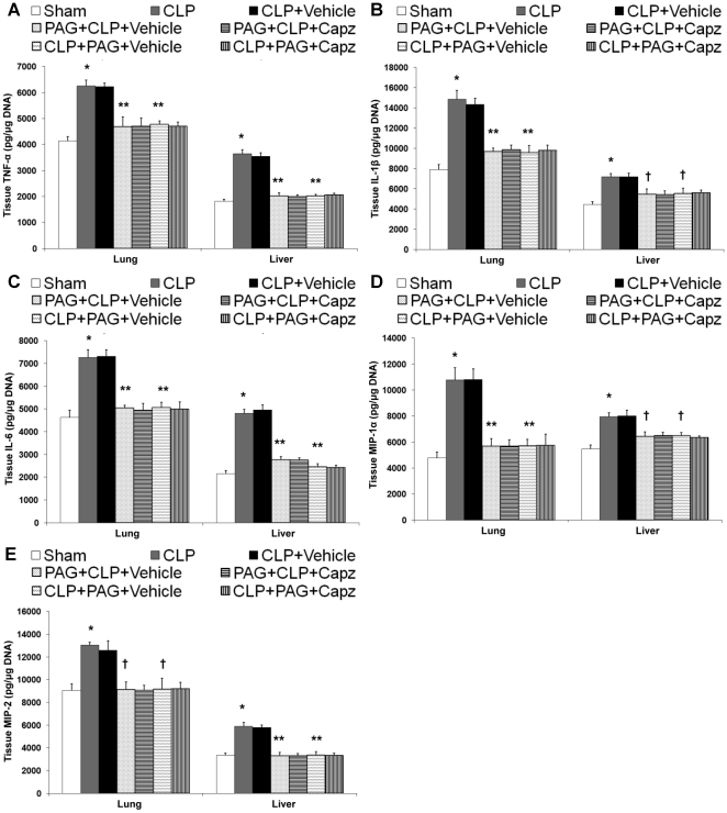
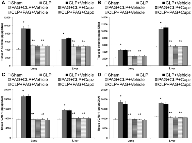
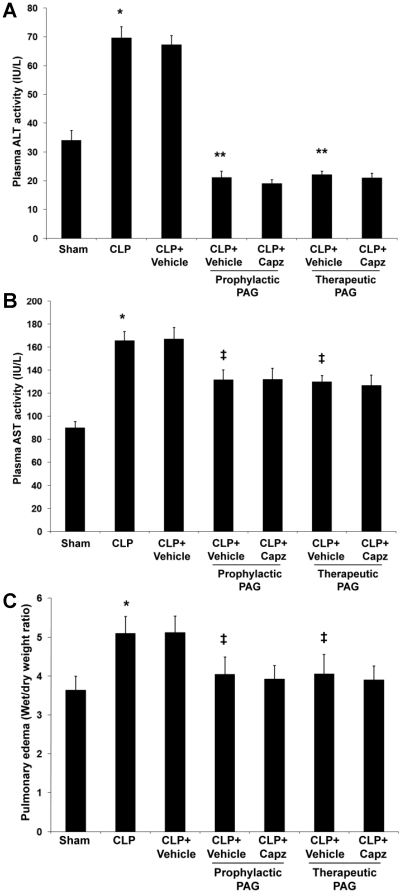
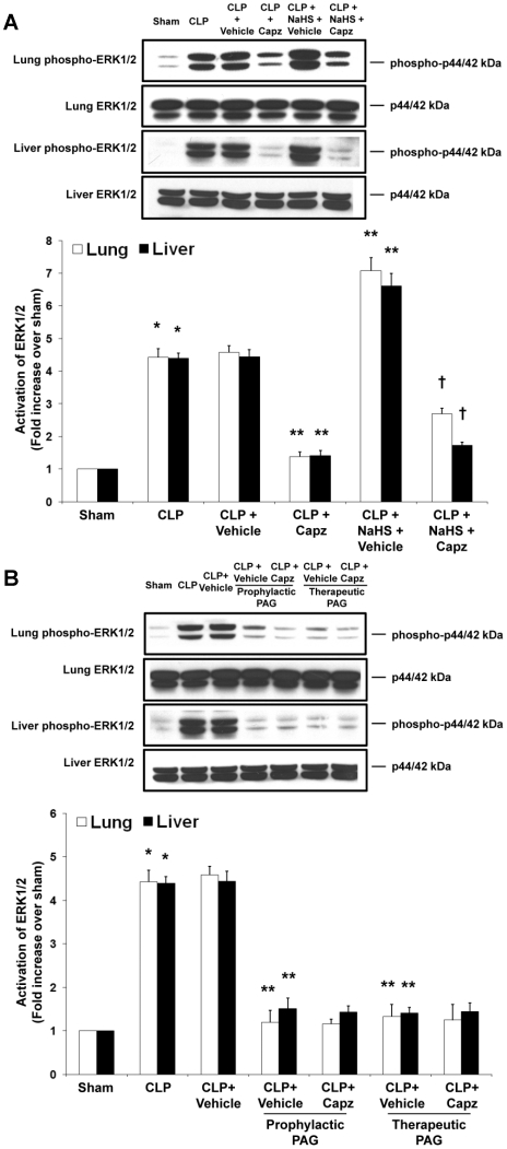
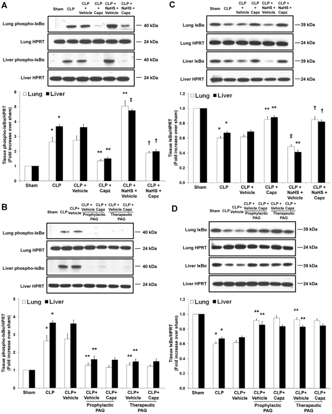
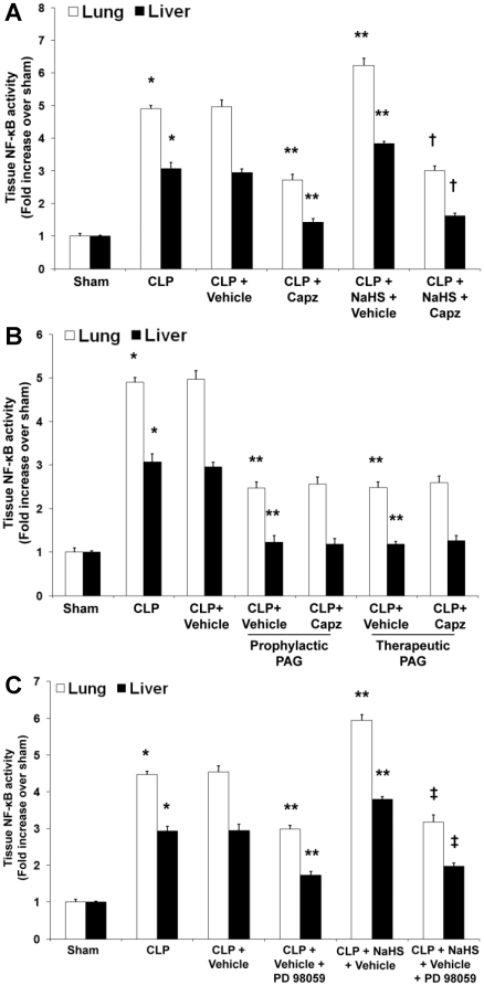
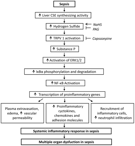
Hydrogen sulfide (H(2)S) has been shown to induce transient receptor potential vanilloid 1 (TRPV1)-mediated neurogenic inflammation in polymicrobial sepsis. However, endogenous neural factors that modulate this event and the molecular mechanism by which this occurs remain unclear. Therefore, this study tested the hypothesis that whether substance P (SP) is one important neural element that implicates in H(2)S-induced neurogenic inflammation in sepsis in a TRPV1-dependent manner, and if so, whether H(2)S regulates this response through activation of the extracellular signal-regulated kinase-nuclear factor-κB (ERK-NF-κB) pathway. Male Swiss mice were subjected to cecal ligation and puncture (CLP)-induced sepsis and treated with TRPV1 antagonist capsazepine 30 minutes before CLP. DL-propargylglycine (PAG), an inhibitor of H(2)S formation, was administrated 1 hour before or 1 hour after sepsis, whereas sodium hydrosulfide (NaHS), an H(2)S donor, was given at the same time as CLP. Capsazepine significantly attenuated H(2)S-induced SP production, inflammatory cytokines, chemokines, and adhesion molecules levels, and protected against lung and liver dysfunction in sepsis. In the absence of H(2)S, capsazepine caused no significant changes to the PAG-mediated attenuation of lung and plasma SP levels, sepsis-associated systemic inflammatory response and multiple organ dysfunction. In addition, capsazepine greatly inhibited phosphorylation of ERK(1/2) and inhibitory κBα, concurrent with suppression of NF-κB activation even in the presence of NaHS. Furthermore, capsazepine had no effect on PAG-mediated abrogation of these levels in sepsis. Taken together, the present findings show that H(2)S regulates TRPV1-mediated neurogenic inflammation in polymicrobial sepsis through enhancement of SP production and activation of the ERK-NF-κB pathway.


Conflict of interest statement

Competing Interests: The authors have declared that no competing interests exist.

Figures

Figure 1
Figure 1. Effect of NaHS and capsazepine on protein and mRNA levels of SP in septic mice.
Mice were randomly given NaHS (10 mg/kg, i.p.) or vehicle (DMSO) at the same time of CLP; and capsazepine (Capz) (15 mg/kg, s.c.) or vehicle (DMSO) 30 minutes before CLP. Sham mice were used as controls. Eight hours after CLP or sham operation, (A) lung and (B) plasma SP levels, and (C) lung PPT-A mRNA levels were measured. Results shown are the mean values ± SEM (n = 8–12 mice per group). *P<0.01 versus sham; **P<0.01 versus CLP+vehicle; ‡P<0.05 versus CLP+vehicle; †P<0.01 versus CLP+NaHS+vehicle.
Figure 2
Figure 2. Effect of NaHS and capsazepine on protein levels of cytokines and chemokines in the lungs and liver of septic mice.
Mice were randomly given NaHS (10 mg/kg, i.p.) or vehicle (DMSO) at the same time of CLP; and capsazepine (Capz) (15 mg/kg, s.c.) or vehicle (DMSO) 30 minutes before CLP. Sham mice were used as controls. Eight hours after CLP or sham operation, protein levels of (A) TNF-α, (B) IL-1β, (C) IL-6, (D) MIP-1α and (E) MIP-2 were measured. Results shown are the mean values ± SEM (n = 8–12 mice per group). *P<0.01 versus sham; **P<0.01 versus CLP+vehicle; ‡P<0.05 versus CLP+vehicle; †P<0.01 versus CLP+NaHS+vehicle.
Figure 3
Figure 3. Effect of NaHS and capsazepine on protein levels of adhesion molecules in the lungs and liver of septic mice.
Mice were randomly given NaHS (10 mg/kg, i.p.) or vehicle (DMSO) at the same time of CLP; and capsazepine (Capz) (15 mg/kg, s.c.) or vehicle (DMSO) 30 minutes before CLP. Sham mice were used as controls. Eight hours after CLP or sham operation, protein levels of (A) P-selectin, (B) E-selectin, (C) ICAM-1, and (D) VCAM-1 were measured. Results shown are the mean values ± SEM (n = 8–12 mice per group). *P<0.01 versus sham; **P<0.01 versus CLP+vehicle; ‡P<0.05 versus CLP+vehicle; †P<0.01 versus CLP+NaHS+vehicle.
Figure 4
Figure 4. Effect of NaHS and capsazepine on liver dysfunction and lung edema in septic mice.
Mice were randomly given NaHS (10 mg/kg, i.p.) or vehicle (DMSO) at the same time of CLP; and capsazepine (Capz) (15 mg/kg, s.c.) or vehicle (DMSO) 30 minutes before CLP. Sham mice were used as controls. Eight hours after CLP or sham operation, plasma levels of (A) ALT and (B) AST, and (C) lung wet-to-dry weight ratio were measured. Results shown are the mean values ± SEM (n = 10–15 mice per group). *P<0.01 versus sham; **P<0.01 versus CLP+vehicle; ‡P<0.05 versus CLP+vehicle; †P<0.01 versus CLP+NaHS+vehicle.
Figure 5
Figure 5. Effect of PAG and capsazepine on protein and mRNA levels of SP in septic mice.
Mice were randomly given PAG (50 mg/kg, i.p.) 1 hour before (“prophylactic”) or 1 hour after (“therapeutic”) CLP; and capsazepine (Capz) (15 mg/kg, s.c.) or vehicle (DMSO) 30 minutes before CLP. Sham mice were used as controls. Eight hours after CLP or sham operation, (A) lung and (B) plasma SP levels, and (C) lung PPT-A mRNA levels were measured. Results shown are the mean values ± SEM (n = 8–12 mice per group). *P<0.01 versus sham; **P<0.01 versus CLP+vehicle.
Figure 6
Figure 6. Effect of PAG and capsazepine on protein levels of cytokines and chemokines in the lungs and liver of septic mice.
Mice were randomly given PAG (50 mg/kg, i.p.) 1 hour before (“prophylactic”; PAG+CLP+Vehicle or PAG+CLP+Capz) or 1 hour after (“therapeutic”; CLP+PAG+Vehicle or CLP+PAG+Capz) CLP; and capsazepine (Capz) (15 mg/kg, s.c.) or vehicle (DMSO) 30 minutes before CLP. Sham mice were used as controls. Eight hours after CLP or sham operation, protein levels of (A) TNF-α, (B) IL-1β, (C) IL-6, (D) MIP-1α and (E) MIP-2 were measured. Results shown are the mean values ± SEM (n = 8–12 mice per group). *P<0.01 versus sham; **P<0.01 versus CLP+vehicle; †P<0.05 versus CLP+vehicle.
Figure 7
Figure 7. Effect of PAG and capsazepine on protein levels of adhesion molecules in the lungs and liver of septic mice.
Mice were randomly given PAG (50 mg/kg, i.p.) 1 hour before (“prophylactic”; PAG+CLP+Vehicle or PAG+CLP+Capz) or 1 hour after (“therapeutic”; CLP+PAG+Vehicle or CLP+PAG+Capz) CLP; and capsazepine (Capz) (15 mg/kg, s.c.) or vehicle (DMSO) 30 minutes before CLP. Sham mice were used as controls. Eight hours after CLP or sham operation, protein levels of (A) P-selectin, (B) E-selectin, (C) ICAM-1, and (D) VCAM-1 were measured. Results shown are the mean values ± SEM (n = 8–12 mice per group). *P<0.01 versus sham; **P<0.01 versus CLP+vehicle.
Figure 8
Figure 8. Effect of PAG and capsazepine on liver dysfunction and lung edema in septic mice.
Mice were randomly given PAG (50 mg/kg, i.p.) 1 hour before (“prophylactic”) or 1 hour after (“therapeutic”) CLP; and capsazepine (Capz) (15 mg/kg, s.c.) or vehicle (DMSO) 30 minutes before CLP. Sham mice were used as controls. Eight hours after CLP or sham operation, plasma levels of (A) ALT and (B) AST, and (C) lung wet-to-dry weight ratio were measured. Results shown are the mean values ± SEM (n = 10–15 mice per group). *P<0.01 versus sham; **P<0.01 versus CLP+vehicle; ‡P<0.05 versus CLP+vehicle.
Figure 9
Figure 9. Effect of NaHS or PAG and capsazepine on ERK1/2 activation in the lungs and liver of septic mice.
Mice were randomly given NaHS (10 mg/kg, i.p.) at the same time of CLP or PAG (50 mg/kg, i.p.) 1 hour before (“prophylactic”) or 1 hour after (“therapeutic”) CLP; and capsazepine (Capz) (15 mg/kg, s.c.) or vehicle (DMSO) 30 minutes before CLP. Sham mice were used as controls. Eight hours after CLP or sham operation, effect of (A) NaHS or (B) PAG and capsazepine on phospho-ERK1/2 and total ERK1/2 expression levels were measured. A representative western blot image is shown, with densitometry data expressed as average ratios of phospho-ERK1/2 to total ERK1/2. Results shown are the mean values ± SEM (n = 6 mice per group). *P<0.01 versus sham; **P<0.01 versus CLP+vehicle; †P<0.01 versus CLP+NaHS+vehicle.
Figure 10
Figure 10. Effect of NaHS or PAG and capsazepine on IκBα phosphorylation and degradation levels in the lungs and liver of septic mice.
Mice were randomly given NaHS (10 mg/kg, i.p.) at the same time of CLP or PAG (50 mg/kg, i.p.) 1 hour before (“prophylactic”) or 1 hour after (“therapeutic”) CLP; and capsazepine (Capz) (15 mg/kg, s.c.) or vehicle (DMSO) 30 minutes before CLP. Sham mice were used as controls. Eight hours after CLP or sham operation, effect of NaHS or PAG and capsazepine on (A and B) phospho-IκBα and (C and D) IκBα expression levels were measured. A representative western blot image is shown, with densitometry data expressed as average ratios of phospho-IκBα to HPRT and IκBα to HPRT levels. Results shown are the mean values ± SEM (n = 6 mice per group). *P<0.01 versus sham; **P<0.01 versus CLP+vehicle; ‡P<0.05 versus CLP+vehicle; †P<0.01 versus CLP+NaHS+vehicle.
Figure 11
Figure 11. Effect of NaHS or PAG and capsazepine and effect of NaHS and PD98059 on NF-κB activation in nuclear extracts of lung and liver tissues in septic mice.
Mice were randomly given NaHS (10 mg/kg, i.p.) at the same time of CLP or PAG (50 mg/kg, i.p.) 1 hour before (“prophylactic”) or 1 hour after (“therapeutic”) CLP; and capsazepine (Capz) (15 mg/kg, s.c.) or vehicle (DMSO) 30 minutes before CLP. Some mice were injected PD98059 (10 mg/kg, i.p.) or vehicle (DMSO) 1 hour before CLP. Sham mice were used as controls. Eight hours after CLP or sham operation, effect of (A) NaHS or (B) PAG and capsazepine and (C) effect of NaHS and PD98059 on the DNA-binding activity of NF-κB were measured. Results shown are the mean values ± SEM (n = 8–12 mice per group). *P<0.01 versus sham; **P<0.01 versus CLP+vehicle; †P<0.01 versus CLP+NaHS+vehicle. ‡P<0.01 versus CLP+NaHS+vehicle.
Figure 12
Figure 12. Schematic summary of signaling events in H2S-induced neurogenic inflammation in a murine model of polymicrobial sepsis.
H2S has been demonstrated to be overproduced in sepsis. H2S stimulation of TRPV1 and the downstream release of SP lead to the activation of ERK1/2, which subsequently induces the phosphorylation and degradation of IκBα, as well as the translocation and activation of NF-κB, thereby leading to SIRS and MODS characteristic of severe sepsis. formula image indicates exogenous administration; formula image indicates inhibition.

Similar articles

Cited by

References

    1. Sternberg EM. Neural regulation of innate immunity: a coordinated nonspecific host response to pathogens. Nat Rev Immunol. 2006;6:318–328. - PMC - PubMed
    1. O'Connor TM, O'Connell J, O'Brien DI, Goode T, Bredin CP, et al. The role of substance P in inflammatory disease. J Cell Physiol. 2004;201:167–180. - PubMed
    1. Richardson JD, Vasko MR. Cellular mechanisms of neurogenic inflammation. J Pharmacol Exp Ther. 2002;302:839–845. - PubMed
    1. Caterina MJ, Schumacher MA, Tominaga M, Rosen TA, Levine JD, et al. The capsaicin receptor: a heat-activated ion channel in the pain pathway. Nature. 1997;389:816–824. - PubMed
    1. Trevisani M, Patacchini R, Nicoletti P, Gatti R, Gazzieri D, et al. Hydrogen sulfide causes vanilloid receptor 1-mediated neurogenic inflammation in the airways. Br J Pharmacol. 2005;145:1123–1131. - PMC - PubMed

Publication types