

Defining the earliest transcriptional steps of chondrogenic progenitor specification during the formation of the digits in the embryonic limb
- PMID: 21931747
- PMCID: PMC3172225
- DOI: 10.1371/journal.pone.0024546
Defining the earliest transcriptional steps of chondrogenic progenitor specification during the formation of the digits in the embryonic limb
Abstract
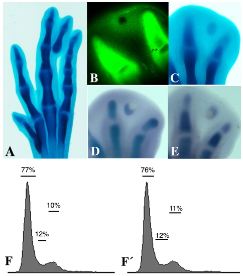
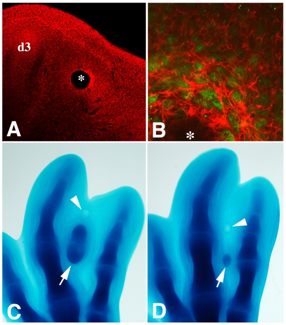
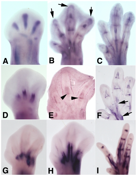
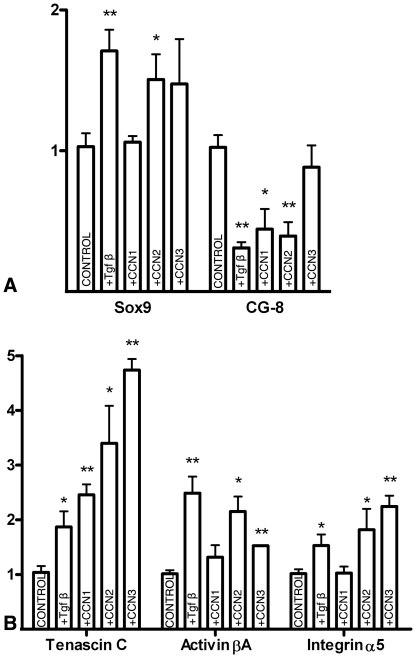
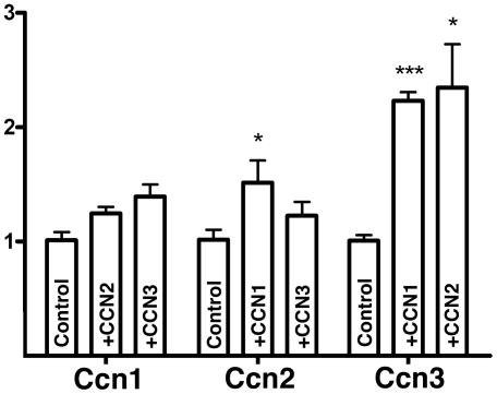
The characterization of genes involved in the formation of cartilage is of key importance to improve cell-based cartilage regenerative therapies. Here, we have developed a suitable experimental model to identify precocious chondrogenic events in vivo by inducing an ectopic digit in the developing embryo. In this model, only 12 hr after the implantation of a Tgfβ bead, in the absence of increased cell proliferation, cartilage forms in undifferentiated interdigital mesoderm and in the course of development, becomes a structurally and morphologically normal digit. Systematic quantitative PCR expression analysis, together with other experimental approaches allowed us to establish 3 successive periods preceding the formation of cartilage. The "pre-condensation stage", occurring within the first 3 hr of treatment, is characterized by the activation of connective tissue identity transcriptional factors (such as Sox9 and Scleraxis) and secreted factors (such as Activin A and the matricellular proteins CCN-1 and CCN-2) and the downregulation of the galectin CG-8. Next, the "condensation stage" is characterized by intense activation of Smad 1/5/8 BMP-signaling and increased expression of extracellular matrix components. During this period, the CCN matricellular proteins promote the expression of extracellular matrix and cell adhesion components. The third period, designated the "pre-cartilage period", precedes the formation of molecularly identifiable cartilage by 2-3 hr and is characterized by the intensification of Sox 9 gene expression, along with the stimulation of other pro-chondrogenic transcription factors, such as HifIa. In summary, this work establishes a temporal hierarchy in the regulation of pro-chondrogenic genes preceding cartilage differentiation and provides new insights into the relative roles of secreted factors and cytoskeletal regulators that direct the first steps of this process in vivo.
Conflict of interest statement
Figures
Similar articles
-
Analysis of the molecular cascade responsible for mesodermal limb chondrogenesis: Sox genes and BMP signaling.Dev Biol. 2003 May 15;257(2):292-301. doi: 10.1016/s0012-1606(03)00066-6. Dev Biol. 2003. PMID: 12729559
-
Reelin/DAB-1 signaling in the embryonic limb regulates the chondrogenic differentiation of digit mesodermal progenitors.J Cell Physiol. 2014 Oct;229(10):1397-404. doi: 10.1002/jcp.24576. J Cell Physiol. 2014. PMID: 24519818
-
Role of TGF beta s and BMPs as signals controlling the position of the digits and the areas of interdigital cell death in the developing chick limb autopod.Development. 1996 Aug;122(8):2349-57. doi: 10.1242/dev.122.8.2349. Development. 1996. PMID: 8756280
-
Divide, accumulate, differentiate: cell condensation in skeletal development revisited.Int J Dev Biol. 1995 Dec;39(6):881-93. Int J Dev Biol. 1995. PMID: 8901191 Review.
-
Cellular interactions and signaling in cartilage development.Osteoarthritis Cartilage. 2000 Sep;8(5):309-34. doi: 10.1053/joca.1999.0306. Osteoarthritis Cartilage. 2000. PMID: 10966838 Review.
Cited by
-
Transcriptional heterochrony in talpid mole autopods.Evodevo. 2012 Aug 9;3(1):16. doi: 10.1186/2041-9139-3-16. Evodevo. 2012. PMID: 22873211 Free PMC article.
-
Understanding the Cellular and Molecular Mechanisms That Control Early Cell Fate Decisions During Appendicular Skeletogenesis.Front Genet. 2019 Oct 11;10:977. doi: 10.3389/fgene.2019.00977. eCollection 2019. Front Genet. 2019. PMID: 31681419 Free PMC article. Review.
-
A Novel EphA4 Signaling-Based Therapeutic Strategy for Osteoarthritis in Mice.J Bone Miner Res. 2022 Apr;37(4):660-674. doi: 10.1002/jbmr.4500. Epub 2022 Jan 29. J Bone Miner Res. 2022. PMID: 34989027 Free PMC article.
-
The role of chondroitin sulfate in regulating hypertrophy during MSC chondrogenesis in a cartilage mimetic hydrogel under dynamic loading.Biomaterials. 2019 Jan;190-191:51-62. doi: 10.1016/j.biomaterials.2018.10.028. Epub 2018 Oct 25. Biomaterials. 2019. PMID: 30391802 Free PMC article.
-
Selection on Phalanx Development in the Evolution of the Bird Wing.Mol Biol Evol. 2021 Sep 27;38(10):4222-4237. doi: 10.1093/molbev/msab150. Mol Biol Evol. 2021. PMID: 34164688 Free PMC article.
References
Publication types
MeSH terms
Substances
LinkOut - more resources
Full Text Sources
Medical
Molecular Biology Databases
Research Materials
Miscellaneous
