. 2011;6(9):e24568.
doi: 10.1371/journal.pone.0024568. Epub 2011 Sep 9.

Hepatocyte growth factor (HGF) inhibits collagen I and IV synthesis in hepatic stellate cells by miRNA-29 induction

Affiliations

Hepatocyte growth factor (HGF) inhibits collagen I and IV synthesis in hepatic stellate cells by miRNA-29 induction

Monika Kwiecinski et al. PLoS One. 2011.

Abstract

Background: In chronic liver disease, hepatic stellate cells (HSC) transdifferentiate into myofibroblasts, promoting extracellular matrix (ECM) synthesis and deposition. Stimulation of HSC by transforming growth factor-β (TGF-β) is a crucial event in liver fibrogenesis due to its impact on myofibroblastic transition and ECM induction. In contrast, hepatocyte growth factor (HGF), exerts antifibrotic activities. Recently, miR-29 has been reported to be involved in ECM synthesis. We therefore studied the influence of HGF and TGF-β on the miR-29 collagen axis in HSC.

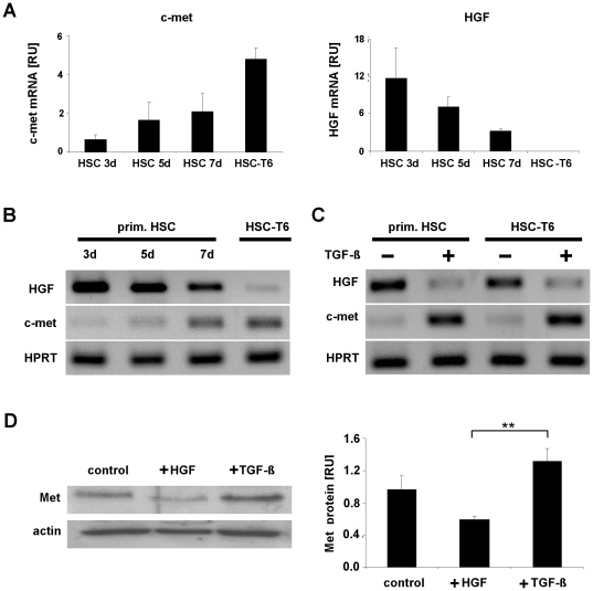
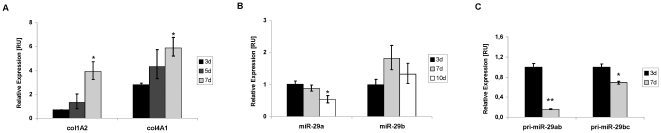
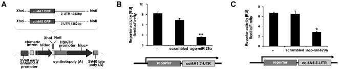
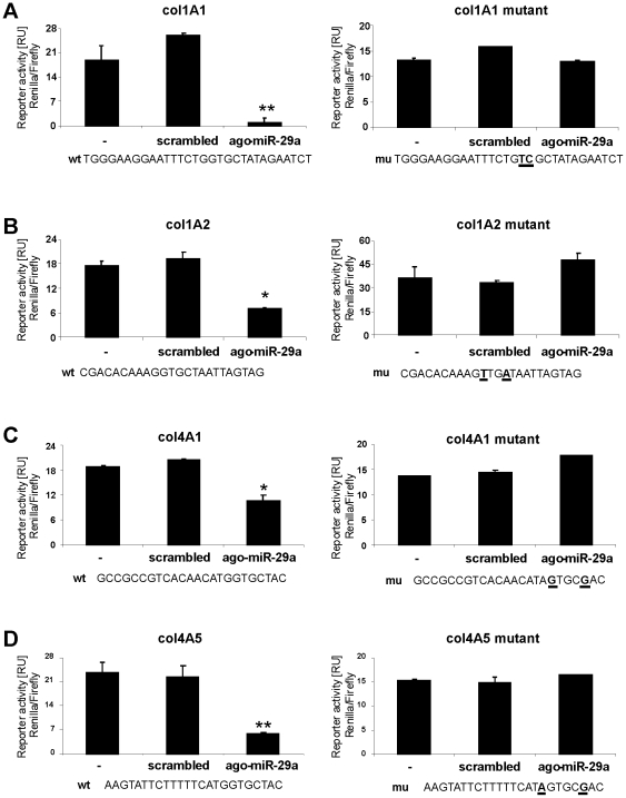
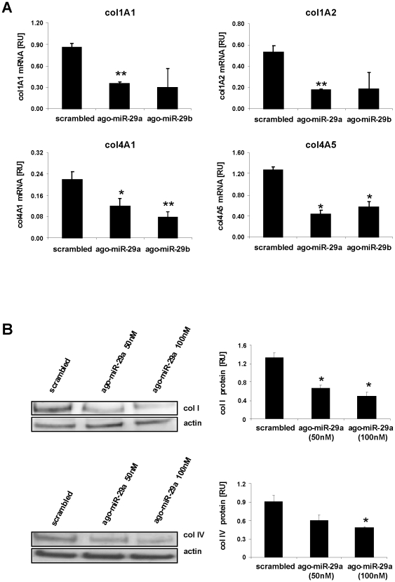
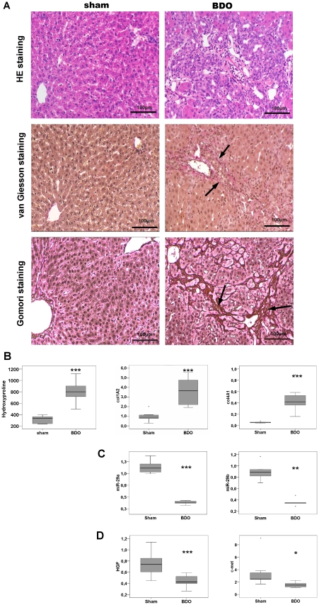
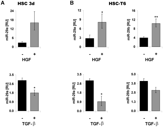
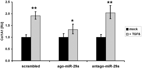
Methodology: HSC, isolated from rats, were characterized for HGF and Met receptor expression by Real-Time PCR and Western blotting during culture induced myofibroblastic transition. Then, the levels of TGF-β, HGF, collagen-I and -IV mRNA, in addition to miR-29a and miR-29b were determined after HGF and TGF-β stimulation of HSC or after experimental fibrosis induced by bile-duct obstruction in rats. The interaction of miR-29 with 3'-untranslated mRNA regions (UTR) was analyzed by reporter assays. The repressive effect of miR-29 on collagen synthesis was studied in HSC treated with miR-29-mimicks by Real-Time PCR and immunoblotting.

Principal findings: The 3'-UTR of the collagen-1 and -4 subtypes were identified to bind miR-29. Hence, miR-29a/b overexpression in HSC resulted in a marked reduction of collagen-I and -IV synthesis. Conversely, a decrease in miR-29 levels is observed during collagen accumulation upon experimental fibrosis, in vivo, and after TGF-β stimulation of HSC, in vitro. Finally, we show that during myofibroblastic transition and TGF-β exposure the HGF-receptor, Met, is upregulated in HSC. Thus, whereas TGF-β stimulation leads to a reduction in miR-29 expression and de-repression of collagen synthesis, stimulation with HGF was definitely associated with highly elevated miR-29 levels and markedly repressed collagen-I and -IV synthesis.

Conclusions: Upregulation of miRNA-29 by HGF and downregulation by TGF-β take part in the anti- or profibrogenic response of HSC, respectively.


Conflict of interest statement

Competing Interests: The authors have declared that no competing interests exist.

Figures

Figure 1
Figure 1. Met and HGF expression in primary and immortalized HSC.
Total RNA was extracted from primary rat HSC after the third, fifth and seventh day of cell culture (3d, 5d, 7d) and from myofibroblastic HSC-T6 cells. Subsequently, c-met and HGF expression was determined (A–C). The c-met and HGF transcript levels were quantified by Real-Time PCR and normalized using HPRT as house-keeping gene (A–C). The expression of c-met increased during the differentiation process of primary HSC and achieved the highest level in the immortalized cell line HSC-T6 representing a myofibroblastic phenotype , whereas HGF levels were shown to be opposite (A, B). Primary HSC at the 3rd culture day (prim. HSC) and immortalized HSC (HSC-T6) were either untreated (−) or stimulated with TGF-β (+) and RNA levels of c-met and HGF were analyzed by Real-Time PCR (C). The Met protein expression in HGF and TGF-β stimulated HSC-T6 (+HGF, +TGF-β) in comparison to untreated HSC-T6 cells (control) was shown by Western blot analysis (D).
Figure 2
Figure 2. Contrary influence of TGF-β and HGF stimulation on collagen, but also on TGF-β and HGF expression, itself.
Collagen 1A1 (col1A1), collagen 1A2 (col1A2), collagen 4A1 (col4A1), and collagen 4A5 (col4A5), HGF and TGF-β mRNA levels were compared by Real-Time PCR in untreated (−) and in HSC-T6 cells stimulated either with TGF- β (+TGF- β) (A) or with HGF (+HGF) (B).
Figure 3
Figure 3. Reciprocal expression of the collagen subunits, col1A1 and col4A2, and primary and mature miR-29.
RNA of primary HSC in the quiescent stage (day 3 of primary culture) and after myofibroblastic activation (day 7 of primary culture) was analyzed for mRNA col1A2 and col4A1 levels (A), as wells as for the levels of mature miR-29a and miR-29b (B), and the primary transcripts of the miR-29a/b and miR-29b/c gene (C).
Figure 4
Figure 4. miR-29 interaction with the 3′-UTR of col4A1 and col4A5 transcripts.
The 3′-UTR of col4A1 (1382 bp) or col4A5 (786 bp) mRNA was subcloned downstream of the Renilla luciferase reporter (hRluc) of the psiCHECKTM-2 vector (A). Reporter plasmids were cotransfected into HSC-T6 in combination with scrambled miRNA or miR-29a mimic (ago-miR-29a), respectively, and luciferase reporter expression was determined by the hRluc luminescence measurement normalized to firefly luminescence (hluc+) (B–C).
Figure 5
Figure 5. Interaction of miR-29a with the binding regions of col1A1, col1A2, col4A1 and col4A5 3′-UTR transcripts.
Putative miR-29 binding sites in the collagen col1A1 (A), col1A2 (B), col4A1 (C) and col4A5 (D) 3′-UTR (wt) and the corresponding mutated sequences (mu) carrying two point mutations (bold and underlined) were cloned into psiCHECKTM-2 vector. The reporter plasmids were co-transfected into HSC-T6 cells with either scrambled miRNA or miR-29a mimic (ago-miR-29a), respectively. Insertion of the miR-29 binding sites (wt), but not with mutated binding site (mu), resulted in reduced reporter gene expression by miR-29a treated HSC (A–D).
Figure 6
Figure 6. Repression of collagen synthesis by miR-29a and miR-29b.
mRNA quantification of collagen subunits in HSC treated either with miR-29a, miR-29b (ago-miR-29a, ago-miR-29b), or scrambled miRNA by Real-Time PCR (A). The col1A1, col1A2, col4A1, and col4A5 mRNA levels were normalized to HPRT expression. Furthermore, collagen I and IV protein levels (col I, col IV) were determined by immunoblotting and subsequent densitometric quantification (B).
Figure 7
Figure 7. Reduced miR-29a and miR-29b expression in livers after BDO.
Liver tissues from bile duct obstructed (BDO) rats showed hepatic inflammation and fibrosis shown by haematoxylin-eosin (HE), van Gieson, and Gomori staining (A). HE histology clearly shows the loss of liver architecture and inflammation after BDO. van Gieson and Gomori stainings revealed the enhancement of connective tissue and the formation of septa (arrows indicate matrix deposition stained in red by van Gieson or in dark brown by Gomori, respectively). Hydroxyproline levels in livers of sham operated (N = 5) and of BDO rats (N = 5) were determined and col1A2 and col4A2 mRNA levels were quantified by Real Time PCR (B). Additionally, miR-29a and miR-29b levels (C) and HGF and c-met transcripts (D) were quantified by Real-Time PCR. While miR-29 and HGF expression was significantly reduced (C,D), hydroxyproline and collagen mRNA were signifcantly increased in BDO livers (B) (*: p<0.05 **:p<0.01; ***: p<0.001).
Figure 8
Figure 8. Contrary effects of HGF and TGF-β on miR-29 expression in HSC.
miR-29a expression in HSC after 3 days (3d) of primary culture (A) and miR-29a and miR-29b expression in myofibroblastic HSC-T6 (B). HSC were not stimulated (−) or treated (+) with either TGF-β or hHGF and miR-29a/b levels were determined by Real Time-PCR analysis.
Figure 9
Figure 9. Diminished effect of TGF-β on col1A1 expression in miR-29 treated HSC.
The myofibroblastic HSC-T6 cells were transfected with scrambled miRNA, miR-29 mimic (ago-miR-29), or with a miR-29 silencer (antago-miR-29). TGF-β treatment resulted in highly increased col1A1 expression in HSC cells treated with scrambled miRNA or with antago-miR-29, but only a moderate col1A1 induction in miR-29 overexpressing HSC after transfection with ago-miR-29.

References

    1. Friedman SL. Mechanisms of hepatic fibrogenesis. Gastroenterology. 2008;134:1655–1669. - PMC - PubMed
    1. Schuppan D, Krebs A, Bauer M, Hahn EG. Hepatitis C and liver fibrosis. Cell Death Differ. 2003;10(Suppl 1):S59–67. - PubMed
    1. Gressner OA, Rizk MS, Kovalenko E, Weiskirchen R, Gressner AM. Changing the pathogenetic roadmap of liver fibrosis? Where did it start; where will it go? J Gastroenterol Hepatol. 2008;23:1024–1035. - PubMed
    1. Gressner AM, Weiskirchen R. Modern pathogenetic concepts of liver fibrosis suggest stellate cells and TGF-beta as major players and therapeutic targets. J Cell Mol Med. 2006;10:76–99. - PMC - PubMed
    1. Geerts A, Schuppan D, Lazeroms S, De Zanger R, Wisse E. Collagen type I and III occur together in hybrid fibrils in the space of Disse of normal rat liver. Hepatology. 1990;12:233–241. - PubMed

Publication types