

Profile of microRNAs following rat sciatic nerve injury by deep sequencing: implication for mechanisms of nerve regeneration
- PMID: 21931774
- PMCID: PMC3172250
- DOI: 10.1371/journal.pone.0024612
Profile of microRNAs following rat sciatic nerve injury by deep sequencing: implication for mechanisms of nerve regeneration
Abstract
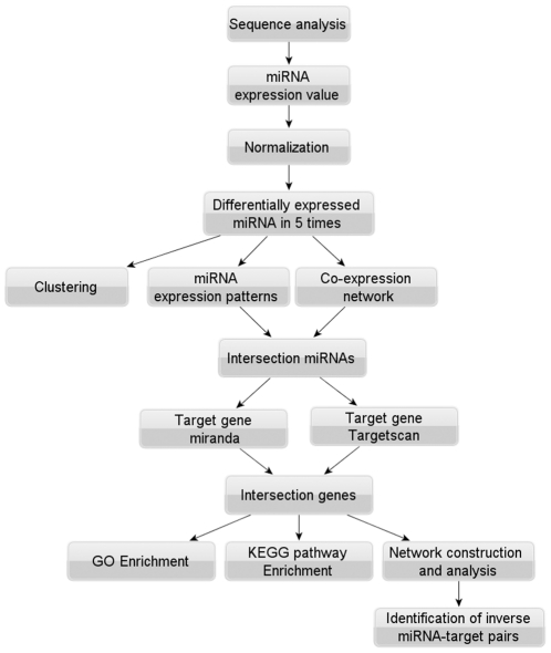
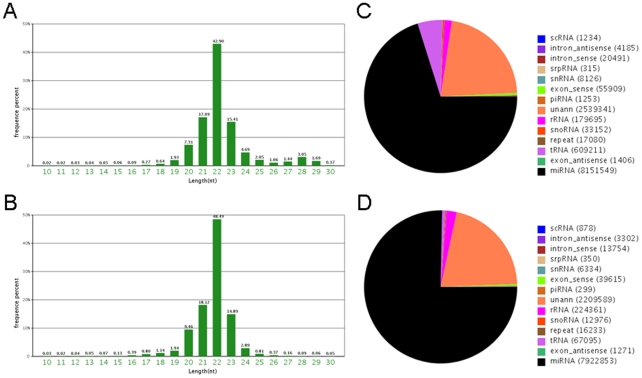
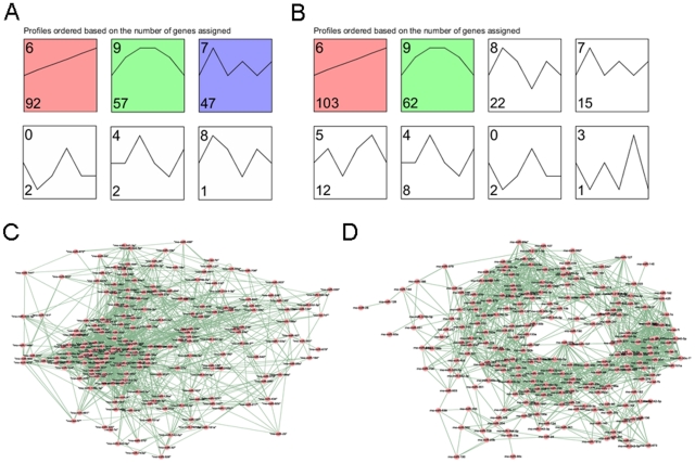
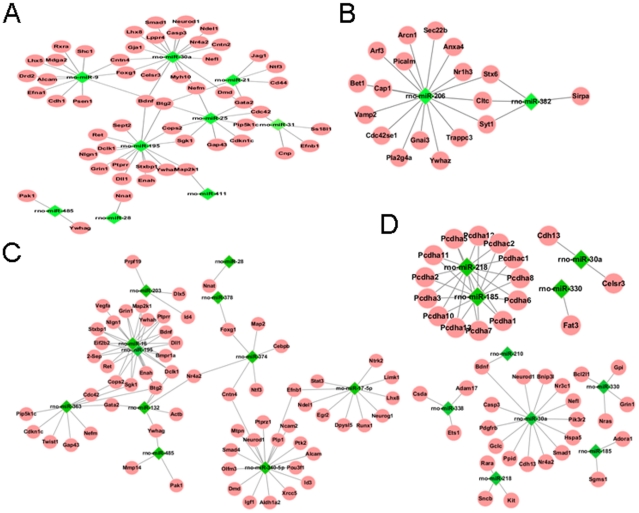
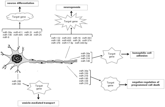
Unlike the central nervous system, peripheral nerves can regenerate when damaged. MicroRNA (miRNA) is a novel class of small, non-coding RNA that regulates gene expression at the post-transcriptional level. Here, we report regular alterations of miRNA expression following rat sciatic nerve injury using deep sequencing. We harvested dorsal root ganglia tissues and the proximal stumps of the nerve, and identified 201 and 225 known miRNAs with significant expression variance at five time points in these tissues after sciatic nerve transaction, respectively. Subsequently, hierarchical clustering, miRNA expression pattern and co-expression network were performed. We screened out specific miRNAs and further obtained the intersection genes through target analysis software (Targetscan and miRanda). Moreover, GO and KEGG enrichment analyses of these intersection genes were performed. The bioinformatics analysis indicated that the potential targets for these miRNAs were involved in nerve regeneration, including neurogenesis, neuron differentiation, vesicle-mediated transport, homophilic cell adhesion and negative regulation of programmed cell death that were known to play important roles in regulating nerve repair. Finally, we combined differentially expressed mRNA with the predicted targets for selecting inverse miRNA-target pairs. Our results show that the abnormal expression of miRNA may contribute to illustrate the molecular mechanisms of nerve regeneration and that miRNAs are potential targets for therapeutic interventions and may enhance intrinsic regenerative ability.
Conflict of interest statement
Figures

References
-
- Gruart A, Streppel M, Guntinas-Lichius O, Angelov DN, Neiss WF, et al. Motoneuron adaptability to new motor tasks following two types of facial-facial anastomosis in cats. Brain. 2003;126:115–133. - PubMed
-
- Rishal I, Fainzilber M. Retrograde signaling in axonal regeneration. Exp Neurol. 2010;223:5–10. - PubMed
-
- Gu X, Ding F, Yang Y, Liu J. Construction of tissue engineered nerve grafts and their application in peripheral nerve regeneration. Prog Neurobiol. 2011;93:204–230. - PubMed
-
- Griffin JW, Pan B, Polley MA, Hoffman PN, Farah MH. Measuring nerve regeneration in the mouse. Exp Neurol. 2010;223:60–71. - PubMed
Publication types
MeSH terms
Substances
LinkOut - more resources
Full Text Sources
