Mutational analysis of photosystem I of Synechocystis sp. PCC 6803: the role of four conserved aromatic residues in the j-helix of PsaB
- PMID: 21931782
- PMCID: PMC3171458
- DOI: 10.1371/journal.pone.0024625
Mutational analysis of photosystem I of Synechocystis sp. PCC 6803: the role of four conserved aromatic residues in the j-helix of PsaB
Abstract
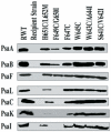
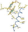
Photosystem I is the light-driven plastocyanin-ferredoxin oxidoreductase in the photosynthetic electron transfer of cyanobacteria and plants. Two histidyl residues in the symmetric transmembrane helices A-j and B-j provide ligands for the P700 chlorophyll molecules of the reaction center of photosystem I. To determine the role of conserved aromatic residues adjacent to the histidyl molecule in the helix of B-j, we generated six site-directed mutants of the psaB gene in Synechocystis sp. PCC 6803. Three mutant strains with W645C, W643C/A644I and S641C/V642I substitutions could grow photoautotrophically and showed no obvious reduction in the photosystem I activity. Kinetics of P700 re-reduction by plastocyanin remained unaltered in these mutants. In contrast, the strains with H651C/L652M, F649C/G650I and F647C substitutions could not grow under photoautotrophic conditions because those mutants had low photosystem I activity, possibly due to low levels of proteins. A procedure to select spontaneous revertants from the mutants that are incapable to photoautotrophic growth resulted in three revertants that were used in this study. The molecular analysis of the spontaneous revertants suggested that an aromatic residue at F647 and a small residue at G650 may be necessary for maintaining the structural integrity of photosystem I. The (P700⁺-P700) steady-state absorption difference spectrum of the revertant F647Y has a ∼5 nm narrower peak than the recovered wild-type, suggesting that additional hydroxyl group of this revertant may participate in the interaction with the special pair while the photosystem I complexes of the F649C/G650T and H651Q mutants closely resemble the wild-type spectrum. The results presented here demonstrate that the highly conserved residues W645, W643 and F649 are not critical for maintaining the integrity and in mediating electron transport from plastocyanin to photosystem I. Our data suggest that an aromatic residue is required at position of 647 for structural integrity and/or function of photosystem I.
Conflict of interest statement
Figures


Similar articles
-
Functional consequences of modification of the photosystem I/photosystem II ratio in the cyanobacterium Synechocystis sp. PCC 6803.J Bacteriol. 2024 May 23;206(5):e0045423. doi: 10.1128/jb.00454-23. Epub 2024 May 2. J Bacteriol. 2024. PMID: 38695523 Free PMC article.
-
Trimeric organization of photosystem I is required to maintain the balanced photosynthetic electron flow in cyanobacterium Synechocystis sp. PCC 6803.Photosynth Res. 2020 Mar;143(3):251-262. doi: 10.1007/s11120-019-00696-9. Epub 2019 Dec 17. Photosynth Res. 2020. PMID: 31848802
-
Photosystem activity and state transitions of the photosynthetic apparatus in cyanobacterium Synechocystis PCC 6803 mutants with different redox state of the plastoquinone pool.Biochemistry (Mosc). 2015 Jan;80(1):50-60. doi: 10.1134/S000629791501006X. Biochemistry (Mosc). 2015. PMID: 25754039
-
Biochemical and biophysical characterization of photosystem I from phytoene desaturase and zeta-carotene desaturase deletion mutants of Synechocystis Sp. PCC 6803: evidence for PsaA- and PsaB-side electron transport in cyanobacteria.J Biol Chem. 2005 May 20;280(20):20030-41. doi: 10.1074/jbc.M500809200. Epub 2005 Mar 9. J Biol Chem. 2005. PMID: 15760840
-
Bidirectional electron transfer in photosystem I: electron transfer on the PsaA side is not essential for phototrophic growth in Chlamydomonas.Biochim Biophys Acta. 2003 Sep 30;1606(1-3):43-55. doi: 10.1016/s0005-2728(03)00083-5. Biochim Biophys Acta. 2003. PMID: 14507426 Review.
Cited by
-
Development of a TSR-based method for understanding structural relationships of cofactors and local environments in photosystem I.BMC Bioinformatics. 2025 Jan 14;26(1):15. doi: 10.1186/s12859-025-06038-y. BMC Bioinformatics. 2025. PMID: 39810075 Free PMC article.
-
Conserved residue PsaB-Trp673 is essential for high-efficiency electron transfer between the phylloquinones and the iron-sulfur clusters in Photosystem I.Photosynth Res. 2021 Jun;148(3):161-180. doi: 10.1007/s11120-021-00839-x. Epub 2021 May 15. Photosynth Res. 2021. PMID: 33991284
-
A single vector-based strategy for marker-less gene replacement in Synechocystis sp. PCC 6803.Microb Cell Fact. 2014 Jan 8;13:4. doi: 10.1186/1475-2859-13-4. Microb Cell Fact. 2014. PMID: 24401024 Free PMC article.
References
-
- Chitnis PR. PHOTOSYSTEM I: Function and Physiology. Annu Rev Plant Physiol Plant Mol Biol. 2001;52:593–626. - PubMed
-
- Jordan P, Fromme P, Witt HT, Klukas O, Saenger W, et al. Three-dimensional structure of cyanobacterial photosystem I at 2.5 A resolution. Nature. 2001;411:909–917. - PubMed
-
- Ben-Shem A, Frolow F, Nelson N. Crystal structure of plant photosystem I. Nature. 2003;426:630–635. - PubMed
-
- Amunts A, Drory O, Nelson N. The structure of a plant photosystem I supercomplex at 3.4 A resolution. Nature. 2007;447:58–63. - PubMed
-
- Grotjohann I, Fromme P. Structure of cyanobacterial photosystem I. Photosynthesis research. 2005;85:51–72. - PubMed
Publication types
MeSH terms
Substances
LinkOut - more resources
Full Text Sources

