. 2011;6(9):e24640.
doi: 10.1371/journal.pone.0024640. Epub 2011 Sep 8.

Defining the molecular character of the developing and adult kidney podocyte

Affiliations

Defining the molecular character of the developing and adult kidney podocyte

Eric W Brunskill et al. PLoS One. 2011.

Abstract

Background: The podocyte is a remarkable cell type, which encases the capillaries of the kidney glomerulus. Although mesodermal in origin it sends out axonal like projections that wrap around the capillaries. These extend yet finer projections, the foot processes, which interdigitate, leaving between them the slit diaphragms, through which the glomerular filtrate must pass. The podocytes are a subject of keen interest because of their key roles in kidney development and disease.

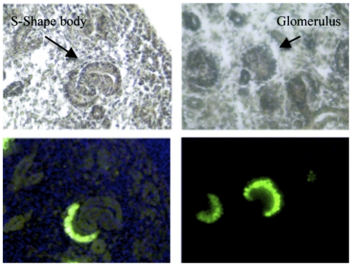
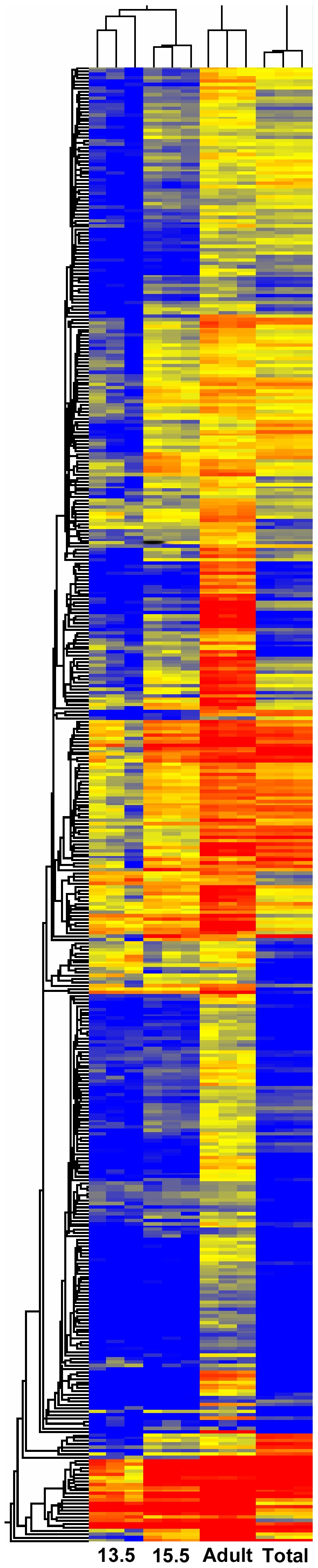
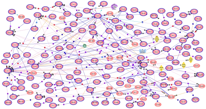
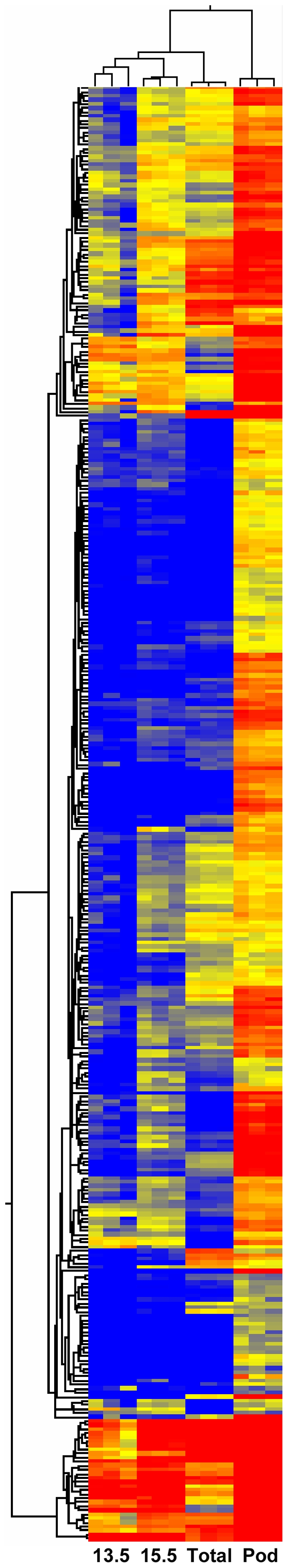
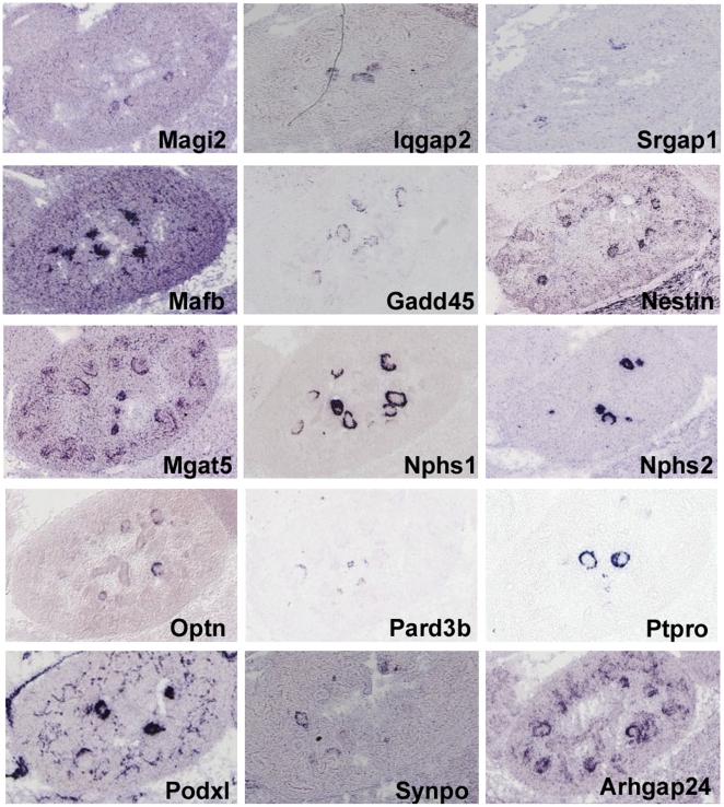
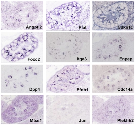
Methodology/principal findings: In this report we identified and characterized a novel transgenic mouse line, MafB-GFP, which specifically marked the kidney podocytes from a very early stage of development. These mice were then used to facilitate the fluorescent activated cell sorting based purification of podocytes from embryos at E13.5 and E15.5, as well as adults. Microarrays were then used to globally define the gene expression states of podocytes at these different developmental stages. A remarkable picture emerged, identifying the multiple sets of genes that establish the neuronal, muscle, and phagocytic properties of podocytes. The complete combinatorial code of transcription factors that create the podocyte was characterized, and the global lists of growth factors and receptors they express were defined.

Conclusions/significance: The complete molecular character of the in vivo podocyte is established for the first time. The active molecular functions and biological processes further define their unique combination of features. The results provide a resource atlas of gene expression patterns of developing and adult podocytes that will help to guide further research of these incredible cells.


Conflict of interest statement

Competing Interests: The authors have declared that no competing interests exist.

Figures

Figure 1
Figure 1. MafB-GFP mice show restricted expression of GFP in podocytes.
Cryostat sections of E15.5 MafB-GFP transgenic kidneys with visible (above panels) and fluorescent (below) illumination. An S-shaped body and glomerulus are marked in above panels (arrows), and prospective podocytes in these structures are shown to be GFP positive in lower panels.
Figure 2
Figure 2. Heatmap of 894 probesets with elevated expression in adult podocytes.
The 894 probset list was created by combining genes with three fold greater expression in adult podocytes compared to total adult kidney cortex (Total) or compared to E13.5 embryonic podocytes (13.5). Adult indicates adult podocytes, and 15.5 indicated E15.5 podocytes. Red marks strong expression levels, and blue shows weak expression. Note that for many probesets there is intermediate expression in E15.5 podocytes, compared to E13.5 and adult podocytes, suggesting that many podocyte specific marker genes show elevated expression as a function of developmental time. Most of the 894 probesets show increased expression in adult podocytes compared to both E13.5 podocytes and total kidney cortex, but some are only elevated relative to one or the other.
Figure 3
Figure 3. GeneSpring pathways analysis of the 894 genes with elevated podocyte expression.
This diagram illustrates many of the known direct interactions between the proteins encoded by the 894 list of podocyte enriched genes. JUN, EGF, RHOA, as well as ITGB1, H2-K1, FYN, ACTR2, VEGFA, APP, ANGPTL2 and HSPA1B, show strong interaction centers. Ovals surrounded by blue lines represent proteins included in the 894 list.
Figure 4
Figure 4. Heatmap of 365 probesets with five fold elevated expression in adult podocytes.
A relatively stringent screen, requiring five fold enrichment in adult podocytes (Pod) compared to either total adult kidney cortex (Total), or E13.5 embryonic podocytes (13.5). Red indicates strong expression and blue weak expression.
Figure 5
Figure 5. Functional groupings of genes with elevated adult podocyte expression.
Cytoscape diagrams of a subset of genes from 365 probeset list of adult podocyte elevated genes (hexagons in center). Key biological processes and molecular functions (green), as well as upstream regulatory transcription factors (purple), are shown as surrounding rectangles, connected to associated genes by lines. A. Neuron related genes, B. Calcium binding related genes, and C. Cytoskeletal protein binding related genes, are highlighted in yellow.
Figure 6
Figure 6. Comparison of podocyte expressed genes across many kidney cell types.
Heatmap showing relative expression levels of most stringently selected 171 adult podocyte probesets. A. E13.5 podocytes. B. Renal vesicles from P4 mice. C. P1 Cap mesenchyme. D. E15.5 podocytes. E. E15.5 endothelial cells. F. Adult total kidney cortex. G. Adult Meis1 expressing mesangial cells. H. Adult cortex endothelial cells. I. Adult glomerular endothelial cells. J. Adult medullary endothelial cells. K. Adult renal capsule. L. Adult podocytes. M. Adult total glomerulus.
Figure 7
Figure 7. Expression of adult podocyte genes in the developing kidney.
In situ hybridizations showing expression in the podocytes of E14.5 kidneys (GenePaint and Eurexpress). Nphs1 and Nphs2 serve as positive controls illustrating the circular or crescent appearance of podocyte specific expression, depending on the angle of the section.
Figure 8
Figure 8. In situ hubridizations suggesting podocyte expression.
Expression patterns for genes from the 144 list of genes with highly enriched expression in adult podocytes. For these genes the hybridization patterns suggest expression in even very early E14.5 podocytes. Data from GenePaint and Eurexpress.
Figure 9
Figure 9. Expression of podocyte genes in E15.5 embryos, as well as P0 and adult.
Genes expressed in podocytes of the embryonic (E15.5) and postnatal kidney (P0 or adult) detected by section in situ hybridization. The entire kidney is shown with a region enlarged to show podocyte expression in the renal corpuscle. For Eya4, in addition to the obvious strong cap mesenchyme expression, weak gene expression is detectable in podocytes (arrow).

Similar articles

Cited by

References

    1. Pavenstadt H, Kriz W, Kretzler M. Cell biology of the glomerular podocyte. Physiol Rev. 2003;83:253–307. - PubMed
    1. Abrahamson DR. Origin of the glomerular basement membrane visualized after in vivo labeling of laminin in newborn rat kidneys. J Cell Biol. 1985;100:1988–2000. - PMC - PubMed
    1. Akilesh S, Huber TB, Wu H, Wang G, Hartleben B, et al. Podocytes use FcRn to clear IgG from the glomerular basement membrane. Proc Natl Acad Sci U S A. 2008;105:967–972. - PMC - PubMed
    1. Costantini F, Kopan R. Patterning a complex organ: branching morphogenesis and nephron segmentation in kidney development. Dev Cell. 2010;18:698–712. - PMC - PubMed
    1. Dijkman H, Smeets B, van der Laak J, Steenbergen E, Wetzels J. The parietal epithelial cell is crucially involved in human idiopathic focal segmental glomerulosclerosis. Kidney Int. 2005;68:1562–1572. - PubMed

Publication types