. 2011;6(9):e24654.
doi: 10.1371/journal.pone.0024654. Epub 2011 Sep 13.

Pneumonic tularemia in rabbits resembles the human disease as illustrated by radiographic and hematological changes after infection

Affiliations

Pneumonic tularemia in rabbits resembles the human disease as illustrated by radiographic and hematological changes after infection

Douglas S Reed et al. PLoS One. 2011.

Abstract

Background: Pneumonic tularemia is caused by inhalation of the gram negative bacterium, Francisella tularensis. Because of concerns that tularemia could be used as a bioterrorism agent, vaccines and therapeutics are urgently needed. Animal models of pneumonic tularemia with a pathophysiology similar to the human disease are needed to evaluate the efficacy of these potential medical countermeasures.

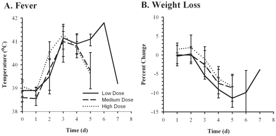
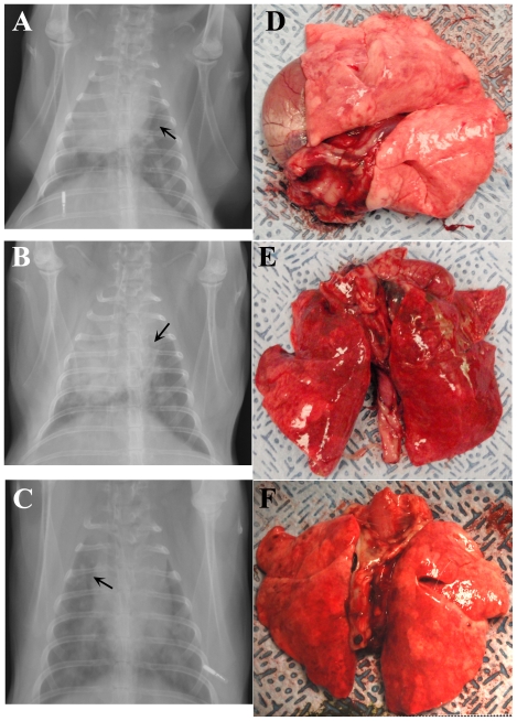
Principal findings: Rabbits exposed to aerosols containing Francisella tularensis strain SCHU S4 developed a rapidly progressive fatal pneumonic disease. Clinical signs became evident on the third day after exposure with development of a fever (>40.5°C) and a sharp decline in both food and water intake. Blood samples collected on day 4 found lymphopenia and a decrease in platelet counts coupled with elevations in erythrocyte sedimentation rate, alanine aminotransferase, cholesterol, granulocytes and monocytes. Radiographs demonstrated the development of pneumonia and abnormalities of intestinal gas consistent with ileus. On average, rabbits were moribund 5.1 days after exposure; no rabbits survived exposure at any dose (190-54,000 cfu). Gross evaluation of tissues taken at necropsy showed evidence of pathology in the lungs, spleen, liver, kidney and intestines. Bacterial counts confirmed bacterial dissemination from the lungs to the liver and spleen.

Conclusions/significance: The pathophysiology of pneumonic tularemia in rabbits resembles what has been reported for humans. Rabbits therefore are a relevant model of the human disease caused by type A strains of F. tularensis.


Conflict of interest statement

Competing Interests: The authors have declared that no competing interests exist.

Figures

Figure 1
Figure 1. Fever and weight loss after inhalation of F. tularensis.
NZW rabbits were exposed to low (<3000 cfu, solid lines), medium (3000–9000 cfu, dashed lines), or high (>9000 cfu, dotted lines) presented doses of F. tularensis SCHU S4 and monitored for changes in body temperature (A) and weight (B). Graphs show averaged values for each timepoint postexposure with error bars representing the standard deviation.
Figure 2
Figure 2. Rabbits reduce food and water intake after inhalation of F. tularensis.
Food (A) and water (B) intake was measured daily for 6 NZW rabbits exposed to aerosolized SCHU S4. Graphs show values for individual rabbits (gray circles) and averaged data (black bars) for each day after exposure.
Figure 3
Figure 3. Lymphopenia after inhalation of F. tularensis.
Blood samples collected pre- and post-exposure were analyzed for changes in WBC (A), lymphocytes (B), monocytes (C), granulocytes (D), and platelets (E). Graphs show counts for individual rabbits (gray circles) and averaged data (black bars) for each time point after exposure when blood was drawn (days −2, 2, 4, 6, and at euthanasia).
Figure 4
Figure 4. Increases in erythrocyte sedimentation rate and liver function markers after inhalation of F. tularensis.
Blood samples collected pre- and post-exposure were analyzed for changes in ESR (A), cholesterol level (B), and ALT (C). Graphs show values for individual rabbits (gray circles) and averaged data (black bars) for each time point after exposure when blood was drawn (days −2, 2, 4, 6, and at euthanasia).
Figure 5
Figure 5. Radiographs and gross pathological changes in lungs demonstrating development of pneumonic tularemia in rabbits.
Anterioposterior radiographs (A–C) taken of three anesthetized rabbits four days after infection with F. tularensis illustrating development of bilaterial pneumonia. Arrows on radiographs indicate presence of air bronchograms. Pictures of the lungs removed from those same three rabbits at necropsy on day 5 (D,E) or day 6 (F) after infection.
Figure 6
Figure 6. Gastrointestinal pathology and hepatosplenomegaly associated with tularemia in rabbits.
Anterioposterior radiographs showing the abdomen of a single representative rabbit two days prior (A) and four days after (B) infection with F. tularensis, illustrating gas production and distension of the intestines and stomach. Pictures of the small intestines (C), colon (D), spleen (E) and liver (F) examined at necropsy from a single representative rabbit.
Figure 7
Figure 7. Bacterial load in organs of rabbits that succumbed to pneumonic tularemia.
Samples of the spleen, liver, and lungs were homogenized, diluted in PBS and plated on CHA to determine bacterial load in rabbits. Graph shows log10 data for individual rabbits that were either euthanized (filled triangles) or found dead (open triangles).

References

    1. Dennis DT, Inglesby TV, Henderson DA, Bartlett JG, Ascher MS, et al. Tularemia as a biological weapon: medical and public health management. JAMA. 2001;285:2763–2773. - PubMed
    1. Pechous RD, McCarthy TR, Zahrt TC. Working toward the future: insights into Francisella tularensis pathogenesis and vaccine development. Microbiol Mol Biol Rev. 2009;73:684–711. - PMC - PubMed
    1. Saslaw S, Eigelsbach HT, Prior JA, Wilson HE, Carhart S. Tularemia vaccine study. II. Respiratory challenge. Arch Intern Med. 1961;107:702–714. - PubMed
    1. Burke DS. Immunization against tularemia: analysis of the effectiveness of live Francisella tularensis vaccine in prevention of laboratory-acquired tularemia. J Infect Dis. 1977;135:55–60. - PubMed
    1. National Select Agent Registry. Available: http://www.selectagents.gov/Select%20Agents%20and%20Toxins%20List.html.Last accessed 2011 Aug 23.

Publication types