

Statins enhance clonal growth of late outgrowth endothelial progenitors and increase myocardial capillary density in the chronically ischemic heart
- PMID: 21931862
- PMCID: PMC3172310
- DOI: 10.1371/journal.pone.0024868
Statins enhance clonal growth of late outgrowth endothelial progenitors and increase myocardial capillary density in the chronically ischemic heart
Abstract
Background: Coronary artery disease and ischemic heart disease are leading causes of heart failure and death. Reduced blood flow to heart tissue leads to decreased heart function and symptoms of heart failure. Therapies to improve heart function in chronic coronary artery disease are important to identify. HMG-CoA reductase inhibitors (statins) are an important therapy for prevention of coronary artery disease, but also have non-cholesterol lowering effects. Our prior work showed that pravastatin improves contractile function in the chronically ischemic heart in pigs. Endothelial progenitor cells are a potential source of new blood vessels in ischemic tissues. While statins are known to increase the number of early outgrowth endothelial progenitor cells, their effects on late outgrowth endothelial progenitor cells (LOEPCs) and capillary density in ischemic heart tissue are not known. We hypothesized that statins exert positive effects on the mobilization and growth of late outgrowth EPCs, and capillary density in ischemic heart tissue.
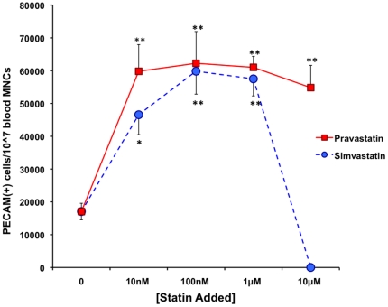
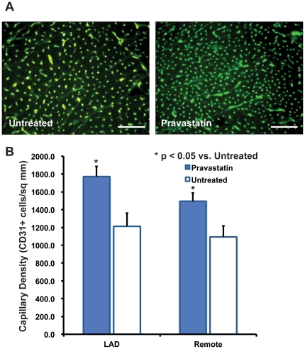
Methodology/principal findings: We determined the effects of statins on the mobilization and growth of late outgrowth endothelial progenitor cells from pigs. We also determined the density of capillaries in myocardial tissue in pigs with chronic myocardial ischemia with or without treatment with pravastatin. Pravastatin therapy resulted in greater than two-fold increase in CD31+ LOEPCs versus untreated animals. Addition of pravastatin or simvastatin to blood mononuclear cells increased the number of LOEPCs greater than three fold in culture. Finally, in animals with chronic myocardial ischemia, pravastatin increased capillary density 46%.
Conclusions: Statins promote the derivation, mobilization, and clonal growth of LOEPCs. Pravastatin therapy in vivo increases myocardial capillary density in chronically ischemic myocardium, providing an in vivo correlate for the effects of statins on LOEPC growth in vitro. Our findings provide evidence that statin therapy can increase the density of capillaries in the chronically ischemic heart.
Conflict of interest statement
Figures


Similar articles
-
Pravastatin improves function in hibernating myocardium by mobilizing CD133+ and cKit+ bone marrow progenitor cells and promoting myocytes to reenter the growth phase of the cardiac cell cycle.Circ Res. 2009 Jan 30;104(2):255-64, 10p following 264. doi: 10.1161/CIRCRESAHA.108.188730. Epub 2008 Dec 18. Circ Res. 2009. PMID: 19096024 Free PMC article.
-
Statins, HMG-CoA Reductase Inhibitors, Improve Neovascularization by Increasing the Expression Density of CXCR4 in Endothelial Progenitor Cells.PLoS One. 2015 Aug 26;10(8):e0136405. doi: 10.1371/journal.pone.0136405. eCollection 2015. PLoS One. 2015. PMID: 26309120 Free PMC article.
-
Pharmacodynamics and pharmacokinetics of the HMG-CoA reductase inhibitors. Similarities and differences.Clin Pharmacokinet. 1997 May;32(5):403-25. doi: 10.2165/00003088-199732050-00005. Clin Pharmacokinet. 1997. PMID: 9160173 Review.
-
The effects of HMG-CoA reductase inhibitor on vascular progenitor cells.J Pharmacol Sci. 2006 Aug;101(4):344-9. doi: 10.1254/jphs.fp0060102. Epub 2006 Aug 5. J Pharmacol Sci. 2006. PMID: 16891763
-
Evidence for the benefit of early intervention with pravastatin for secondary prevention of cardiovascular events.Atherosclerosis. 1999 Sep 9;147 Suppl 1:S17-21. doi: 10.1016/s0021-9150(99)00251-8. Atherosclerosis. 1999. PMID: 10575058 Review.
Cited by
-
Endothelial progenitor cells: therapeutic perspective for ischemic stroke.CNS Neurosci Ther. 2013 Feb;19(2):67-75. doi: 10.1111/cns.12040. Epub 2012 Dec 11. CNS Neurosci Ther. 2013. PMID: 23230897 Free PMC article. Review.
-
Mesenchymal stem cells from sternum: the type of heart disease, ischemic or valvular, does not influence the cell culture establishment and growth kinetics.J Transl Med. 2017 Jul 25;15(1):161. doi: 10.1186/s12967-017-1262-0. J Transl Med. 2017. PMID: 28743269 Free PMC article.
-
Single-dose local simvastatin injection improves implant fixation via increased angiogenesis and bone formation in an ovariectomized rat model.Med Sci Monit. 2015 May 18;21:1428-39. doi: 10.12659/MSM.892247. Med Sci Monit. 2015. PMID: 25982481 Free PMC article.
-
Endothelial Progenitor Cells in Diabetic Microvascular Complications: Friends or Foes?Stem Cells Int. 2016;2016:1803989. doi: 10.1155/2016/1803989. Epub 2016 May 29. Stem Cells Int. 2016. PMID: 27313624 Free PMC article. Review.
-
Comparison of Fibronectin and Collagen in Supporting the Isolation and Expansion of Endothelial Progenitor Cells from Human Adult Peripheral Blood.PLoS One. 2013 Jun 18;8(6):e66734. doi: 10.1371/journal.pone.0066734. Print 2013. PLoS One. 2013. PMID: 23824996 Free PMC article.
References
-
- Gheorghiade M, Bonow RO. Chronic heart failure in the United States. Circulation. 1998;97:282–289. - PubMed
-
- Boodhwani M, Mieno S, Voisine P, Feng J, Sodha N, et al. High-dose atorvastatin is associated with impaired myocardial angiogenesis in response to vascular endothelial growth factor in hypercholesterolemic swine. J Thorac Cardiovasc Surg. 2006;132:1299–1306. - PubMed
Publication types
MeSH terms
Substances
Grants and funding
LinkOut - more resources
Full Text Sources
Medical
