. 2011;6(9):e24957.
doi: 10.1371/journal.pone.0024957. Epub 2011 Sep 13.

Hepatitis C virus proteins activate NRF2/ARE pathway by distinct ROS-dependent and independent mechanisms in HUH7 cells

Affiliations

Hepatitis C virus proteins activate NRF2/ARE pathway by distinct ROS-dependent and independent mechanisms in HUH7 cells

Alexander V Ivanov et al. PLoS One. 2011.

Abstract

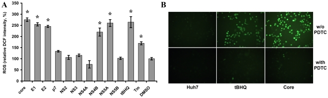
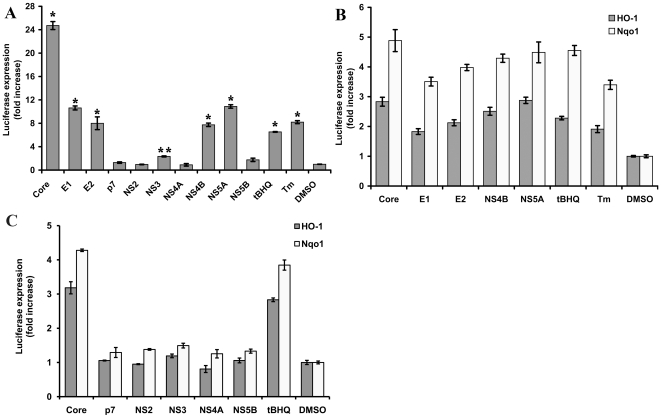
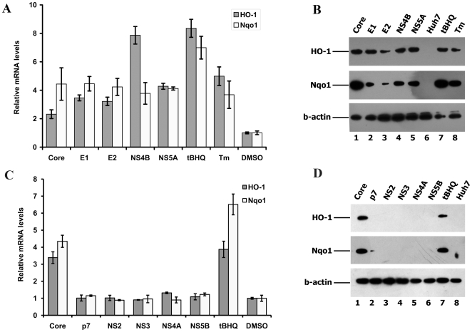
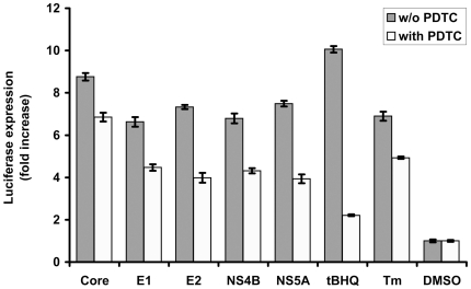
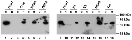
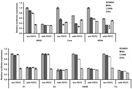
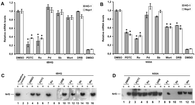
Hepatitis C virus (HCV) is a highly pathogenic human virus associated with liver fibrosis, steatosis, and cancer. In infected cells HCV induces oxidative stress. Here, we show that HCV proteins core, E1, E2, NS4B, and NS5A activate antioxidant defense Nrf2/ARE pathway via several independent mechanisms. This was demonstrated by the analysis of transient co-expression in Huh7 cells of HCV proteins and luciferase reporters. Expression, controlled by the promoters of stress-response genes or their minimal Nrf2-responsive elements, was studied using luminescence assay, RT-qPCR and/or Western-blot analysis. All five proteins induced Nrf2 activation by protein kinase C in response to accumulation of reactive oxygen species (ROS). In addition, expression of core, E1, E2, NS4B, and NS5A proteins resulted in the activation of Nrf2 in a ROS-independent manner. The effect of core and NS5A was mediated through casein kinase 2 and phosphoinositide-3 kinase, whereas those of NS4B, E1, and E2, were not mediated by either PKC, CK2, PI3K, p38, or ERK. Altogether, on the earliest stage of expression HCV proteins induced a strong up-regulation of the antioxidant defense system. These events may underlie the harmful effects of HCV-induced oxidative stress during acute stage of hepatitis C.


Conflict of interest statement

Competing Interests: The authors have declared that no competing interests exist.

Figures

Figure 1
Figure 1. Effect of transient expression of HCV proteins on accumulation of reactive oxygen species (ROS).
Transient expression of HCV core, E1, E2, NS4B, and NS5A proteins in Huh7 cells induced oxidative stress registered by ROS formation detected by fluorometry of DCF-DA-stained cells (A). Alternatively, ROS were visualized by fluorescence microscopy, as is shown for cells expressing core protein or treated with tBHQ or/and 40 µM ROS scavenger PDTC (B). Cells treated with 100 µM tBHQ (A,B) or Tm (A) are given as positive and DMSO-treated as negative controls. Error bars indicate SD. *P<0.01 versus DMSO (Tukey-Kramer test).
Figure 2
Figure 2. Impact of HCV proteins on ARE-dependent luciferase expression.
Histograms of the relative luciferase activities detected for Huh7 cells transfected with plasmids encoding HCV protein and reporter plasmids bearing minimal ARE (*P<0.01 versus DMSO; **P = 0.03 versus DMSO; Tukey-Kramer test) (A) or the full-length promoters of HO-1 or Nqo1 genes (B and C) in the presence of HCV proteins expressed transiently: HCV proteins activating (B) or not influencing (C) transcription from HO-1 and Nqo1; HCV core is given as positive control of transcription activation. Error bars indicate SD.
Figure 3
Figure 3. Up-regulation of HO-1 and Nqo1 gene expression by HCV proteins.
Histograms of the relative ho-1 and nqo1 mRNA levels in Huh7 cells expressing HCV proteins, as quantified by RT-qPCR (Error bars indicate SD) (A and C). Western-blot analysis of the expression of HO-1 and Nqo1 of samples presented in panels A (panel B) and C (panel D). In both panels, Huh7 cells treated with oxidative stress inducer (tBHQ) or ER stress inducer (Tm) are given as positive and DMSO-treated cells as negative controls, and b-actin was used as an internal control. On panels C and D HCV core was also used as a positive control.
Figure 4
Figure 4. Effect of ROS scavenger PDTC on ARE-dependent luciferase expression induced by HCV proteins.
Luciferase activity was measured in lysates of Huh7 cells expressing core, E1, E2, NS4B, or NS5A proteins, pre-treated with PDTC. The cells treated with oxidative stress inducer (tBHQ) or ER stress inducer (Tm) are given as positive and DMSO-treated cells as negative controls. Error bars indicate SD.
Figure 5
Figure 5. Influence of HCV proteins on Nrf2 subcellular localization.
Nrf2 localization was determined by separation of cytoplasmic (c) and nuclear (n) protein fractions as described in “Materials and Methods” section with subsequent detection of the transcription factor by Western blot analysis. tBHQ and Tm were used as control stress inducers.
Figure 6
Figure 6. Influence of protein kinases inhibitors on ARE-dependent luciferase expression induced by HCV proteins.
Histograms of relative luciferase activity in Huh7 cells expressing HCV core, E1, E2, NS4B, or NS5A proteins, treated with commercially available inhibitors of PI3K (wortmannin, Wo), CK2 (DRB), or PKC (Ro 31-8220, Ro). In a parallel experiment the cells were pre-treated with ROS scavenger PDTC. The control cells were treated with DMSO. Error bars indicate SD.
Figure 7
Figure 7. Effect of protein kinase inhibitors and PDTC on HO-1/Nqo1 gene expression, and Nrf2 localization.
(A,B). Histograms of relative HO-1 and Nqo1 mRNA levels in Huh7 cells treated with tBHQ (A) or expressing NS5A protein (B), treated with the inhibitors of PI3K (wortmannin, Wo), p38 (SB 239063, Sb), ERK1/2 (PD98,059, Pd), CK2 (DRB), or PKC (Ro 31-8220, Ro), or with ROS scavenger PDTC. Quantification of HO-1 and Nqo1 mRNA levels was performed by RT-qPCR using b-actin as a loading control (*P<0.01 versus DMSO, and tBHQ-treated cells or DMSO-treated cells expressing NS5A, respectively). (C,D). Western blot analysis of Nrf2 subcellular localization in the same samples used in panels (A) and (B).

Similar articles

Cited by

References

    1. Shepard CW, Finelli L, Alter MJ. Global epidemiology of hepatitis C virus infection. Lancet Infect Dis. 2005;5:558–567. - PubMed
    1. Nocente R, Ceccanti M, Bertazzoni G, Cammarota G, Silveri NG, et al. HCV infection and extrahepatic manifestations. Hepatogastroenterology. 2003;50:1149–1154. - PubMed
    1. Jacobson IM, Davis GL, El-Serag H, Negro F, Trepo C. Prevalence and challenges of liver diseases in patients with chronic hepatitis C virus infection. Clin Gastroenterol Hepatol. 2010;8:924–933; quiz e117. - PubMed
    1. National Institutes of Health. NIH Consensus Statement on Management of Hepatitis C: 2002. NIH Consens State Sci Statements. 2002;19:1–46. - PubMed
    1. Levrero M. Viral hepatitis and liver cancer: the case of hepatitis C. Oncogene. 2006;25:3834–3847. - PubMed

Publication types

MeSH terms

Substances