

Luciferase-expressing Leishmania infantum allows the monitoring of amastigote population size, in vivo, ex vivo and in vitro
- PMID: 21931877
- PMCID: PMC3172198
- DOI: 10.1371/journal.pntd.0001323
Luciferase-expressing Leishmania infantum allows the monitoring of amastigote population size, in vivo, ex vivo and in vitro
Abstract
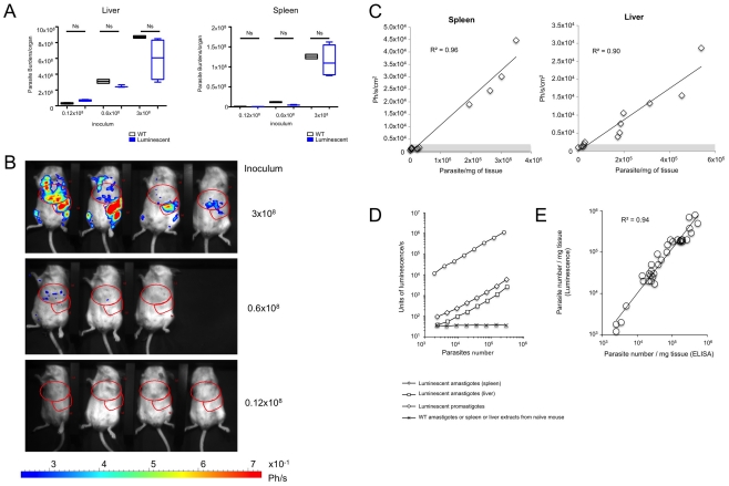
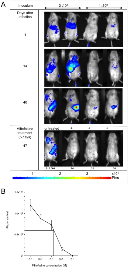
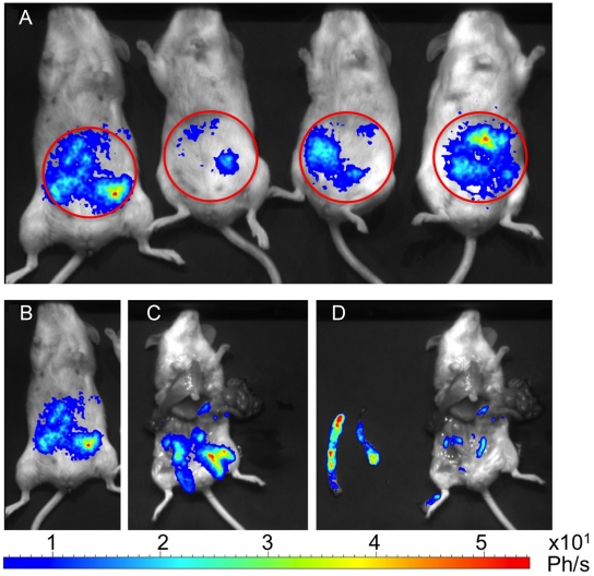
Here we engineered transgenic Leishmania infantum that express luciferase, the objectives being to more easily monitor in real time their establishment either in BALB/c mice--the liver and spleen being mainly studied-or in vitro. Whatever stationary phase L. infantum promastigotes population--wild type or engineered to express luciferase-the parasite burden was similar in the liver and the spleen at day 30 post the intravenous inoculation of BALB/c mice. Imaging of L. infantum hosting BALB/C mice provided sensitivity in the range of 20,000 to 40,000 amastigotes/mg tissue, two tissues-liver and spleen-being monitored. Once sampled and processed ex vivo for their luciferin-dependent bioluminescence the threshold sensitivity was shown to range from 1,000 to 6,000 amastigotes/mg tissue. This model further proved to be valuable for in vivo measurement of the efficiency of drugs such as miltefosine and may, therefore, additionally be used to evaluate vaccine-induced protection.
Conflict of interest statement
The authors have declared that no competing interests exist.
Figures
References
-
- Ridley RG. Evaluating diagnostics: VL. Nat Rev Micro 2007
-
- Sereno D, Cordeiro da Silva A, Mathieu-Daude F, Ouaissi A. Advances and perspectives in Leishmania cell based drug-screening procedures. Parasitol Int. 2007;56:3–7. - PubMed
-
- Chan MM, Bulinski JC, Chang KP, Fong D. A microplate assay for Leishmania amazonensis promastigotes expressing multimeric green fluorescent protein. Parasitol Res. 2003;89:266–271. - PubMed
Publication types
MeSH terms
Substances
LinkOut - more resources
Full Text Sources
Other Literature Sources
