. 2011 Mar 7;2(1):260-79.
doi: 10.3390/genes2010260.

NEK1 Facilitates Cohesin Removal during Mammalian Spermatogenesis

Affiliations

NEK1 Facilitates Cohesin Removal during Mammalian Spermatogenesis

Kim Holloway et al. Genes (Basel). .

Abstract

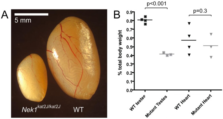
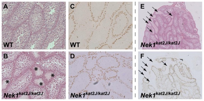
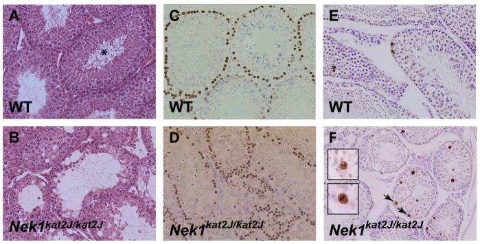
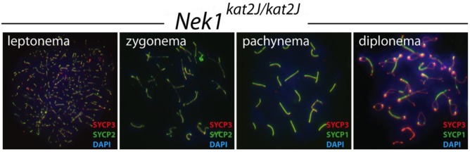
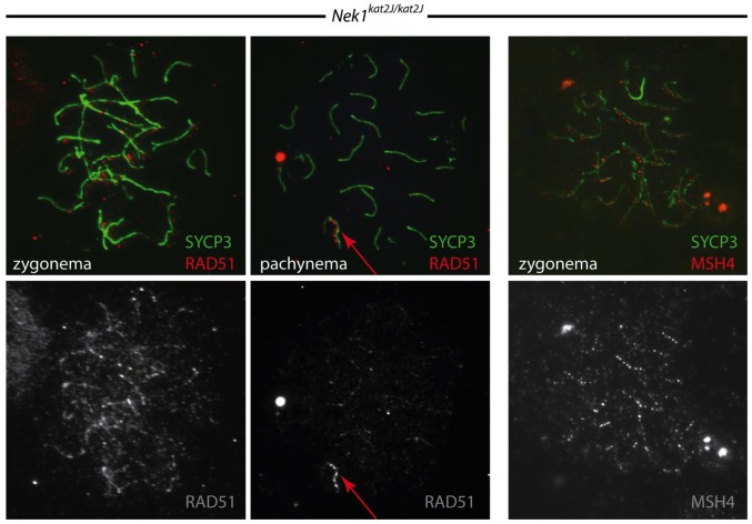
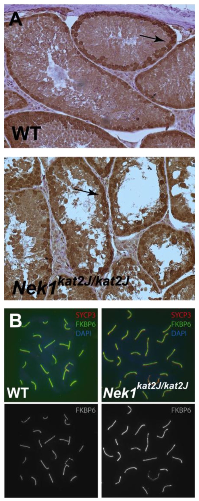
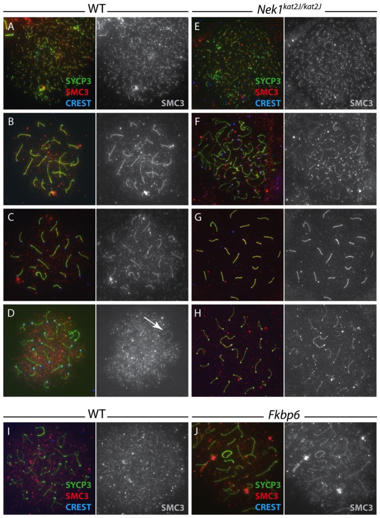
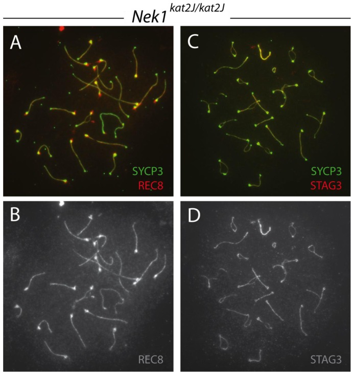
Meiosis is a highly conserved process, which is stringently regulated in all organisms, from fungi through to humans. Two major events define meiosis in eukaryotes. The first is the pairing, or synapsis, of homologous chromosomes and the second is the exchange of genetic information in a process called meiotic recombination. Synapsis is mediated by the meiosis-specific synaptonemal complex structure in combination with the cohesins that tether sister chromatids together along chromosome arms through prophase I. Previously, we identified FKBP6 as a novel component of the mammalian synaptonemal complex. Further studies demonstrated an interaction between FKBP6 and the NIMA-related kinase-1, NEK1. To further investigate the role of NEK1 in mammalian meiosis, we have examined gametogenesis in the spontaneous mutant, Nek1kat2J. Homozygous mutant animals show decreased testis size, defects in testis morphology, and in cohesin removal at late prophase I of meiosis, causing complete male infertility. Cohesin protein SMC3 remains localized to the meiotic chromosome cores at diplonema in the Nek1 mutant, and also in the related Fkbp6 mutant, while in wild type cells SMC3 is removed from the cores at the end of prophase I and becomes more diffuse throughout the DAPI stained region of the nucleus. These data implicate NEK1 as a possible kinase involved in cohesin redistribution in murine spermatocytes.


Figures

Figure 1
Figure 1
Nek1kat2J/kat2J mice show decreased testes size. (A) Photograph of both Nek1kat2J/kat2J and wild type littermate testis; (B) Wild type (black) and mutant Nek1kat2J/kat2J (gray) testes and heart sizes, shown as a percentage of total body weights.
Figure 2
Figure 2
3 week old Nek1kat2J/kat2J mice show disorganized testes morphology. 3 week old wild type (A, C) and Nek1kat2J/kat2J testes (B, D-F) were stained with H&E (A, B, E) or TRA-98 antibody (C, D, F). Empty tubules are shown by the asterisks and empty tubules corresponding to those with no germ cell staining by the arrows.
Figure 3
Figure 3
8 week old Nek1kat2J/kat2J mice show disorganized testes morphology and an increase in apoptotic cells. 8 week old wild type (A, C, E) and Nek1kat2J/kat2J (B, D, F) testes were stained with H&E (A, B), TRA-98 (C, D) or TUNEL (E, F). The sperm tails in the wild type seminiferous tubules are shown with an asterisk, and the TUNEL positive cells in metaphase by the arrows and in the insets.
Figure 4
Figure 4
Nek1kat2J/kat2J spermatocytes undergo normal synapsis. Wild type (not shown) and Nek1kat2J/kat2J testes were stained with a variety of antibodies against meiotic prophase I proteins. Shown here is staining with SYCP3 (red), either SYCP1 or SYCP2 (green) and DAPI (blue) during the first four stages of prophase I; leptonema, zygonema, pachynema and diplonema.
Figure 5
Figure 5
Nek1kat2J/kat2J spermatocytes undergo normal double-strand break (DSB) initiation and repair. Wild type (not shown) and Nek1kat2J/kat2J testes were stained with a variety of antibodies against meiotic prophase I proteins. Shown here is staining with SYCP3 (green), and either RAD51 or MHSH4 (red) during the zygotene and pachytene stages of prophase I.
Figure 6
Figure 6
NEK1 protein acts later than FKBP6 in prophase I cells. (A) Wild type and Nek1kat2J/kat2J testes sections were stained with an antibody to FKBP6 (brown stain) and counter stained with Hematoxylin; (B) Wild type and Nek1kat2J/kat2J pachytene spermatocytes stained with anti-SYCP3 (red), anti-FKBP6 (green) and DAPI (blue).
Figure 7
Figure 7
Cohesin protein SMC3 is not removed in the proper temporal manner in Nek1kat2J/kat2J spermatocytes. Wild type (A–D) and Nek1kat2J/kat2J (E–H) spermatocytes were stained with anti-SYCP3 (green), anti-SMC3 (red) and CREST autoimmune serum (blue). Cells in leptonema (A, E), zygonema (B, F), pachynema (C, G) and diplonema (D, H) were assessed for SMC3 localization (gray panels show SMC3 staining alone). The persistent XY chromosome staining of SMC3 in wild type cells is shown by the arrow. Wild type (I) and Fkbp6−/− (J) spermatocytes were also stained with anti-SYCP3 (green), anti-SMC3 (red) and CREST autoimmune serum (blue), however only the diplotene stage is shown here.
Figure 8
Figure 8
REC8 and STAG3 cohesin localization in Nek1kat2J/kat2J spermatocytes. Nek1kat2J/kat2J prophase I spermatocytes were stained with anti-SYCP3 (green) and either anti-REC8 (A, B), or anti-STAG3 (C, D) (red or gray).
Figure 9
Figure 9
Chromosomes fail to align on the metaphase spindle correctly in Nek1kat2J/kat2J spermatocytes. Nek1kat2J/kat2J and wild type testis sections were stained with H&E, and cells undergoing metaphase I imaged. Wild type cells generally show proper alignment of chromosomes on the spindle (left), whereas the majority of cells in Nek1kat2J/kat2J testis look abnormally aligned or have lagging chromosomes (arrow).

Similar articles

Cited by

References

    1. Handel M.A., Schimenti J.C. Genetics of mammalian meiosis: Regulation, dynamics and impact on fertility. Nat. Rev. Genet. 2010;11:124–136. - PubMed
    1. Hassold T., Hunt P. To err (meiotically) is human: The genesis of human aneuploidy. Nat. Rev. Genet. 2001;2:280–291. - PubMed
    1. Hassold T.J., Burrage L.C., Chan E.R., Judis L.M., Schwartz S., James S.J., Jacobs P.A., Thomas N.S. Maternal folate polymorphisms and the etiology of human nondisjunction. Am. J. Hum. Genet. 2001;69:434–439. - PMC - PubMed
    1. Hassold T., Hunt P.A., Sherman S. Trisomy in humans: Incidence, origin and etiology. Curr. Opin. Genet. Dev. 1993;3:398–403. - PubMed
    1. Hassold T., Abruzzo M., Adkins K., Griffin D., Merrill M., Millie E., Saker D., Shen J., Zaragoza M. Human aneuploidy: Incidence, origin, and etiology. Environ. Mol. Mutagen. 1996;28:167–175. - PubMed

LinkOut - more resources