

The John Charnley Award: an accurate and extremely sensitive method to separate, display, and characterize wear debris: part 2: metal and ceramic particles
- PMID: 21932105
- PMCID: PMC3254749
- DOI: 10.1007/s11999-011-2058-9
The John Charnley Award: an accurate and extremely sensitive method to separate, display, and characterize wear debris: part 2: metal and ceramic particles
Abstract
Background: Metal-on-metal and ceramic-on-ceramic bearings were introduced as alternatives to conventional polyethylene in hip arthroplasties to reduce wear. Characterization of wear particles has been particularly challenging due to the low amount and small size of wear particles. Current methods of analysis of such particles have shortcomings, including particle loss, clumping, and inaccurate morphologic and chemical characterization.
Questions/purposes: We describe a method to recover and characterize metal and ceramic particles that (1) improves particle purification, separation, and display; (2) allows for precise particle shape characterization; (3) allows accurate chemical identification; and (4) minimizes particle loss.
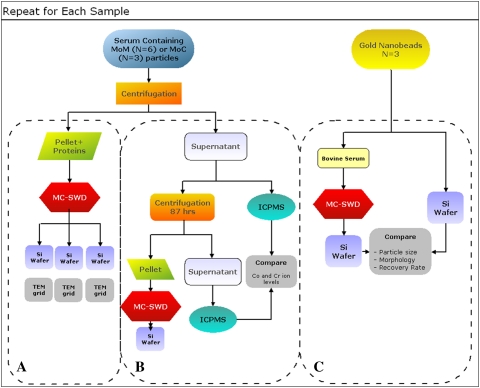
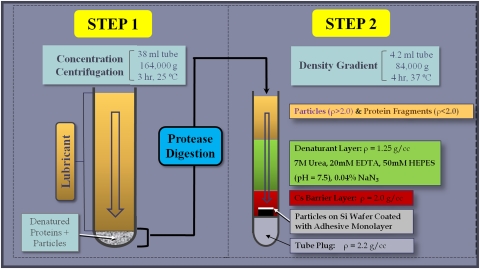
Methods: After enzymatic digestion, a single pass of ultracentrifugation cleaned and deposited particles onto silicon wafers or grids for imaging analysis. During centrifugation, particles were passed through multiple layers of denaturants and a metal-selective high-density layer that minimized protein and nucleic acid contamination. The protocol prevented aggregation, providing well-dispersed particles for chemical and morphologic analysis. We evaluated the efficacy and accuracy of this protocol by recovering gold nanobeads and metal and ceramic particles from joint simulator wear tests.
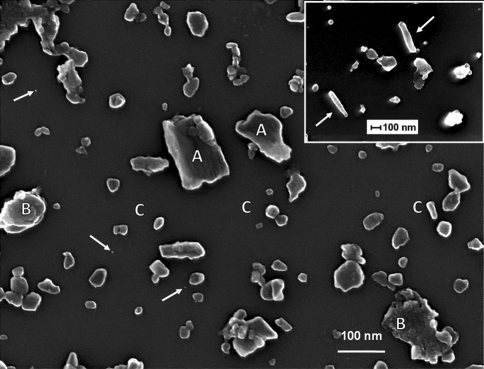
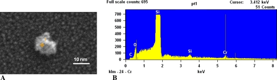
Results: The new protocol recovered particles ranging in size from nanometers to micrometers and enabled accurate morphologic and chemical characterization of individual particles.
Conclusion: Both polyethylene and metal wear debris can be simultaneously analyzed from the same sample by combining a silicon wafer display protocol for polyethylene and the metal and ceramics silicon wafer display protocol.
Clinical relevance: Accurate analysis of wear debris is essential in understanding the processes that produce debris and a key step in development of more durable and biocompatible implants.
Figures
Similar articles
-
The John Charnley Award: an accurate and sensitive method to separate, display, and characterize wear debris: part 1: polyethylene particles.Clin Orthop Relat Res. 2012 Feb;470(2):329-38. doi: 10.1007/s11999-011-2057-x. Clin Orthop Relat Res. 2012. PMID: 21997780 Free PMC article.
-
A novel method for isolation and recovery of ceramic nanoparticles and metal wear debris from serum lubricants at ultra-low wear rates.Acta Biomater. 2016 Sep 15;42:420-428. doi: 10.1016/j.actbio.2016.07.004. Epub 2016 Jul 6. Acta Biomater. 2016. PMID: 27395827
-
Scanning Electron Microscopy and Energy-Dispersive X-Ray Spectroscopy as a Valuable Tool to Investigate the Ultra-High-Molecular-Weight Polyethylene Wear Mechanisms and Debris in Hip Implants.J Arthroplasty. 2018 Jan;33(1):258-262. doi: 10.1016/j.arth.2017.07.039. Epub 2017 Aug 1. J Arthroplasty. 2018. PMID: 28844766
-
How has the introduction of new bearing surfaces altered the biological reactions to byproducts of wear and modularity?Clin Orthop Relat Res. 2014 Dec;472(12):3699-708. doi: 10.1007/s11999-014-3725-4. Clin Orthop Relat Res. 2014. PMID: 24942963 Free PMC article. Review.
-
Biological reactions to wear debris in total joint replacement.Proc Inst Mech Eng H. 2000;214(1):21-37. doi: 10.1243/0954411001535219. Proc Inst Mech Eng H. 2000. PMID: 10718048 Review.
Cited by
-
Arthroscopic surgical tools: a source of metal particles and possible joint damage.Arthroscopy. 2013 Sep;29(9):1559-65. doi: 10.1016/j.arthro.2013.05.030. Epub 2013 Jul 30. Arthroscopy. 2013. PMID: 23910000 Free PMC article.
-
Pulsed electromagnetic field (PEMF) transiently stimulates the rate of mineralization in a 3-dimensional ring culture model of osteogenesis.PLoS One. 2021 Feb 4;16(2):e0244223. doi: 10.1371/journal.pone.0244223. eCollection 2021. PLoS One. 2021. PMID: 33539401 Free PMC article.
-
Metal-on-metal hip prostheses: correlation between debris in the synovial fluid and levels of cobalt and chromium ions in the bloodstream.Int Orthop. 2014 Mar;38(3):469-75. doi: 10.1007/s00264-013-2137-5. Int Orthop. 2014. PMID: 24122048 Free PMC article.
-
Simple isolation method for the bulk isolation of wear particles from metal on metal bearing surfaces generated in a hip simulator test.J Mater Sci Mater Med. 2012 Apr;23(4):891-901. doi: 10.1007/s10856-012-4573-y. Epub 2012 Mar 6. J Mater Sci Mater Med. 2012. PMID: 22391991
-
Investigation of CoCrMo material loss in a novel bio-tribometer designed to study direct cell reaction to wear and corrosion products.Biotribology (Oxf). 2019 Jun;18:100090. doi: 10.1016/j.biotri.2019.100090. Epub 2019 Mar 27. Biotribology (Oxf). 2019. PMID: 30984811 Free PMC article.
References
-
- ASTM International. ASTM F1877-05. Standard Practice for Characterization of Particles: West Conshohocken, PA: ASTM International; 2005.
MeSH terms
Substances
LinkOut - more resources
Full Text Sources
