The fusiform response to faces: explicit versus implicit processing of emotion
- PMID: 21932258
- PMCID: PMC6870213
- DOI: 10.1002/hbm.21406
The fusiform response to faces: explicit versus implicit processing of emotion
Abstract
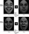
Regions of the fusiform gyrus (FG) respond preferentially to faces over other classes of visual stimuli. It remains unclear whether emotional face information modulates FG activity. In the present study, whole-head magnetoencephalography (MEG) was obtained from fifteen healthy adults who viewed emotionally expressive faces and made button responses based upon emotion (explicit condition) or age (implicit condition). Dipole source modeling produced source waveforms for left and right primary visual and left and right fusiform areas. Stronger left FG activity (M170) to fearful than happy or neutral faces was observed only in the explicit task, suggesting that directed attention to the emotional content of faces facilitates observation of M170 valence modulation. A strong association between M170 FG activity and reaction times in the explicit task provided additional evidence for a role of the fusiform gyrus in processing emotional information.
Copyright © 2011 Wiley Periodicals, Inc.
Figures


Similar articles
-
Attention inhibition of early cortical activation to fearful faces.Brain Res. 2010 Feb 8;1313:113-23. doi: 10.1016/j.brainres.2009.11.060. Epub 2009 Dec 11. Brain Res. 2010. PMID: 20004181
-
Visual processing of facial affect.Neuroreport. 2003 Oct 6;14(14):1841-5. doi: 10.1097/00001756-200310060-00017. Neuroreport. 2003. PMID: 14534432
-
Emotional attention in acquired prosopagnosia.Soc Cogn Affect Neurosci. 2009 Sep;4(3):268-77. doi: 10.1093/scan/nsp014. Epub 2009 Apr 28. Soc Cogn Affect Neurosci. 2009. PMID: 19401380 Free PMC article.
-
Fusiform gyrus responses to neutral and emotional faces in children with autism spectrum disorders: a high density ERP study.Behav Brain Res. 2013 Aug 15;251:155-62. doi: 10.1016/j.bbr.2012.10.040. Epub 2012 Nov 1. Behav Brain Res. 2013. PMID: 23124137
-
EEG-MEG evidence for early differential repetition effects for fearful, happy and neutral faces.Brain Res. 2009 Feb 13;1254:84-98. doi: 10.1016/j.brainres.2008.11.079. Epub 2008 Dec 3. Brain Res. 2009. PMID: 19073158
Cited by
-
Gut microbiota profiles are associated with different spontaneous cortical activity in healthy older people.Sci Rep. 2025 Aug 27;15(1):31590. doi: 10.1038/s41598-025-16090-6. Sci Rep. 2025. PMID: 40866493 Free PMC article.
-
Progesterone and Cerebral Function during Emotion Processing in Men and Women with Schizophrenia.Schizophr Res Treatment. 2012;2012:917901. doi: 10.1155/2012/917901. Epub 2012 Feb 15. Schizophr Res Treatment. 2012. PMID: 22966452 Free PMC article.
-
Exogenous attention to facial vs non-facial emotional visual stimuli.Soc Cogn Affect Neurosci. 2013 Oct;8(7):764-73. doi: 10.1093/scan/nss068. Epub 2012 Jun 11. Soc Cogn Affect Neurosci. 2013. PMID: 22689218 Free PMC article.
-
Maturational trajectory of fusiform gyrus neural activity when viewing faces: From 4 months to 4 years old.Front Hum Neurosci. 2022 Aug 12;16:917851. doi: 10.3389/fnhum.2022.917851. eCollection 2022. Front Hum Neurosci. 2022. PMID: 36034116 Free PMC article.
-
Attention Bias of Avoidant Individuals to Attachment Emotion Pictures.Sci Rep. 2017 Jan 27;7:41631. doi: 10.1038/srep41631. Sci Rep. 2017. PMID: 28128347 Free PMC article.
References
-
- Adolphs R, Tranel D, Damasio H, Damasio A ( 1994): Impaired recognition of emotion in facial expressions following bilateral damage to the human amygdala. Nature 372: 669–672. - PubMed
-
- Allison T, Ginter H, McCarthy G, Nobre AC, Puce A, Luby M, Spencer DD ( 1994): Face recognition in human extrastriate cortex. J Neurophysiol 71: 821–825. - PubMed
-
- Ashley V, Vuilleumier P, Swick D ( 2004): Time course and specificity of event‐related potentials to emotional expressions. Neuroreport 15: 211–216. - PubMed
-
- Baas D, Aleman A, Kahn RS ( 2004): Lateralization of amygdala activation: A systematic review of functional neuroimaging studies. Brain Res Brain Res Rev 45: 96–103. - PubMed
Publication types
MeSH terms
Grants and funding
LinkOut - more resources
Full Text Sources

