. 2011 Dec;26(12):2948-58.
doi: 10.1002/jbmr.511.

Skeletal unloading-induced insulin-like growth factor 1 (IGF-1) nonresponsiveness is not shared by platelet-derived growth factor: the selective role of integrins in IGF-1 signaling

Affiliations

Skeletal unloading-induced insulin-like growth factor 1 (IGF-1) nonresponsiveness is not shared by platelet-derived growth factor: the selective role of integrins in IGF-1 signaling

Roger K Long et al. J Bone Miner Res. 2011 Dec.

Abstract

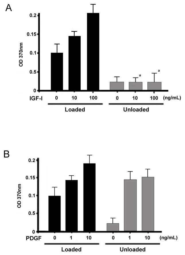
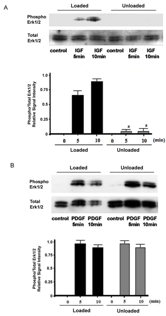
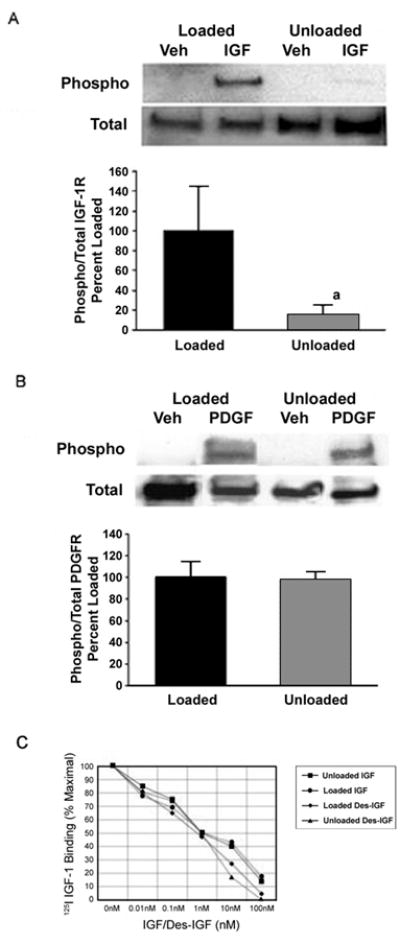
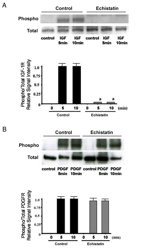
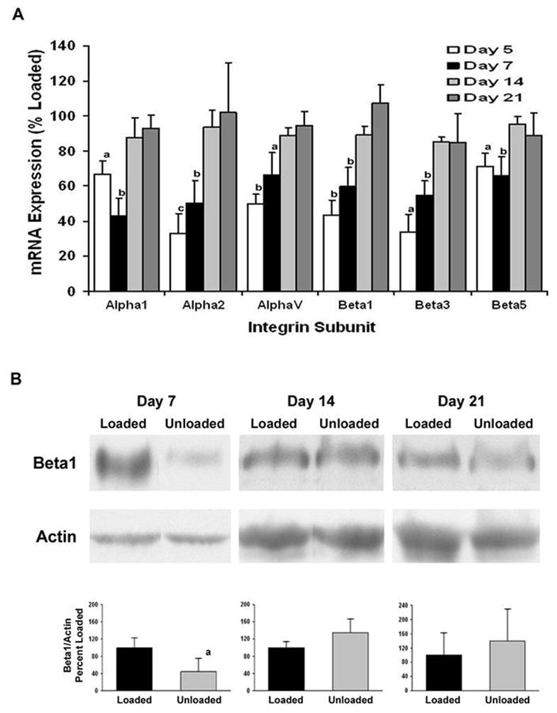
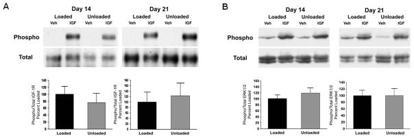
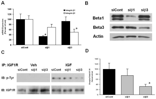
Integrin receptors bind extracellular matrix proteins, and this link between the cell membrane and the surrounding matrix may translate skeletal loading to biologic activity in osteoprogenitor cells. The interaction between integrin and growth factor receptors allows for mechanically induced regulation of growth factor signaling. Skeletal unloading leads to decreased bone formation and osteoblast proliferation that can be explained in part by a failure of insulin-like growth factor 1 (IGF-1) to activate its signaling pathways in unloaded bone. The aim of this study is to determine whether unloading-induced resistance is specific for IGF-1 or common to other skeletal growth factors, and to examine the regulatory role of integrins in IGF-1 signaling. Bone marrow osteoprogenitor (BMOp) cells were isolated from control or hindlimb suspended rats. Unloaded BMOp cells treated with IGF-1 failed to respond with increased proliferation, receptor phosphorylation, or signaling activation in the setting of intact ligand binding, whereas the platelet-derived growth factor (PDGF) response was fully intact. Pretreatment of control BMOp cells with an integrin inhibitor, echistatin, failed to disrupt PDGF signaling but blocked IGF-1 signaling. Recovery of IGF-1 signaling in unloaded BMOp cells followed the recovery of marked reduction in integrin expression induced by skeletal unloading. Selective targeting of integrin subunits with siRNA oligonucleotides revealed that integrin β1 and β3 are required for normal IGF-1 receptor phosphorylation. We conclude that integrins, in particular integrin β3, are regulators of IGF-1, but not PDGF, signaling in osteoblasts, suggesting that PDGF could be considered for investigation in prevention and/or treatment of bone loss during immobilization and other forms of skeletal unloading.


Conflict of interest statement

Disclosures:

All authors state that they have no conflicts of interest.

Figures

Figure 1
Figure 1. Skeletal unloading affects proliferation of BMOp cells in response to IGF-I and PDGF in vitro
BMOp cells from loaded and unloaded bones were incubated with IGF-I (A) or PDGF (B) for 24 hrs at day 7 in culture. During the last 4 hours, the cultures were labeled with BrdU and absorbance quantifies the BrdU incorporation and proliferation. Means ± SD, n = 3. a p < 0.05 vs. Loaded + IGF-I.
Figure 2
Figure 2. Effect of skeletal unloading on growth factor stimulated phosphorylation of ERK1/2
BMOp cells from loaded and unloaded bones were serum deprived at day 7 in culture, and then treated with IGF-I (A) or PDGF (B). Representative immunoblots illustrate skeletal unloading induced impairment of IGF-I stimulated ERK1/2 phosphorylation. Relative signal intensities of the ratio of phosphorylated to total ERK1/2 were evaluated. Means ± SD, n = 3. a p < 0.05 vs. Loaded + IGF-I.
Figure 3
Figure 3. Effect of skeletal unloading on the activation of PDGF and IGF-I receptors
BMOp cells from loaded and unloaded bones were serum deprived at day 7 in culture, and then treated with IGF-I (A) or PDGF (B). Representative immunoblots illustrate skeletal unloading induced impairment of IGF-I receptor phosphorylation. Relative signal intensities of the ratio of phosphorylated to total IGF-I or PDGF receptors were evaluated. Means ± SD, n = 3. a p < 0.05 vs. Loaded. (C) BMOp cells from loaded and unloaded bones were serum deprived at day 7 in culture, and then exposed to 25pM 125I-IGF and various doses of unlabeled IGF-I or des-IGF-I for 10 minutes. Data are means of triplicates. Error bars are not shown to simplify presentation, but in all cases enclosed an SD of less than 10% of the mean. These values are expressed as a percentage of maximal specific 125I-IGF binding that was comparable for BMOp cells isolated from loaded and unloaded bones.
Figure 4
Figure 4. Effect of echistatin on the activation of the PDGF and IGF-I receptors
BMOp cells from loaded bones were serum deprived at day 7 in culture, and then treated with IGF-I (A) or PDGF (B). Twelve hours prior, echistatin (100nM) or vehicle was added. Representative immunoblots illustrate echistatin-induced impairment of IGF-I receptor phosphorylation. Relative signal intensities of the ratio of phosphorylated to total IGF-I and PDGF receptors were evaluated. Means ± SD, n = 3. a p < 0.05 vs. Control.
Figure 5
Figure 5. Recovery of skeletal unloading induced reduction in expression of integrin subunits
The BMOp cells from loaded and unloaded bones were grown in culture. Total RNA was harvested at days 5, 7, 14, and 21 in culture and cell lysates collected at days 7, 14, and 21 in culture. A) Relative mRNA expression of the integrin subunits normalized to L19 of the unloaded BMOp cells as a percentage of that of loaded BMOp are displayed. Means ± SEM, of four independent BMOp cell pools at day 5, thirteen at day 7, six at day 14 and four at day 21. a p < 0.05, b p < 0.01, c p = 0.055. B) A representative immunoblot illustrates that expression of the β1 integrin subunit was significantly less than that in loaded BMOp cells after 7 days but not 14 or 21 days in culture. Relative signal intensities of the ratio of β1 integrin to actin were evaluated. Means ± SD, n = 3. a p < 0.05 vs. Loaded.
Figure 6
Figure 6. Recovery of IGF-I signaling in BMOp cells from unloaded bone followed longitudinally
BMOp cells from loaded and unloaded bones were grown in culture over 21 days. At days 14 and 21, cultures were serum deprived and then treated with IGF-I. Representative immunoblots illustrate recovery of ligand induced IGF-I receptor (A) and ERK1/2 (B) phosphorylation at 14 and 21 days in culture. Relative signal intensities of the ratio of phosphorylated to total IGF-I receptor and ERK1/2 were evaluated. Means ± SD, n = 4. There were no significant differences.
Figure 7
Figure 7. Effects of targeted integrin subunit knockdown on IGF-I signaling
(A) BMOp cells from loaded bones were treated with specific (siβ1 or siβ3) or non-targeting control (siCont) siRNA oligonucleotides on day 4 in culture. At day 7, cultures were serum deprived and then treated with IGF-I. RNA expression of β1 or β3 integrin subunits following targeted siRNA treatment was specifically and significantly reduced. Means ± SEM, n = 4, a p < 0.05 vs. siCont. (B) A representative immunoblot confirms the diminished expression of β1 and β3 subunit following siRNA treatment. (C) IGF-IR activation is impaired by β1 or β3 integrin subunit knockdown. (D) Relative signal intensities of the ratio of phosphorylated to total IGF-I receptor were evaluated. Means ± SEM, n = 3, a p < 0.05 vs. siCont.

Similar articles

Cited by

References

    1. Bikle DD, Halloran BP. The response of bone to unloading. J Bone Miner Metab. 1999;17(4):233–44. - PubMed
    1. Globus RK, Bikle DD, Morey-Holton E. The temporal response of bone to unloading. Endocrinology. 1986;118(2):733–42. - PubMed
    1. Halloran BP, Bikle DD, Harris J, Foskett HC, Morey-Holton E. Skeletal unloading decreases production of 1,25-dihydroxyvitamin D. Am J Physiol. 1993;264(5 Pt 1):E712–6. - PubMed
    1. Halloran BP, Bikle DD, Harris J, Tanner S, Curren T, Morey-Holton E. Regional responsiveness of the tibia to intermittent administration of parathyroid hormone as affected by skeletal unloading. J Bone Miner Res. 1997;12(7):1068–74. - PubMed
    1. Sakata T, Halloran BP, Elalieh HZ, Munson SJ, Rudner L, Venton L, Ginzinger D, Rosen CJ, Bikle DD. Skeletal unloading induces resistance to insulin-like growth factor I on bone formation. Bone. 2003;32(6):669–80. - PubMed

Publication types

MeSH terms