. 2012 Jan;55(1):267-76.
doi: 10.1002/hep.24681. Epub 2011 Nov 30.

Conjugated bile acids activate the sphingosine-1-phosphate receptor 2 in primary rodent hepatocytes

Affiliations

Conjugated bile acids activate the sphingosine-1-phosphate receptor 2 in primary rodent hepatocytes

Elaine Studer et al. Hepatology. 2012 Jan.

Abstract

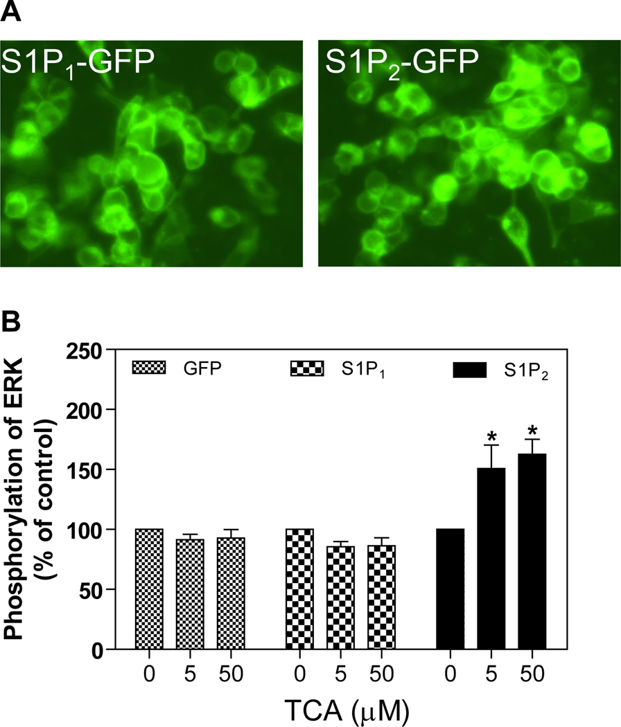
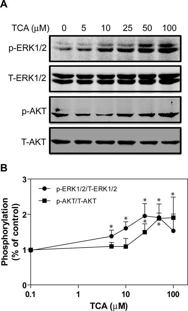
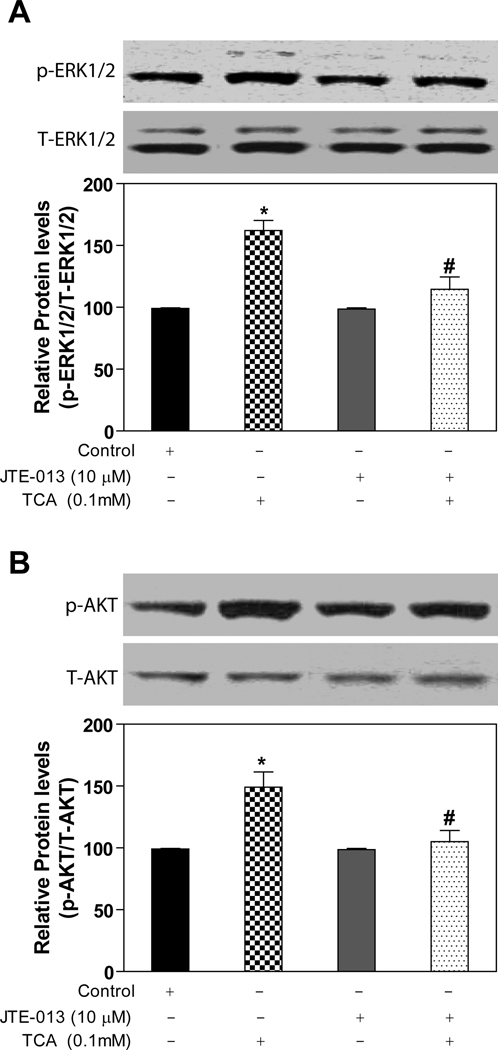
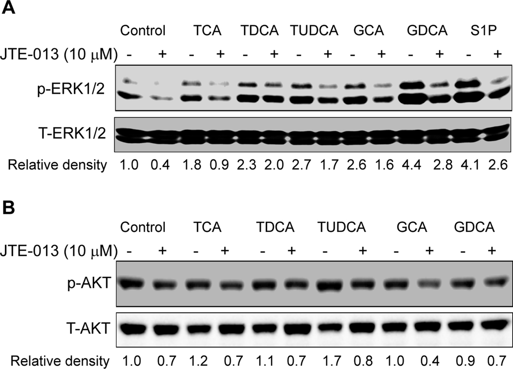
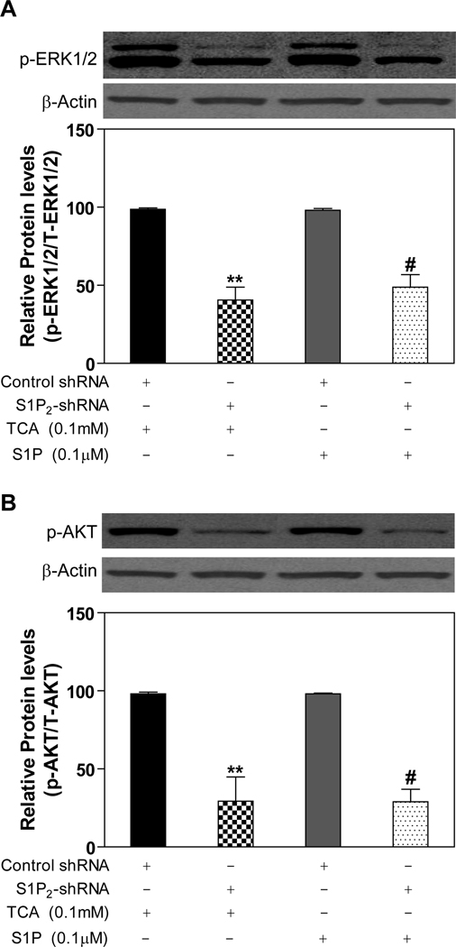
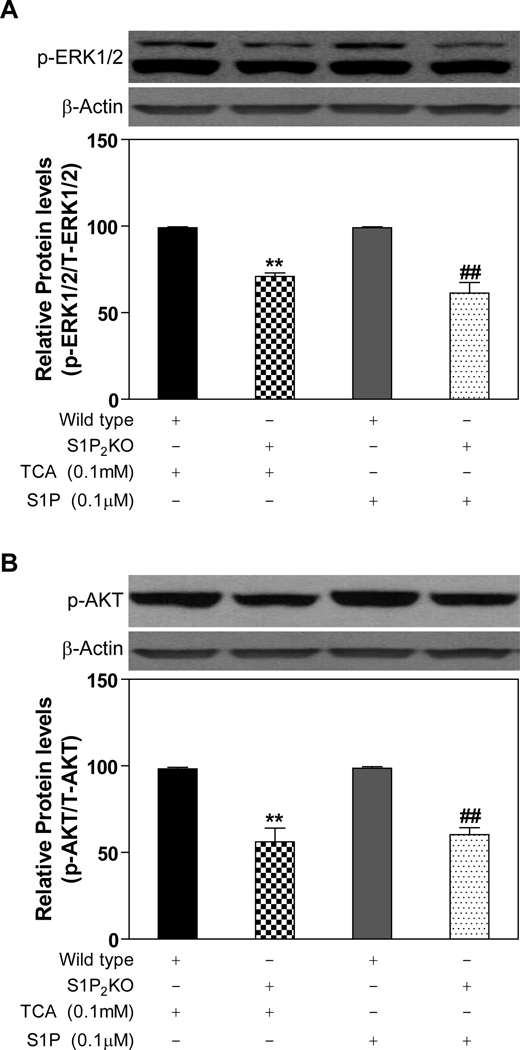
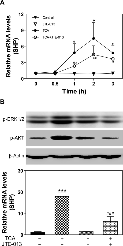
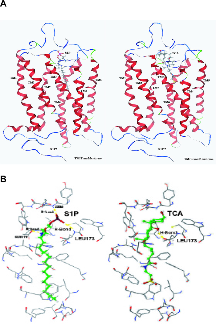
Bile acids have been shown to be important regulatory molecules for cells in the liver and gastrointestinal tract. They can activate various cell signaling pathways including extracellular regulated kinase (ERK)1/2 and protein kinase B (AKT) as well as the G-protein-coupled receptor (GPCR) membrane-type bile acid receptor (TGR5/M-BAR). Activation of the ERK1/2 and AKT signaling pathways by conjugated bile acids has been reported to be sensitive to pertussis toxin (PTX) and dominant-negative Gα(i) in primary rodent hepatocytes. However, the GPCRs responsible for activation of these pathways have not been identified. Screening GPCRs in the lipid-activated phylogenetic family (expressed in HEK293 cells) identified sphingosine-1-phosphate receptor 2 (S1P(2) ) as being activated by taurocholate (TCA). TCA, taurodeoxycholic acid (TDCA), tauroursodeoxycholic acid (TUDCA), glycocholic acid (GCA), glycodeoxycholic acid (GDCA), and S1P-induced activation of ERK1/2 and AKT were significantly inhibited by JTE-013, a S1P(2) antagonist, in primary rat hepatocytes. JTE-013 significantly inhibited hepatic ERK1/2 and AKT activation as well as short heterodimeric partner (SHP) mRNA induction by TCA in the chronic bile fistula rat. Knockdown of the expression of S1P(2) by a recombinant lentivirus encoding S1P(2) shRNA markedly inhibited the activation of ERK1/2 and AKT by TCA and S1P in rat primary hepatocytes. Primary hepatocytes prepared from S1P(2) knock out (S1P(2) (-/-) ) mice were significantly blunted in the activation of the ERK1/2 and AKT pathways by TCA. Structural modeling of the S1P receptors indicated that only S1P(2) can accommodate TCA binding. In summary, all these data support the hypothesis that conjugated bile acids activate the ERK1/2 and AKT signaling pathways primarily through S1P(2) in primary rodent hepatocytes.


Figures

Fig. 1
Fig. 1. Activation by TCA of S1P2 expressed in HEK293 cells
A. Representative images of the GFP-tagged S1P1 and S1P2 stably expressed in HEK293 cells. B. Effect of TCA on ERK1/2 activation. HEK293 cells expressing GFP-tagged S1P1 or S1P2 or GFP only were treated with TCA (5 or 50 µM) or vehicle control for 20 minutes at 37°C. The protein levels of phosphor-ERK1/2 and total-ERK1/2 were determined by Western blot analysis. *p<0.05 compared to vehicle control, n=3.
Fig. 2
Fig. 2. Saturation curves of TCA-induced ERK1/2 and AKT activation in primary rat hepatocytes
Primary rat hepatocytes were treated with different amounts of TCA (0, 5, 10, 25, 50 and 100 µM) for 30 minutes. The cells were harvested to prepare total cell lysates. The protein levels of phosphorylated ERK1/2 (p-ERK1/2) and AKT (p-AKT) were determined by Western blot analysis and normalized with total ERK1/2 (T-ERK1/2) and total AKT (T-AKT) as described in Materials and Methods. A. Representative images of Western blots of p-ERK1/2, T-ERK1/2, p-AKT, and T-AKT. B. Saturation curves of TCA-induced ERK1/2 and AKT activation. *p<0.05 compared to control, n=4.
Fig. 3
Fig. 3. Effect of JTE-013 on TCA-induced activation of ERK1/2 and AKT in primary rat hepatocytes
Cells were pre-incubated with JTE-013 (10 µM) for 30 minutes and then treated with 100 µM TCA for 20 minutes at 37°C. The protein levels of phosphorylated ERK1/2 (p-ERK1/2) and AKT (p-AKT) were determined by Western blot analysis and normalized with total ERK1/2 (T-ERK1/2) and total AKT (T-AKT) as described in Materials and Methods. *p<0.05 compared to vehicle control, #p<0.05 compared to TCA group, n=3.
Fig. 4
Fig. 4. Effect of JTE-013 on conjugated bile acid-induced activation of ERK1/2 and AKT in primary rat hepatocytes
Cells were pre-incubated with JTE-013 (10 µM) for 30 minutes and then treated with individual conjugated bile acids (TCA, TDCA, TUDCA, GCA, GDCA, 50 µM) or S1P (100 nM) for 30 minutes at 37°C. The protein levels of phosphorylated ERK1/2 (p-ERK1/2) and AKT (p-AKT) were determined by Western blot analysis and normalized with total ERK1/2 (T-ERK1/2) and total AKT (T-AKT) as described in Materials and Methods. The representative Western blot images are shown. The relative density was determined by Image J software.
Fig. 5
Fig. 5. Effect of S1P2 shRNA on activation of pAKT and pERK by TCA and S1P
Three hours after plating, rat primary hepatocytes were transduced with appropriate lentivirus encoding scramble control shRNA sequence or specific shRNA to S1P2 for 40 h and then treated with TCA (100 µM) or S1P (100 nM) for 20 minutes. The protein levels of phosphorylated ERK1/2 (p-ERK1/2) and AKT (p-AKT) were determined by Western blot analysis and normalized with actin as described in Materials and Methods. **p<0.01 compared to control shRNA group treated with TCA, n=3; #p<0.05 compared to control shRNA group treated with S1P, n=3.
Fig. 6
Fig. 6. Activation of the ERK1/2 and AKT pathways in primary mouse hepatocytes prepared from S1P2−/− and control mice
Primary mouse hepatocytes were prepared from the S1P2−/− mouse and wild type littermate and treated with TCA (100 µM) for 20 minutes. The protein levels of phosphorylated ERK1/2 (p-ERK1/2) and AKT (p-AKT) were determined by Western blot analysis and normalized with actin as described in Materials and Methods. **p<0.01 compared to wild type treated with TCA, n=3; ##p<0.01 compared to wild type treated with S1P, n=3.
Fig. 7
Fig. 7. Effect of JTE-013 on TCA-induced SHP expression
A. Primary rat hepatocytes were pre-incubated with JTE-013 (10 µM) for 30 minutes and then treated with TCA (50 µM) for 0.5, 1, 2, and 3 h at 37°C. The mRNA levels of SHP were determined by real-time RT-PCR as described in Materials and Methods. *p<0.05 compared to control group, n=3; #p<0.05 compared to TCA group, n=3. B. Bile fistula rats were i.p injected with JTE-013 (20 mg/kg) 2 h before TCA infusion. Rat livers were harvested after infusion with TCA for 3h. The protein levels of p-ERK1/2 and p-AKT were determined by Western blot analysis and normalized with actin and the mRNA levels of SHP were determined by real-time RT-PCR as described in Materials and Methods. Representative Western blot images are shown. ***p<0.001 compared to vehicle control with TCA, n=3; ###p<0.001 compared to TCA-treated group, n=3.
Fig. 8
Fig. 8. Modeling of S1P and TCA in the binding site of S1P2
(A) Model S1P2 with bound S1P or TCA. (B) Specific amino acid residues interacting with bound S1P and TCA.

Similar articles

Cited by

References

    1. Scotti E, Gilardi F, Godio C, Gers E, Krneta J, Mitro N, De Fabiani E, et al. Bile acids and their signaling pathways: eclectic regulators of diverse cellular functions. Cell Mol Life Sci. 2007;64:2477–2491. - PMC - PubMed
    1. Thomas C, Pellicciari R, Pruzanski M, Auwerx J, Schoonjans K. Targeting bile-acid signalling for metabolic diseases. Nat Rev Drug Discov. 2008;7:678–693. - PubMed
    1. Nguyen A, Bouscarel B. Bile acids and signal transduction: role in glucose homeostasis. Cell Signal. 2008;20:2180–2197. - PubMed
    1. Lefebvre P, Cariou B, Lien F, Kuipers F, Staels B. Role of bile acids and bile acid receptors in metabolic regulation. Physiol Rev. 2009;89:147–191. - PubMed
    1. Zhang Y, Edwards PA. FXR signaling in metabolic disease. FEBS Lett. 2008;582:10–18. - PubMed

Publication types

MeSH terms