. 2012 Feb;55(2):354-63.
doi: 10.1002/hep.24686. Epub 2011 Dec 16.

CD59 incorporation protects hepatitis C virus against complement-mediated destruction

Affiliations

CD59 incorporation protects hepatitis C virus against complement-mediated destruction

Tohti Amet et al. Hepatology. 2012 Feb.

Abstract

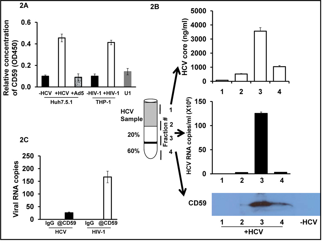
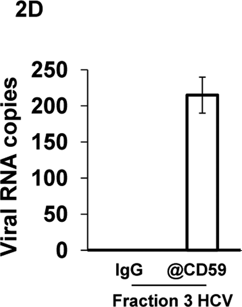
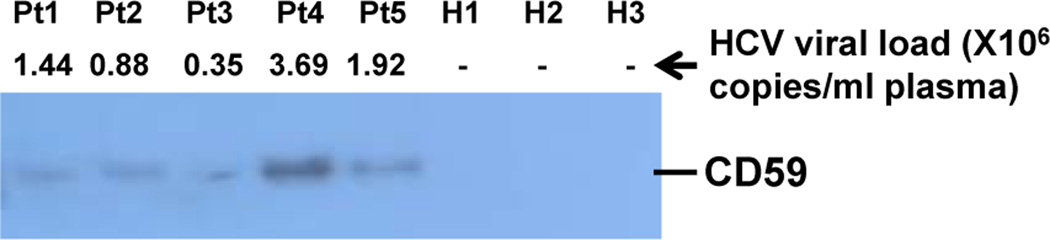
Several enveloped viruses including human immunodeficiency virus type 1 (HIV-1), cytomegalovirus (CMV), herpes simplex virus 1 (HSV-1), Ebola virus, vaccinia virus, and influenza virus have been found to incorporate host regulators of complement activation (RCA) into their viral envelopes and, as a result, escape antibody-dependent complement-mediated lysis (ADCML). Hepatitis C virus (HCV) is an enveloped virus of the family Flaviviridae and incorporates more than 10 host lipoproteins. Patients chronically infected with HCV develop high-titer and crossreactive neutralizing antibodies (nAbs), yet fail to clear the virus, raising the possibility that HCV may also use the similar strategy of RCA incorporation to escape ADCML. The current study was therefore undertaken to determine whether HCV virions incorporate biologically functional CD59, a key member of RCA. Our experiments provided several lines of evidence demonstrating that CD59 was associated with the external membrane of HCV particles derived from either Huh7.5.1 cells or plasma samples from HCV-infected patients. First, HCV particles were captured by CD59-specific Abs. Second, CD59 was detected in purified HCV particles by immunoblot analysis and in the cell-free supernatant from HCV-infected Huh7.5.1 cells, but not from uninfected or adenovirus serotype 5 (Ad5) (a nonenveloped cytolytic virus)-infected Huh7.5.1 cells by enzyme-linked immunosorbent assay. Last, abrogation of CD59 function with its blockers increased the sensitivity of HCV virions to ADCML, resulting in a significant reduction of HCV infectivity. Additionally, direct addition of CD59 blockers into plasma samples from HCV-infected patients increased autologous virolysis.

Conclusion: Our study, for the first time, demonstrates that CD59 is incorporated into both cell line-derived and plasma primary HCV virions at levels that protect against ADCML. This is also the first report to show that direct addition of RCA blockers into plasma from HCV-infected patients renders endogenous plasma virions sensitive to ADCML.


Figures

Fig. 1
Fig. 1. Intracellular and surface CD59 expression by human hepatocytes
(A) FACS analysis of CD59 surface expression on human hepatocytes (PHHs and Huh7.5.1 cells). (B) PHHs and Huh7.5.1 cells were treated with PI-PLC, and followed by surface and intracellular staining of CD59. (C) Cells were treated (Intra) or untreated (Total) with PI-PLC, and followed by protein extraction for Western blot analysis of CD59 expression. Histograms in red and blue lines show control (isotype) and CD59 staining, respectively. Intra: intracellular CD59; Total: total CD59; Huh7: Huh7.5.1 cells; PHHs: primary human hepatocytes.
Fig. 2
Fig. 2. Identification of CD59 association with exterior of HCV virions
(A) ELISA detection of CD59 in cell-free supernatant from: uninfected Huh7.5.1 cells (−HCV); Huh7.5.1 cells infected with 1 MOI of HCV (+HCV) for 5 days; or Huh7.5.1 cells infected with 2 MOI of Ad5 (+Ad5) for 3 days; uninfected THP-1 cells (−HIV-1); THP-1 cells infected with HIV-1 (+HIV-1); and activated U1 cells. (B) Viral particles collected from fractions 1 to 4 were subjected to ELISA, qPCR and Western blot to detect HCV core protein, HCV RNA copies and CD59 expression, respectively. Supernatant from uninfected (−HCV) Huh7.5.1 cells were parallelly tested. (C) Immunocapture of HCV and HIV-1 from cell-free culture supernatant of infected cells by anti-CD59 pAbs. (D) Immunocapture of HCV from the purified HCV virus fraction 3 by anti-CD59 pAbs. Viral RNA copies were quantified by qPCR. All experiments were repeated twice with similar results. @CD59: rabbit anti-human CD59 pAbs.
Fig. 3
Fig. 3. Identification of CD59 association with plasma primary HCV virions
Five ml of plasma from each of 5 HCV-infected individuals (Pt1 to Pt5, Table 1) and 3 healthy donors (H1 to H3) was subjected to virus purification for measuring CD59 by Western blot. Pt1 to Pt 5: HCV-infected patients #1 to #5; H1 to H3: healthy donors #1 to #3.
Fig. 4
Fig. 4. Sensitivity of cell line-derived HCV to complement-mediated virolysis
Cell-free supernatant from HCV (JFH-1)-infected Huh7.5.1 cells was preincubated with IgG, BRIC229 or rILYd4 at 1.25 – 20 µg/ml, followed by addition of anti-HCV E2 pAbs plus complement or heat-inactivated complement. Viral preparations were also treated with PBS and Triton X-100 to determine 0 and 100% lysis, respectively. Virolysis of HCV was quantified by measuring HCV core release using the QuickTiter™ HCV Core ELISA Kit. (A) Dose-dependent analysis of HCV core released from lysed HCV virions in response to BRIC229 or rILYd4 treatment in the presence (solid lines with solid symbols) or absence (dot lines with open symbols) of potent complement plus anti-HCV E2 pAbs. (B) Effects of anti-HCV E2 pAbs on complement-mediated virolysis of HCV virions in response to IgG, BRIC229 or rILYd4 treatment at 20 µg/ml. Complement was added in all conditions with either anti-HCV E2 pAbs (+anti-HCV E2) or anti-HIV-1 gp120/160 pAbs (-anti-HCV E2). (C) Statistical analysis of HCV lysis by ADCML from the experiments described in Fig. 4B. (D) Reduction assay of HCV infectivity. Following the virolysis assays, a reduction assay of viral infectivity was performed by inoculating fresh Huh7.5.1 cells with the conditioned solutions from experiments depicted in the Fig. 4B (+anti-HCV E2). The number of infectious HCV virions remaining in the virolysis solution samples of the ADCML experiments was quantitated by a HCV focus-forming assay. HCV foci were visualized by an immunofluorescence microscopic analysis. Each value represents the mean ± SD of triplicate determinations. ** indicates p<0.01 and ≈ indicates p>0.05, respectively, by paired Student’s t test.
Fig. 5
Fig. 5. Direct addition of CD59 blockers enhanced virolysis of primary HCV virions
(A) Virolysis of primary HCV virions. Plasma from HCV-infected patients was directly treated with PBS, IgG (20 µg/ml, BRIC229 (20 µg/ml), rILYd4 (20 µg/ml), or Triton X-100 to trigger autologous ADCML. HCV core released from lysed HCV virions was detected by ELISA. HCV virions treated with Triton X-100 and PBS were used as 100% and blank of virolysis, respectively. (B) The percentage of virolysis was calculated as follows: (core released by CD59 blocker - core released by PBS)/(core released by Triton X-100 - core released by PBS) X 100%. (C) Virolysis data pooled from all HCV plasma samples. Horizontal bars represent mean of virolysis values. Each value represents the mean ± SD of triplicate determinations. The experiments were repeated twice with similar results. **, *, and ≈ indicate p<0.01, p<0.05, and p>0.05, respectively, by the paired Student’s t test.

Similar articles

Cited by

References

    1. Ricklin D, Hajishengallis G, Yang K, Lambris JD. Complement: a key system for immune surveillance and homeostasis. Nat Immunol. 2010;11:785–797. - PMC - PubMed
    1. Saifuddin M, et al. Role of virion-associated glycosylphosphatidylinositol-linked proteins CD55 and CD59 in complement resistance of cell line-derived and primary isolates of HIV-1. J Exp Med. 1995;182:501–509. - PMC - PubMed
    1. Nguyen DH, Hildreth JE. Evidence for budding of human immunodeficiency virus type 1 selectively from glycolipid-enriched membrane lipid rafts. J Virol. 2000;74:3264–3272. - PMC - PubMed
    1. Vanderplasschen A, Mathew E, Hollinshead M, Sim RB, Smith GL. Extracellular enveloped vaccinia virus is resistant to complement because of incorporation of host complement control proteins into its envelope. Proc Natl Acad Sci U S A. 1998;95:7544–7549. - PMC - PubMed
    1. Spear GT, et al. Host cell-derived complement control proteins CD55 and CD59 are incorporated into the virions of two unrelated enveloped viruses. Human T cell leukemia/lymphoma virus type I (HTLV-I) and human cytomegalovirus (HCMV) J Immunol. 1995;155:4376–4381. - PubMed

Publication types